Diffrential diagnosis of common teeth complaints
•Download as PPTX, PDF•
5 likes•327 views

This document provides an overview of common dental complaints and conditions seen in a dental clinic. It discusses developmental disturbances of teeth, dental pain, halitosis, gingival enlargement, gingival recession, dental deposits, stains and discoloration, and dental caries. For each condition, it describes the etiology, signs and symptoms, classification where relevant, and examples. The document aims to comprehensively cover a wide range of oral health issues that patients may present with.

Recommended
More Related Content
What's hot
What's hot (20)
Similar to Diffrential diagnosis of common teeth complaints
Similar to Diffrential diagnosis of common teeth complaints (20)
More from Shraddha Joshi
More from Shraddha Joshi (20)
Recently uploaded
Recently uploaded (20)
Diffrential diagnosis of common teeth complaints
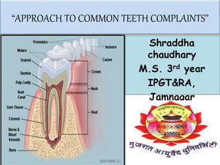
- 1. “APPROACH TO COMMON TEETH COMPLAINTS” Shraddha chaudhary M.S. 3rd year IPGT&RA, Jamnagar
- 2. Common chief complaints we get in dental OPD….. Developmental disturbances Pain Halitosis Gingival enlargement Gingival recession Dental deposits Dental stains and discoloration Dental caries
- 3. Developmental Disturbances Developmental alteration in size of teeth: Microdontia: When teeth are physically smaller than usual. Maxillary lateral incisor or maxillary 3rd molar are commonly involved. Macrodontia: One or more teeth are physically larger than normal. Commonly in mandibular canines.
- 4. Developmental alteration in number of teeth: Anodontia: Congenital absence of all teeth. Partial anodontia / Hypodontia : Congential absence of one or more teeth. Oligodontia: Congenital missing 6 or more teeth. Hyperdontia/ Superneumrary teeth: Frequently in permanent dentition. Commonly in maxillary incisor. Mesiodens:- Between two central incisors. Distomolar:-Superneumrary 4th molar. These terms cannot be applied to teeth that have developed but have failed to errupt. Enviroment also influences the final outcome or in some cases may be responsible completely for the lack of tooth formation. (eg. Trauma, infection, radiation, chemotherapy,endocrine disturbances, etc.)
- 6. Developmental alteration in shape of teeth: Double teeth:- Refers to two conjoined teeth. This term should not be applied to teeth joined only through their root cementum. Fusion:- Refers to union of two saperate tooth germs during their formation. They may be joined only by enamel or dentin. Gemination:- refers to a double teeth originated from one tooth bud.
- 7. DENTAL PAIN Unpleasent sensation felt by the patient in relation to odontological cause. Causes:- Dentine sensitivity- Caries , Trauma. Pulpitis- Reversible, Irreversible. Abcess- Periapical, Periodontal. Pericoronitis (An inflammation of the oerculum around the crown of partially erupted tooth, usually 3rd molar) Dry Socket :- Faliure of formation or maintenance of blood clot in extraction socket leaving bare bone.
- 8. HALITOSIS It is a general term used to define an unpleasant odour from the breath. It should not be confused with general temporary oral odour caused by intake of certain foods, tobacco or medications. Classification:- Genuine halitosis- Physiologic and pathologic halitosis Pseudo halitosis Halitophobia
- 9. GENUINE HALITOSIS Physiological Halitosis:- Morning breath odour, tobacco, smoking, certain foods and medications. Pathological Halitosis:- Intraoral origin:- Poor OH, Dental caries, Periodontal disease, Dry socket, other oral infections,tongue coating and Oral Ca. Extraoral origin:- GIT diseases, infection or malignancy in respiratory tract, chronic sinusitis and tonsillitis, etc. Some systemic conditions DM (Acetone, sweet fruity); Renal Faliure (Ammonia); Liver Faliure (Fresh cadaver),etc. Tongue coatings include desquamated epithelial cells, food debris, bacteria and salivary protiens and provide an ideal enviroment for the genration of VSCs (Volatile sulphur compound) and other compounds that contribute malodour.
- 10. Pseudo Halitosis:- Apparently healthy individuals. When an obvious breath malodour cannot be perceived, but the patients is convinced that he or she suffers from it. Halitophobia:- Exaggerated fear of having halitosis. Also known as Delusional halitosis. If the patient still believes that there is a bad breath after treatment of genuine halitosis or diagnosis of pseudo halitosis, is recognized psychiatric condition. Cont …
- 11. GINGIVAL ENLARGEMENT Increase in size of gingiva or gingival overgrowth. On the basis of location and distribution:- Localized, Generalised, Marginal. Inflammatory enlargement:- Acute and Chronic. Drug induced (Anticonvulsants, Immunosuppressants, Ca channel blocker) Enlargement associated with systemic diseases:- a. Conditioned enlargement- Pregnancy, Vit. C, Plasma cell gingivitis. b. Systemic diseases causing gingival enlargement-Leukameia, Sarcodosis,etc. Neoplastic enlargement:- Benign and malignant tumors.
- 12. 0-No signs of gingival enlargement 3-Covers 3 quarters or most of the crown. 2-Involves papilla and marginal gingiva 1-Enlargement confined to interdental paillae Scoring of gingival enlargement
- 13. LOCALISED GENERALIZED Drug induced Plasma cell gingivitis
- 14. Leukemic gingival enlargement Severe deficiency of Vit. C Cont …
- 15. It is a exposure of root surface by an apical shift in the position of gingiva. The recession is determined by actual position of the gingiva not by its apparent position. Can be:- Localised and Generalised. Gingival Recession
- 17. Dental Depositions Soft Deposits Hard Deposits Aquired Pellicle Microbial Plaque Materia Alba Calculus Stains
- 18. Aquired Pellicle:- Following tooth eruption or a dental prophylaxis, a thin, saliva- deried layer called aquired pellicle,covers the tooth surface. Dental Plaque:- Resulting from sequential colonization and growth of microorganisms on the surfaces of teeth and restorations. Materia Alba:- Refers to soft accumulations of bacteria and tissue cells that lack the organized structure of dental plaque.
- 19. Calculus:- It is a hard concretion that forms on teeth or dental prostheses through calcification of bacterial plaque. Classified as:-
- 20. Healthy tooth:- Smooth and shiny surface (Pearl white to pale yellow). Discoloration can be:- • Stains limited to external surfaces. • Stains incoperated within tooth structure. • Stains present due to formation of calculus and soft deposits. External stains can be removed by scaling or polishing. Stains occurring within tooth substances are hard to remove. Dental Stains and Discoloration
- 21. Etiology External Source Internal Source Poor oral hygiene Plaque Trauma Food pigmments Smoking Tobacco and Pan Chlorohexidine Stannous fluroide Metallic Salts Chromogenic bacteria Drugs Developmental defect Non vital tooth Metals Internal Resorption Flurosis Dental Materials
- 22. Yellow Stains:- Dull light yellow appearance where personal OH is neglected. It occurs from food pigments and improper brushing techniques. Green Stains:- These stains are generally embedded in bacterial plaque extending from facial to proximal surfaces of tooth. Facial 1/3rd of maxillary anterior teeth are involved. Primarily in early childhood due to improper brushing and plaque accumulation. D/D:- Drugs, Industrial metallic dust.
- 23. Black Stains:- Highly retentive Calcalus. More common in childhood, females and found in clean mouth. Commonly cervical region is involved. Tobbaco stains:- Can be in narrow crest, diffused, from cervical 3rd to middle 3rd of tooth. May cover fissure, pits, lingual surface. May penetrate enamel.
- 24. Metallic Stains:- Occurs due to- Metal present in drugs (May affect all surfaces of tooth) (consumption of iron- black stain or brown stain). Metals from metal industry (Inhalation of metallic dust). (Copper- Bluish green stains; Nickle- Green stain; Cadmium- Yellow Brown stains). Stannous fluroide:- Formed after the prolonged use of fluroide product results in formation of brownish stain. This results from increased level of fluroides in drinking water during the period of teeth development. (with level >1.2 ppm)
- 25. Chlorohexidine mouth rinses:- Due to repeated use. Brownish stains. Betel Leaf Stains:- Brownish color stains. Resemble to subgingival calcalus.
- 26. Tetracycline Stain:- This antibiotic has the affinity towards the mineralized tissue and gets absorbed by hard tissue of body. It can easily cross the placenta and enters the fetal circulation. So, when expecting mother is administered this drug during 3rd trimester it gets deposited into forming bones and teeth in fetus. Can also occur when the drug is administered to the child in infancy and early childhood. It may be generalized or localized. Color- Light green to dark yellow to grey brown.
- 28. Pulpless Tooth/ Non-Vital Tooth:- Not all pulpless tooth shows discoloration; the one which discolor shows black color. Pulp chamber is non complaiant chamber Decomposed Hb stagnantes within the pulp chamber Seeps into dentinal tubules of tooth Results into intrinsic discoloration.
- 29. Stains from systemic disease:- Porphyria:- It is a group of disorder caused by an over accumulation of porphyrin that helps make Haemoglobin. Occur in decidious or permanent tooth. Appear pink or lavender in colour. Erythroblastosis fetalis:- It is a grave haemolytic anemia results from development of Rh –ve antibody in response to Rh –ve factor in the fetal blood . Colour:- Bluish, blackish or greenish in colour.
- 30. Others:- (Amelogenesis Imperfecta, def. of vit. C, D) Opaque white patches which may be stained in the cases of hypo mineralized enamel. Pitted and grooved tooth surface in the cases of hypoplastic enamel. Cont …
- 31. Aging:- With the age there will be a change in the color of teeth due to enamel will be thin and dentin will be thicker due to deposition of secondary dentin so the tooth will appear darker, also there will be staining of teeth and this will depend on individual variations of consumption of tea, coffee, beverages,, alcohol, smoking. Colour:- More yellowish, darker.
- 32. DENTAL CARIES Dental caries is a multifactorial microbial infections disease characterized by demineralization of the inorganic and destruction of the organic substance of the tooth. It is a progressive bacterial damage to teeth exposed to saliva. It is one of the most major causes of all disease and major cause of tooth loss. It has ultimate effect to breakdown enamel and dentin and open a path for bacteria to reach pulp. Consequences- Inflammation of pulp and periapical tissues.
- 33. S &S:- Caries initially involves only the enamel and produces no symptoms. Cavity that invades the dentin causes pain, first when hot, cold, sweet foods or beverages contact the involved tooth and later with chewing or percussion. Pain can be intense and persistent when the pulp is severely involved. Etiology :-
- 34. G.V. Black Classification of dental caries Caries affecting proximal surfaces of central incisor, lateral incisor and cuspids without involving the incisal angles. Caries affecting gingival 1/3rd of facial or lingual surfaces of anterior or posterior teeth. Caries affecting pits and fissures on occlusal 3rd of molars and premolars; occlusal 2/3rd of molars and premolars; lingual part of ant. teeth Caries affecting proximal including incisal angles of anterior teeth. Caries affecting proximal surfaces of molars and premolars. Caries affecting cusp tips of molars, premolars and cuspids