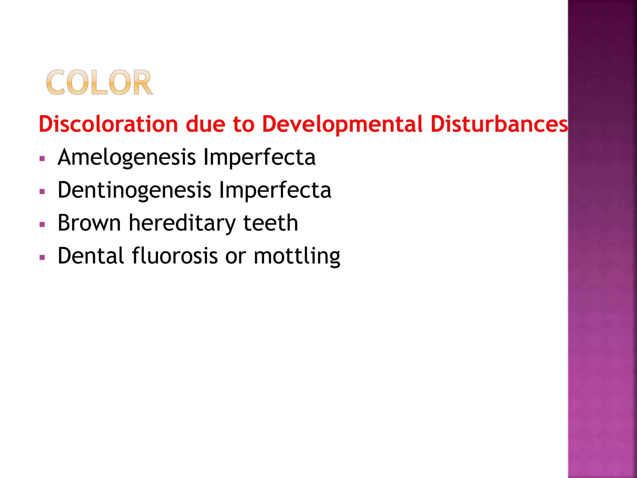
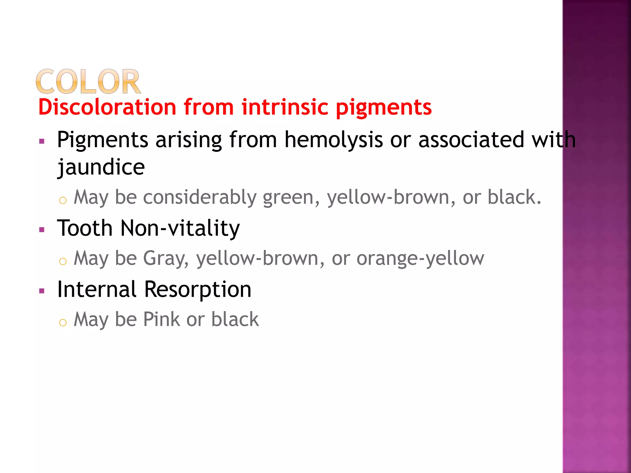
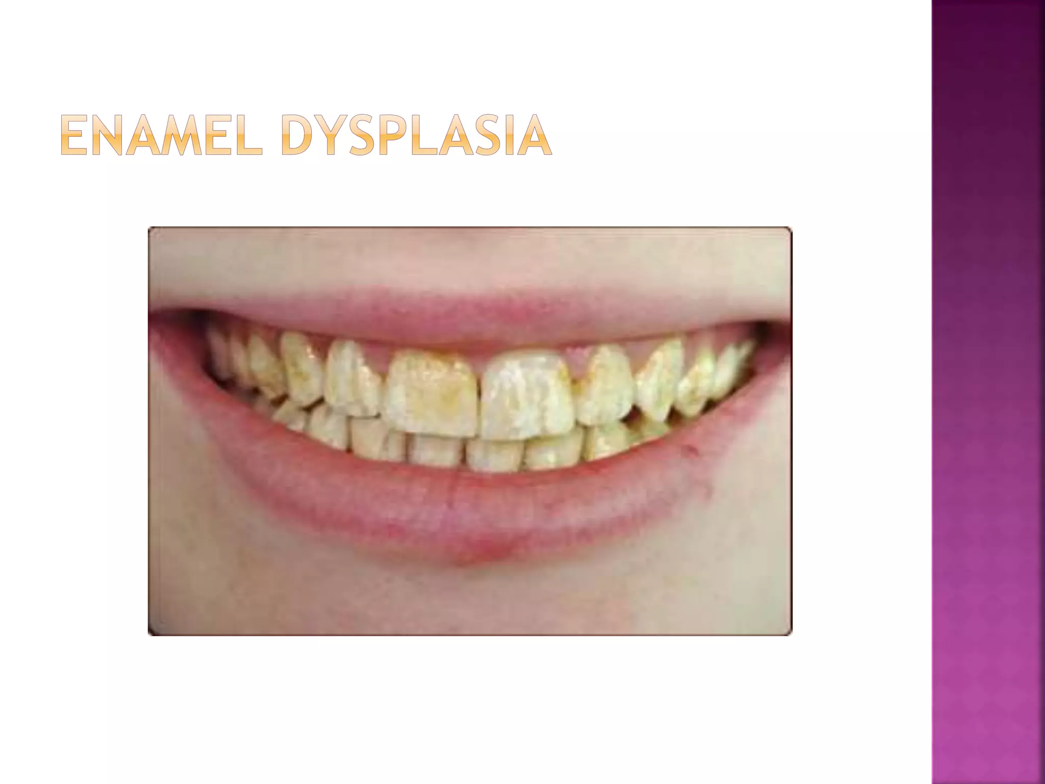
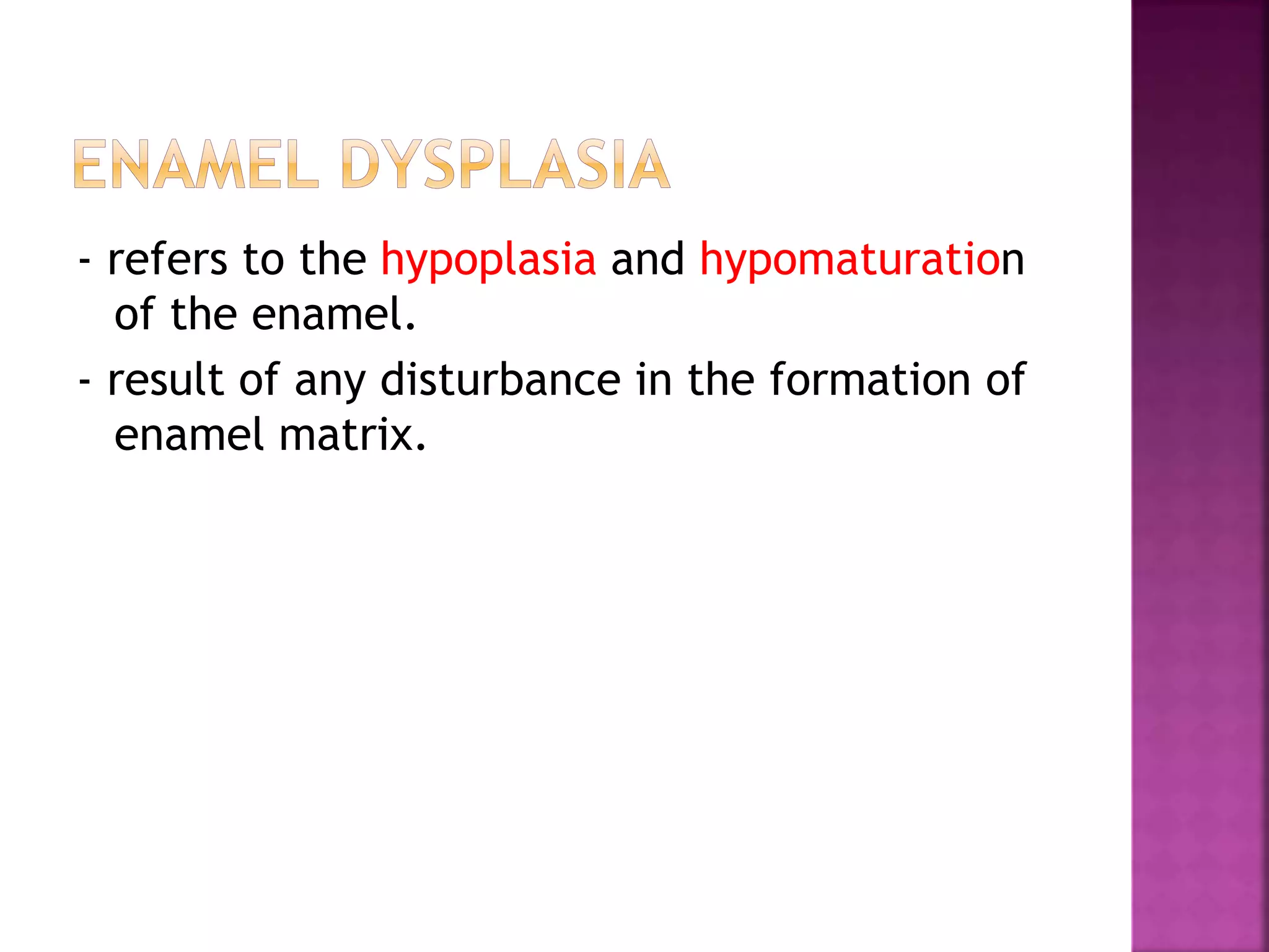
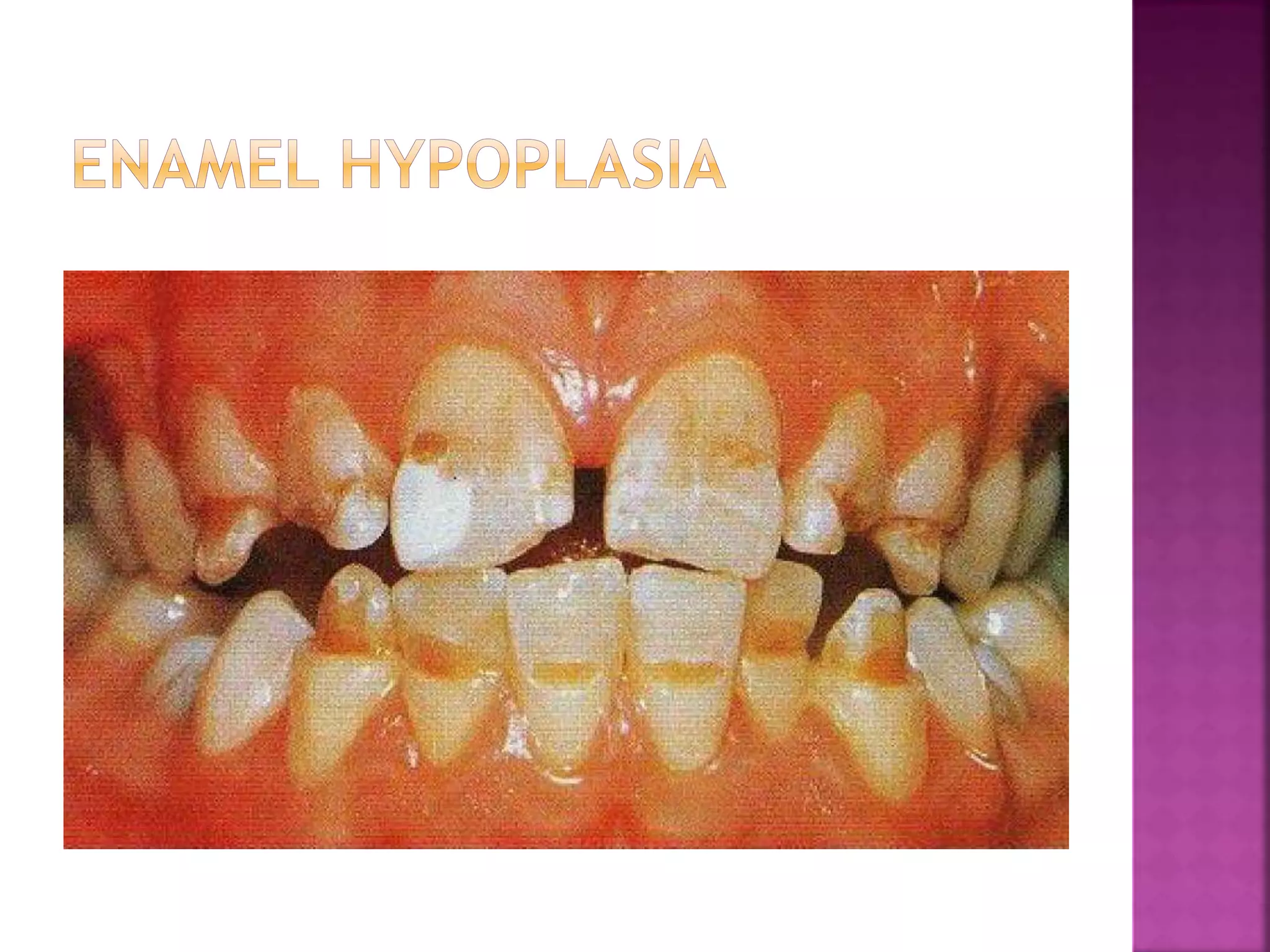
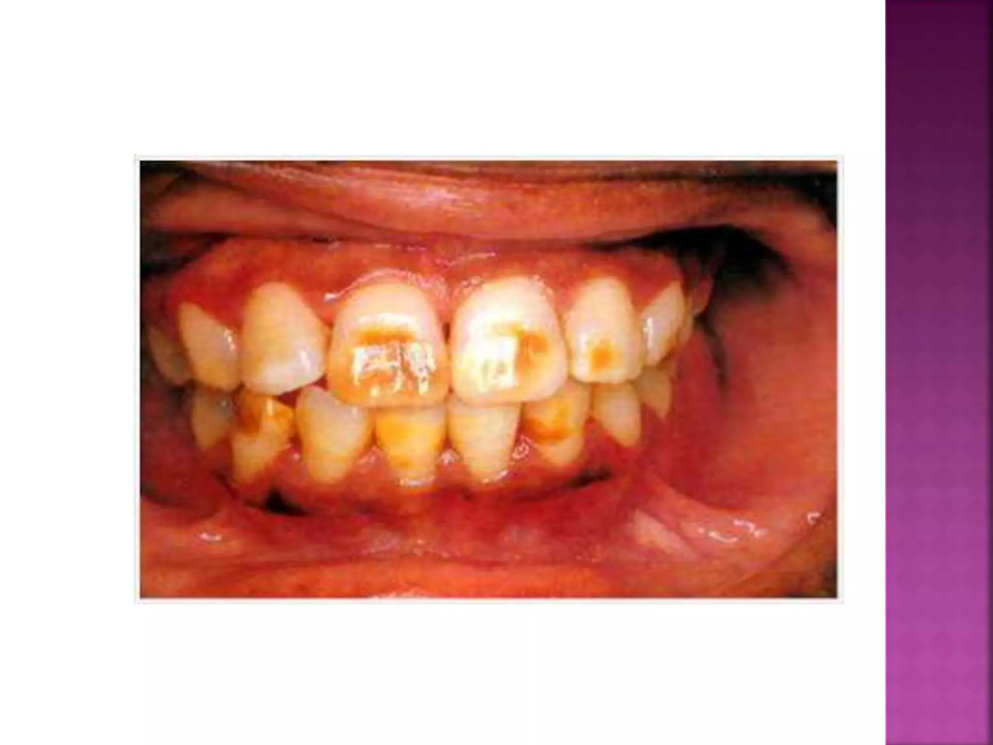
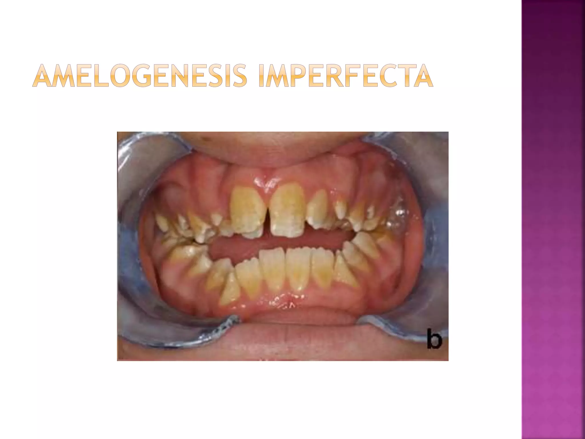
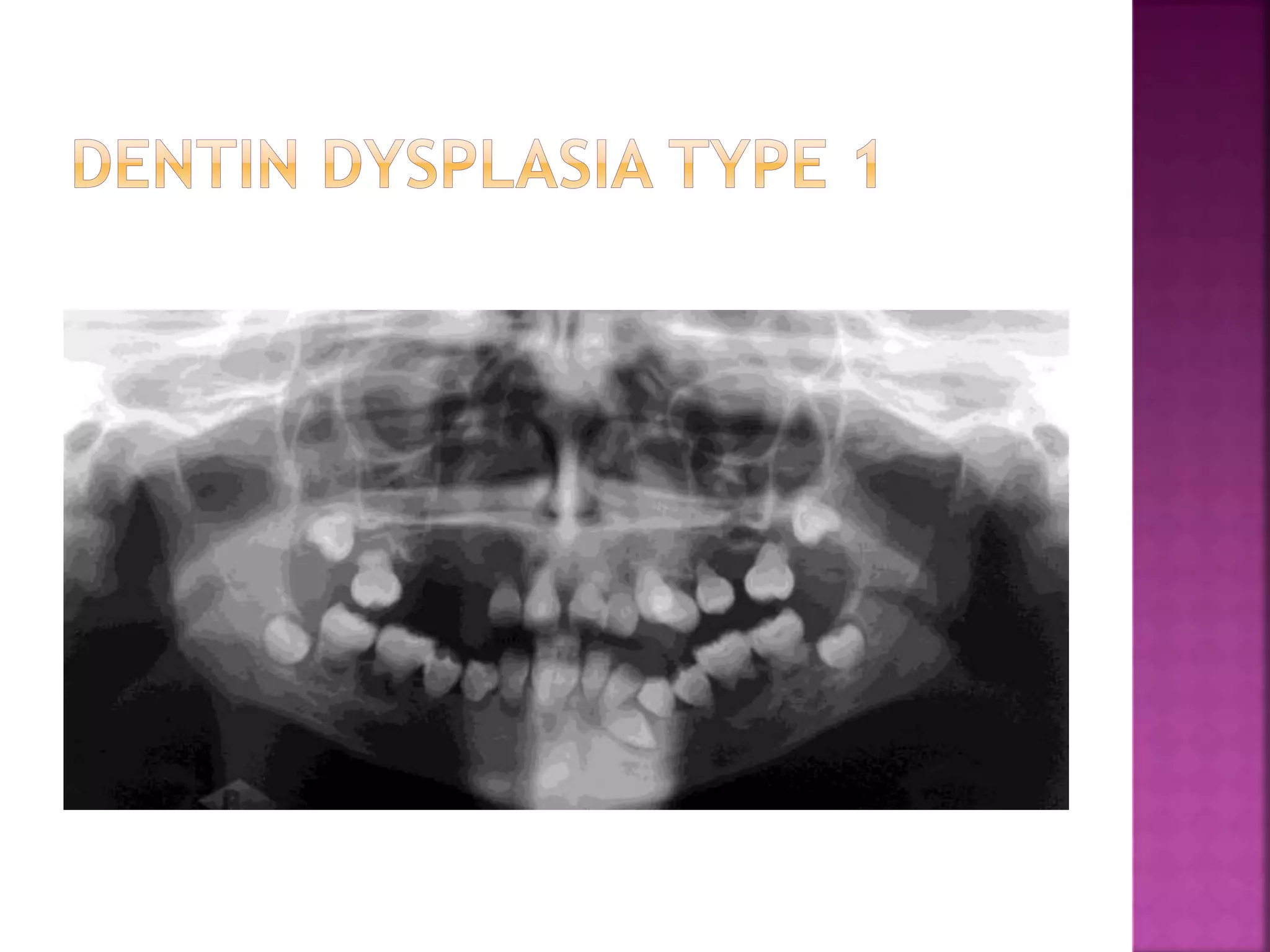
 Color 
 Color is determined by translucency and thickness of the 
enamel and by thickness and color of the underlying 
dentin 
 Discoloration may result from developmental 
disturbances whereby the normal pattern of enamel 
prisms and dental tubules is disturbed.
Discoloration due to Developmental Disturbances 
 Amelogenesis Imperfecta 
 Dentinogenesis Imperfecta 
 Brown hereditary teeth 
 Dental fluorosis or mottling
Discoloration from intrinsic pigments 
 Pigments arising from hemolysis or associated with 
jaundice 
o May be considerably green, yellow-brown, or black. 
 Tooth Non-vitality 
o May be Gray, yellow-brown, or orange-yellow 
 Internal Resorption 
o May be Pink or black
Discoloration from medicaments during 
Endodontic therapy 
 Staining 
 From permeation of medicaments into dentinal 
tubules during sterilization of cavity prep 
 Metallic staining 
 From ingestion or inhalation of metals or their salts 
 Tobacco use 
 Most frequent cause of staining
 Macrodontia 
 microdontia
 Supernumerary teeth 
 Complete or partial anodontia
 Developmental disturbances of form: 
 Geminated teeth 
 Fused teeth 
 Concrescence 
 Enamel pearls 
 Dens in dente
 Developmental disturbances of form: 
 Turner’s teeth 
 Odontomas 
 Hutchinson’s teeth 
 Mulberry molar 
 Accessory cusps 
 Hypoplastic defects
 Hereditary disturbances of form: 
 Enamel hypoplasia 
 Dentinogenesis imperfecta 
 Dentin dysplasia 
 Amelogenesis imperfecta 
 Extrinsic or environmental alterations 
responsible for variations after teeth have 
fully formed are manifested as erosion, 
abrasion, caries or extensive attrition
- refers to the hypoplasia and hypomaturation 
of the enamel. 
- result of any disturbance in the formation of 
enamel matrix.
 Hypomaturation- occurs from incomplete 
crystallization of the enamel. 
 Hypocalcification- result from any 
disturbance that interferes with the 
normal deposition of calcium.
 Local - Periapical inflamation, Truma, 
Surgical procedure. 
 Systemic - Nutritional deficiencies, endocrine 
disturbances, and other systemic 
disease. 
 Hereditary disease – various hereditary 
defects of the enamel are seen, such as 
amelogenisis imperfecta, brown teeth, 
and other hypoplasia of enamel.
Appears as a localized alteration of one or 
several or may involve all the teeth. 
Consist of the absence of enamel and dentin or 
of enamel only.
 Depends on the stage of amelogenisis. 
 Central incisors show well defined horizontal 
lines of pits and grooves. 
 Single or multiple chalky white opaque spots 
may be present on the teeth. 
- opaque may also be caused by faulty 
matrix apposition which changes the 
index of refraction
Hypocalcified Decalcified 
 White and opaque 
 Glazed 
 Smooth 
 White and opaque 
 Granular 
 Rough 
 Soft surface
 Resulting from consumption of water containing an 
excess of flourine. 
 Maybe mild to severe in character.
 Cloudy opaque areas of yellow or brown 
areas if extrinsic material has pigmented the 
areas.
 Results from hypoplasia of the central 
developmental lobe with a collapse of the 
lateral developmental lobe. 
 Screw driver shape appearance.
 Examiner attributes notching of the incisors teeth 
of central developmental lobe rather than loss of 
tooth structure from trauma.
 Mulberry molars are also sugestive of 
congenital syphilis. They are characterized 
by normal buccal and lingual surfaces but 
have occlusal surfaces analogous to a 
mulberry. 
 Localized defects of the occlusal surface of 
the molar teeth have at times been 
incorrectly diagnosed as mulberry molars
Amelogenesis imperfecta in which the enamel 
is apparently of normal thickness and surface 
consistency is hereditary brown teeth. 
In this condition it is not hard as normal and 
tends to chip on the Incisal and occlusal 
surfaces. 
Extension of brown color throughout the 
enamel and involvement of all the teeth.
 Opalescence 
 Abnormal coloration 
 Absence of pulp canals 
 Poorly calcified dentin 
 Constricted roots
 Clinically manifested by: 
- wandering teeth 
- malposed teeth 
 Radiographically 
- absence of pulp canals 
- decreased density of dentin 
- short narrow roots 
- radiolucent apical area
 Refers to the increase in the normal number 
of teeth present in dentition. 
 Supernumerary teeth may interfere with 
normal eruption. 
 Can be seen radiograhically in person with 
cleidocranial dysostosis.
 In addition to the dental defect, defective 
ossification of the clavicles and bones of the 
skull can be noted.
 Result from a chemical process, and the 
defects are usually limited to the labial and 
buccal surfaces of teeth. 
 Vary in shape from suacerlike depression to 
deep wedge-like groves
 May occur anywhere on the enamel surface 
or the cervical area of root 
 Mechanical wearing of the tooth structure by 
physical agents such as toothbrushes, 
abrasive powders, hairpins, nails, clay 
pipestems, glass, toothpick, dental tape, 
sand, and thread.
 Involves both the crown and the roots. 
 Root fractures require radiographic 
evaluation.
Two factors that must be determined in 
evaluation of fractered tooth: 
1. Fracture involves the pulp 
2. Pulp has been secondarily involved by 
injury at the apex.
Pathologic conditions affecting developmental disturbances and anomalies

Pathologic conditions affecting developmental disturbances and anomalies

  • 2.
     Color Color is determined by translucency and thickness of the enamel and by thickness and color of the underlying dentin  Discoloration may result from developmental disturbances whereby the normal pattern of enamel prisms and dental tubules is disturbed.
  • 3.
    Discoloration due toDevelopmental Disturbances  Amelogenesis Imperfecta  Dentinogenesis Imperfecta  Brown hereditary teeth  Dental fluorosis or mottling
  • 4.
    Discoloration from intrinsicpigments  Pigments arising from hemolysis or associated with jaundice o May be considerably green, yellow-brown, or black.  Tooth Non-vitality o May be Gray, yellow-brown, or orange-yellow  Internal Resorption o May be Pink or black
  • 5.
    Discoloration from medicamentsduring Endodontic therapy  Staining  From permeation of medicaments into dentinal tubules during sterilization of cavity prep  Metallic staining  From ingestion or inhalation of metals or their salts  Tobacco use  Most frequent cause of staining
  • 6.
  • 7.
     Supernumerary teeth  Complete or partial anodontia
  • 8.
     Developmental disturbancesof form:  Geminated teeth  Fused teeth  Concrescence  Enamel pearls  Dens in dente
  • 9.
     Developmental disturbancesof form:  Turner’s teeth  Odontomas  Hutchinson’s teeth  Mulberry molar  Accessory cusps  Hypoplastic defects
  • 10.
     Hereditary disturbancesof form:  Enamel hypoplasia  Dentinogenesis imperfecta  Dentin dysplasia  Amelogenesis imperfecta  Extrinsic or environmental alterations responsible for variations after teeth have fully formed are manifested as erosion, abrasion, caries or extensive attrition
  • 12.
    - refers tothe hypoplasia and hypomaturation of the enamel. - result of any disturbance in the formation of enamel matrix.
  • 13.
     Hypomaturation- occursfrom incomplete crystallization of the enamel.  Hypocalcification- result from any disturbance that interferes with the normal deposition of calcium.
  • 15.
     Local -Periapical inflamation, Truma, Surgical procedure.  Systemic - Nutritional deficiencies, endocrine disturbances, and other systemic disease.  Hereditary disease – various hereditary defects of the enamel are seen, such as amelogenisis imperfecta, brown teeth, and other hypoplasia of enamel.
  • 16.
    Appears as alocalized alteration of one or several or may involve all the teeth. Consist of the absence of enamel and dentin or of enamel only.
  • 18.
     Depends onthe stage of amelogenisis.  Central incisors show well defined horizontal lines of pits and grooves.  Single or multiple chalky white opaque spots may be present on the teeth. - opaque may also be caused by faulty matrix apposition which changes the index of refraction
  • 19.
    Hypocalcified Decalcified White and opaque  Glazed  Smooth  White and opaque  Granular  Rough  Soft surface
  • 20.
     Resulting fromconsumption of water containing an excess of flourine.  Maybe mild to severe in character.
  • 22.
     Cloudy opaqueareas of yellow or brown areas if extrinsic material has pigmented the areas.
  • 23.
     Results fromhypoplasia of the central developmental lobe with a collapse of the lateral developmental lobe.  Screw driver shape appearance.
  • 25.
     Examiner attributesnotching of the incisors teeth of central developmental lobe rather than loss of tooth structure from trauma.
  • 26.
     Mulberry molarsare also sugestive of congenital syphilis. They are characterized by normal buccal and lingual surfaces but have occlusal surfaces analogous to a mulberry.  Localized defects of the occlusal surface of the molar teeth have at times been incorrectly diagnosed as mulberry molars
  • 28.
    Amelogenesis imperfecta inwhich the enamel is apparently of normal thickness and surface consistency is hereditary brown teeth. In this condition it is not hard as normal and tends to chip on the Incisal and occlusal surfaces. Extension of brown color throughout the enamel and involvement of all the teeth.
  • 30.
     Opalescence Abnormal coloration  Absence of pulp canals  Poorly calcified dentin  Constricted roots
  • 32.
     Clinically manifestedby: - wandering teeth - malposed teeth  Radiographically - absence of pulp canals - decreased density of dentin - short narrow roots - radiolucent apical area
  • 34.
     Refers tothe increase in the normal number of teeth present in dentition.  Supernumerary teeth may interfere with normal eruption.  Can be seen radiograhically in person with cleidocranial dysostosis.
  • 35.
     In additionto the dental defect, defective ossification of the clavicles and bones of the skull can be noted.
  • 37.
     Result froma chemical process, and the defects are usually limited to the labial and buccal surfaces of teeth.  Vary in shape from suacerlike depression to deep wedge-like groves
  • 39.
     May occuranywhere on the enamel surface or the cervical area of root  Mechanical wearing of the tooth structure by physical agents such as toothbrushes, abrasive powders, hairpins, nails, clay pipestems, glass, toothpick, dental tape, sand, and thread.
  • 41.
     Involves boththe crown and the roots.  Root fractures require radiographic evaluation.
  • 42.
    Two factors thatmust be determined in evaluation of fractered tooth: 1. Fracture involves the pulp 2. Pulp has been secondarily involved by injury at the apex.