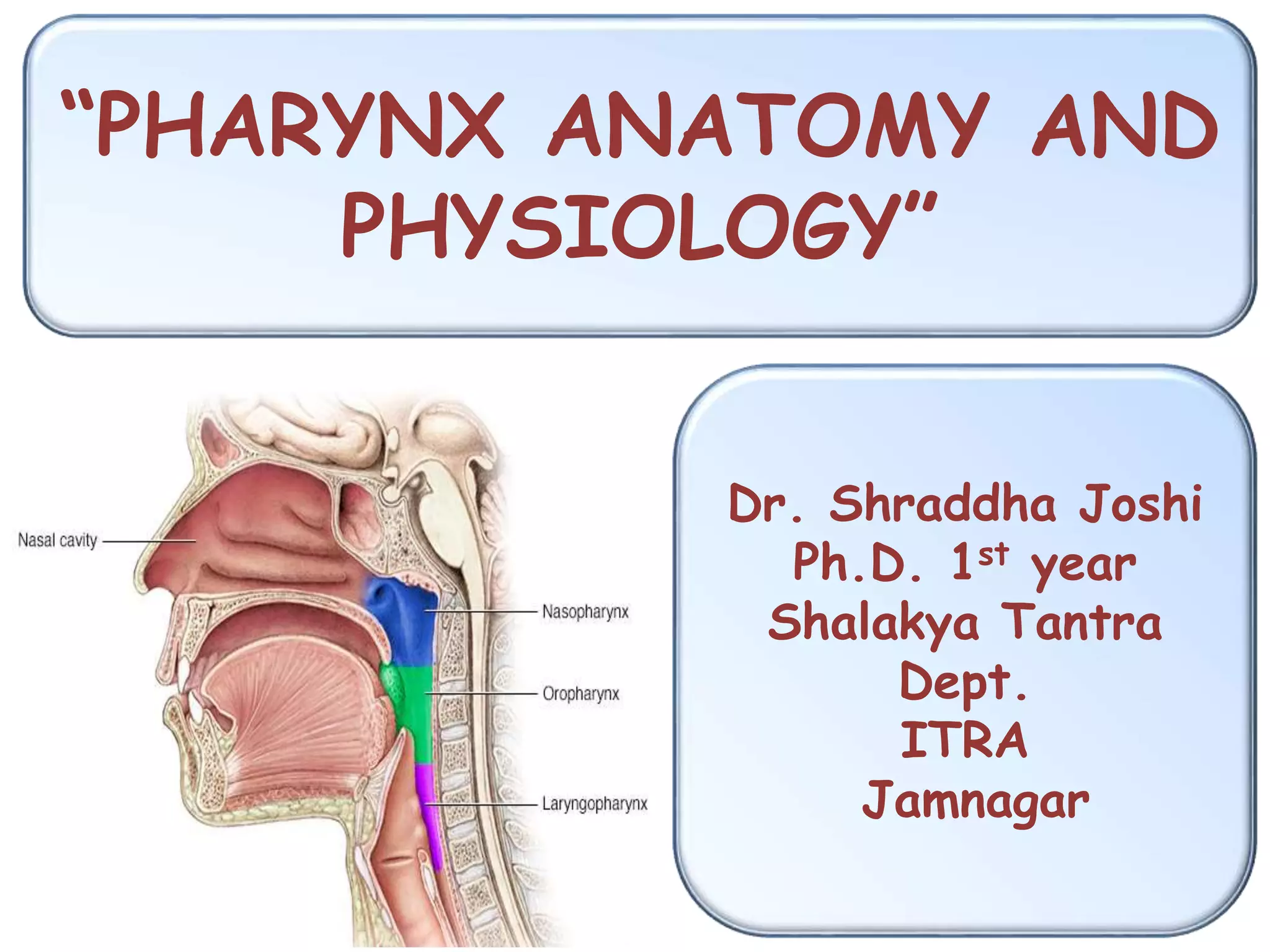
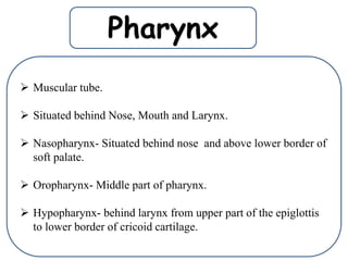
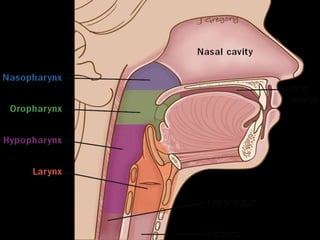
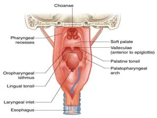
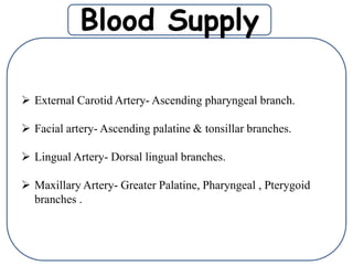
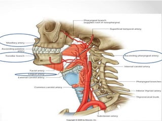
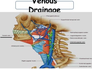
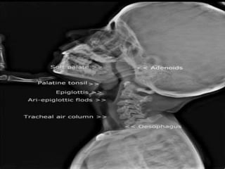
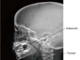
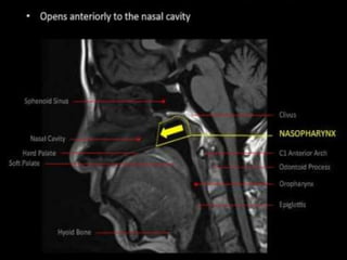
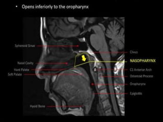
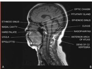
“PHARYNX ANATOMY AND
PHYSIOLOGY”
Dr. Shraddha Joshi
Ph.D. 1st year
Shalakya Tantra
Dept.
ITRA
Jamnagar
 Muscular tube.
 Situated behind Nose, Mouth and Larynx.
 Nasopharynx- Situated behind nose and above lower border of
soft palate.
 Oropharynx- Middle part of pharynx.
 Hypopharynx- behind larynx from upper part of the epiglottis
to lower border of cricoid cartilage.
Pharynx
Naso-
Pharynx
 Situated behind the nose and above the soft palate.
 Bony landmark is Hard palate- at C1 level.
 Rigid wall- Pharyngobasilar fascia and posterior median
pharyngeal ligament.
 Anteriorly- Communicates with conchae.
 Inferiorly- Communicates with oropharynx at pharyngeal
isthmus.
 Nasopharyngeal Isthmus- Passavant’s muscle. (Lower border
of Soft palate & posterior wall of pharynx.
 Lateral wall:
• Pharyngeal opening of the eustachian tube.
• Tubal elevation bounds the tubal opening.
• Salpingopharyngeal fold.
• Leveator palatani.
• Fossa of Rosenmuller.
 Roof and Posterior wall:
• Continuous slope, opposite the posterior part of body of
sphenoid, basiocciput and anterior arch of atlas.
• Adenoids- In the roof and the posterior wall of lymphoid
tissue.
Oro-Pharynx
 Situated behind oral cavity
 Extend from the level of the soft palate to the level of the
hyoid bone.
 Superiorly- communicates to the nasopharynx through the
pharyngeal isthmus.
 Anteriorly- It communicates with the oral cavity
 Posteriorly- opposite to C2 and C3.
 Lateral wall- Palatine tonsils and anterior & posterior pillars.
 Inferior border- Hyoid bone.
Hypo-
Pharynx
 Also known as Laryngopharynx.
 Situated behind the larynx.
 Extends from upper border of epiglottis/ level of hyoid bone to
the lower border of cricoid cartilage.
 Anterior wall-
• Inlet of larynx.
• Posterior surface of cricoid and arytenoid cartilages.
 Posterior wall-
• 4th and 5th cervical vertebrae.
 Lateral wall-
• Pyriform fossa, one on
each side of the inlet of
the larynx.
Pharyngeal wall
1. Mucosa- Lined by squamous epithelium except the
nasopharyx (Nasopharynx- ciliated columnar epithelium)
2. Submucosa
3. Pharyngobasilar fascia-
• Fibrous sheet deep to the pharyngeal muscle.
• Fills the gap between upper border of superior constrictor and
base of skull.
4. Muscular coat-
• Consist of outer circular layer made of 3 constrictors.
• Inner longitudinal layer made up of Stylopharyngeus,
Salpingopharyngeus, Palatopharyngeus muscles.
5. Buccopharyngeal fascia-
• Covers the outer surface of the constrictors.
• Below the buccopharyngeal fascia, on the muscular coat there
lie the pharyngeal plexus of veins and nerves.
 External Carotid Artery- Ascending pharyngeal branch.
 Facial artery- Ascending palatine & tonsillar branches.
 Lingual Artery- Dorsal lingual branches.
 Maxillary Artery- Greater Palatine, Pharyngeal , Pterygoid
branches .
Blood Supply
Venous
Drainage
Lymphatic
Drainage
 Retropharyngeal Lymphnodes.
 Deep Cervical Lymphnodes.
Nerve Supply
 Supplied by pharyngeal plexus of nerves chiefly on
the middle constrictor.
 Plexus is formed by-
• Pharyngeal branch of vagus (cranial accessory).
• Pharyngeal branch of glossopharyngeal.
• Pharyngeal branches of superior cervical sympathetic
ganglion.
Pharynx Anatomy
Pharynx Anatomy
Pharynx Anatomy
Pharynx Anatomy
Pharynx Anatomy
Pharynx Anatomy
Pharynx Anatomy

Pharynx Anatomy

  • 1.
    “PHARYNX ANATOMY AND PHYSIOLOGY” Dr.Shraddha Joshi Ph.D. 1st year Shalakya Tantra Dept. ITRA Jamnagar
  • 2.
     Muscular tube. Situated behind Nose, Mouth and Larynx.  Nasopharynx- Situated behind nose and above lower border of soft palate.  Oropharynx- Middle part of pharynx.  Hypopharynx- behind larynx from upper part of the epiglottis to lower border of cricoid cartilage. Pharynx
  • 5.
    Naso- Pharynx  Situated behindthe nose and above the soft palate.  Bony landmark is Hard palate- at C1 level.  Rigid wall- Pharyngobasilar fascia and posterior median pharyngeal ligament.  Anteriorly- Communicates with conchae.  Inferiorly- Communicates with oropharynx at pharyngeal isthmus.  Nasopharyngeal Isthmus- Passavant’s muscle. (Lower border of Soft palate & posterior wall of pharynx.
  • 6.
     Lateral wall: •Pharyngeal opening of the eustachian tube. • Tubal elevation bounds the tubal opening. • Salpingopharyngeal fold. • Leveator palatani. • Fossa of Rosenmuller.  Roof and Posterior wall: • Continuous slope, opposite the posterior part of body of sphenoid, basiocciput and anterior arch of atlas. • Adenoids- In the roof and the posterior wall of lymphoid tissue.
  • 9.
    Oro-Pharynx  Situated behindoral cavity  Extend from the level of the soft palate to the level of the hyoid bone.  Superiorly- communicates to the nasopharynx through the pharyngeal isthmus.  Anteriorly- It communicates with the oral cavity  Posteriorly- opposite to C2 and C3.  Lateral wall- Palatine tonsils and anterior & posterior pillars.  Inferior border- Hyoid bone.
  • 11.
    Hypo- Pharynx  Also knownas Laryngopharynx.  Situated behind the larynx.  Extends from upper border of epiglottis/ level of hyoid bone to the lower border of cricoid cartilage.  Anterior wall- • Inlet of larynx. • Posterior surface of cricoid and arytenoid cartilages.  Posterior wall- • 4th and 5th cervical vertebrae.
  • 12.
     Lateral wall- •Pyriform fossa, one on each side of the inlet of the larynx.
  • 13.
    Pharyngeal wall 1. Mucosa-Lined by squamous epithelium except the nasopharyx (Nasopharynx- ciliated columnar epithelium) 2. Submucosa 3. Pharyngobasilar fascia- • Fibrous sheet deep to the pharyngeal muscle. • Fills the gap between upper border of superior constrictor and base of skull.
  • 14.
    4. Muscular coat- •Consist of outer circular layer made of 3 constrictors. • Inner longitudinal layer made up of Stylopharyngeus, Salpingopharyngeus, Palatopharyngeus muscles. 5. Buccopharyngeal fascia- • Covers the outer surface of the constrictors. • Below the buccopharyngeal fascia, on the muscular coat there lie the pharyngeal plexus of veins and nerves.
  • 17.
     External CarotidArtery- Ascending pharyngeal branch.  Facial artery- Ascending palatine & tonsillar branches.  Lingual Artery- Dorsal lingual branches.  Maxillary Artery- Greater Palatine, Pharyngeal , Pterygoid branches . Blood Supply
  • 19.
  • 20.
  • 21.
    Nerve Supply  Suppliedby pharyngeal plexus of nerves chiefly on the middle constrictor.  Plexus is formed by- • Pharyngeal branch of vagus (cranial accessory). • Pharyngeal branch of glossopharyngeal. • Pharyngeal branches of superior cervical sympathetic ganglion.