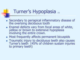
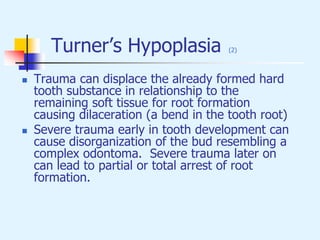
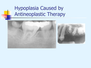
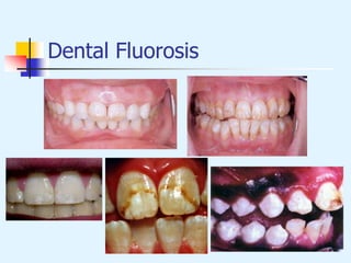
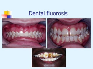
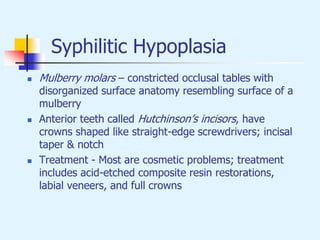
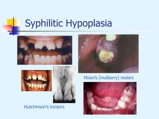
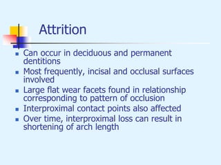
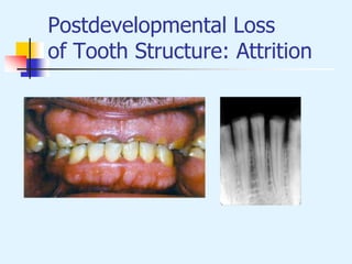
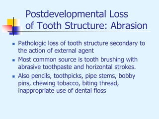
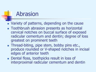
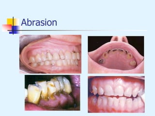
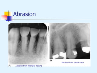
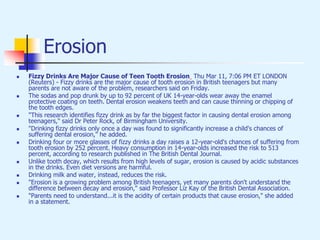
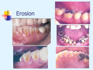
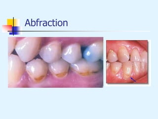
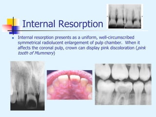
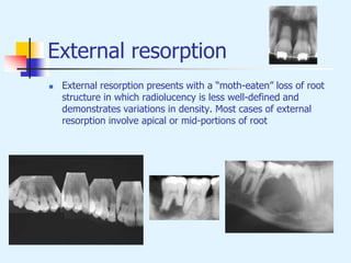
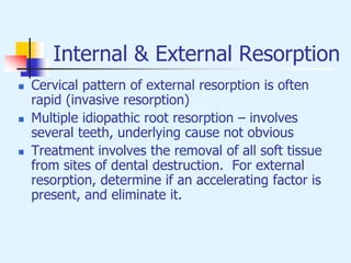
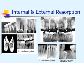
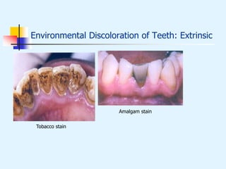
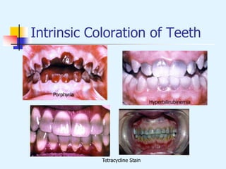
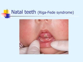
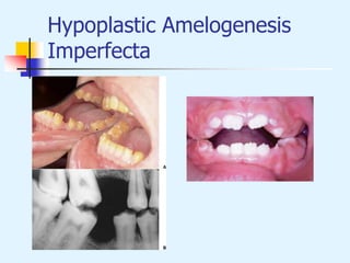
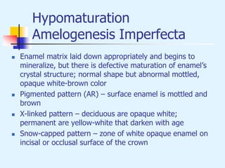
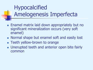
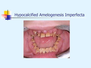
Abnormalities of the Teeth
Advanced Oral Pathology
Environmental Effects on Tooth
Structure Development
Environmental Effects on
Tooth Structure Development
 Visible environmental enamel defects can be classified into one
of three patterns:
 Enamel hypoplasia – pits, grooves, or larger areas of
missing enamel
 Diffuse opacities of enamel – variations in translucency or
normal thickness; increased white opacity with no clear
boundary with adjacent normal enamel
 Demarcated opacities of enamel show areas of decreased
translucence, increased opacity, and a sharp boundary with
adjacent enamel; normal thickness
Environmental Effects on
Tooth Structure Development
 Common pattern, occurs as result of systemic
influences (such as exanthematous fevers) which
occur during the first two years of life; horizontal
rows of pits or diminished enamel on anterior teeth
and first molars; enamel loss is bilateral
 Similar pattern in cuspids, bicuspids, and second
molars when the inciting event occurs at age 4-5
Enamel Hypoplasia associated
with exanthematous fevers
Turner’s Hypoplasia (1)
 Secondary to periapical inflammatory disease of
the overlying deciduous tooth
 Enamel defects vary from focal areas of white,
yellow or brown to extensive hypoplasia
involving the entire crown.
 Most frequently affects permanent bicuspids
 Traumatic injury to deciduous teeth also causes
Turner’s teeth (45% of children sustain injuries
to primary teeth)
Turner’s Hypoplasia (2)
 Trauma can displace the already formed hard
tooth substance in relationship to the
remaining soft tissue for root formation
causing dilaceration (a bend in the tooth root)
 Severe trauma early in tooth development can
cause disorganization of the bud resembling a
complex odontoma. Severe trauma later on
can lead to partial or total arrest of root
formation.
Turner’s Hypoplasia
Turner’s Hypoplasia
Hypoplasia Caused by
Antineoplastic Therapy
 Degree and severity related to age, form of therapy
(chemotherapy/radiotherapy) and dose
 Defects include hypodontia, microdontia, radicular
hypoplasia, enamel hypoplasia and discolorations
 Radiotherapy effects more severe than chemotherapy
alone but sometimes used together
 Dose of radiation as low as 0.72 Gy can cause mild
defects in enamel/dentin
 Mandibular hypoplasia due to direct radiation, alveolar
deficiency or pituitary effects.
Hypoplasia Caused by
Antineoplastic Therapy
Dental Fluorosis
 Critical period is age 2-3, if fluoride levels
greater than 1 part per million are ingested
 Fluoride comes from several sources besides
water: adult-strength fluoride toothpastes,
fluoride supplements, infant foods, soft drinks,
and fruit juices
 Severity is dose dependent (higher intakes
during critical periods associated with more
severe fluorosis)
Dental Fluorosis
Dental fluorosis
Syphilitic Hypoplasia
 Mulberry molars – constricted occlusal tables with
disorganized surface anatomy resembling surface of a
mulberry
 Anterior teeth called Hutchinson’s incisors, have
crowns shaped like straight-edge screwdrivers; incisal
taper & notch
 Treatment - Most are cosmetic problems; treatment
includes acid-etched composite resin restorations,
labial veneers, and full crowns
Hutchinson’s triad
Syphilitic Hypoplasia
Hutchinson’s incisors
Moon’s (mulberry) molars
Postdevelopmental Loss
of Tooth Structure: Attrition
 Loss of tooth structure caused by tooth-to-tooth
contact during occlusion and mastication.
 Pathologic when it affects appearance and
function.
 Process can be accelerated by poor quality or
absent enamel, premature edge-to-edge
occlusion, intraoral abrasives, erosion, and
grinding habits.
Attrition
 Can occur in deciduous and permanent
dentitions
 Most frequently, incisal and occlusal surfaces
involved
 Large flat wear facets found in relationship
corresponding to pattern of occlusion
 Interproximal contact points also affected
 Over time, interproximal loss can result in
shortening of arch length
Postdevelopmental Loss
of Tooth Structure: Attrition
Postdevelopmental Loss
of Tooth Structure: Abrasion
 Pathologic loss of tooth structure secondary to
the action of external agent
 Most common source is tooth brushing with
abrasive toothpaste and horizontal strokes.
 Also pencils, toothpicks, pipe stems, bobby
pins, chewing tobacco, biting thread,
inappropriate use of dental floss
Abrasion
 Variety of patterns, depending on the cause
 Toothbrush abrasion presents as horizontal
cervical notches on buccal surface of exposed
radicular cementum and dentin; degree of loss
greatest on prominent teeth
 Thread-biting, pipe stem, bobby pins etc.,
produce rounded or V-shaped notches in incisal
edges of anterior teeth
 Dental floss, toothpicks result in loss of
interproximal radicular cementum and dentin
Abrasion
Abrasion
Abrasion from improper flossing
Abrasion from partial clasp
Postdevelopmental Loss
of Tooth Structure: Erosion
 Loss of tooth structure by chemical reaction, not
that associated with bacteria (caries)
 Secondary to presence of acid or chelating agent
 Source can be dietary (e.g., vinegar, lemons),
internal (gastric secretions – perimolysis), or
external (e.g., acids, industrial, atmosphere)
 “If it is not abrasion or attrition, it must be
erosion”
Erosion
 Commonly affects facial surface of maxillary
anteriors and appears as shallow spoon-shaped
depressions in cervical portion of the crown
 Posterior teeth exhibit loss of occlusal surface,
where dentin is destroyed more rapidly than
enamel, resulting in concave depression of dentin
surrounded by elevated rim of enamel
 Erosion limited to facial surfaces of maxillary
anterior dentition is usually associated with dietary
acid.
Erosion
 Tooth loss confined to incisal portions of
anterior dentition of both arches indicates
environmental source.
 Erosion on palatal surfaces of maxillary anterior
teeth and occlusal surfaces of posterior teeth of
both dentitions probably caused by regurgitation
of gastric secretions.
Erosion
 Fizzy Drinks Are Major Cause of Teen Tooth Erosion Thu Mar 11, 7:06 PM ET LONDON
(Reuters) - Fizzy drinks are the major cause of tooth erosion in British teenagers but many
parents are not aware of the problem, researchers said on Friday.
 The sodas and pop drunk by up to 92 percent of UK 14-year-olds wear away the enamel
protective coating on teeth. Dental erosion weakens teeth and can cause thinning or chipping of
the tooth edges.
 "This research identifies fizzy drink as by far the biggest factor in causing dental erosion among
teenagers," said Dr Peter Rock, of Birmingham University.
 "Drinking fizzy drinks only once a day was found to significantly increase a child's chances of
suffering dental erosion," he added.
 Drinking four or more glasses of fizzy drinks a day raises a 12-year-old's chances of suffering from
tooth erosion by 252 percent. Heavy consumption in 14-year-olds increased the risk to 513
percent, according to research published in The British Dental Journal.
 Unlike tooth decay, which results from high levels of sugar, erosion is caused by acidic substances
in the drinks. Even diet versions are harmful.
 Drinking milk and water, instead, reduces the risk.
 "Erosion is a growing problem among British teenagers, yet many parents don't understand the
difference between decay and erosion," said Professor Liz Kay of the British Dental Association.
 "Parents need to understand...it is the acidity of certain products that cause erosion," she added
in a statement.
Erosion
Postdevelopmental Loss
of Tooth Structure: Abfraction
 Loss of tooth structure resulting from repeated
tooth (enamel & dentin) flexure produced by
occlusal stresses
 Disruption of chemical bonds at cervical
fulcrum leads to cracked enamel that can be
vulnerable to abrasion and erosion
Abfraction
 Wedge-shaped defects limited to cervical area
 Deep, narrow, V-shaped
 Sometimes single tooth or subgingival
 More common in mandibular dentition and
among those with bruxism
Abfraction
Treatment of Postdevelopmental
Loss of Tooth Structure
 Early diagnosis and intervention to restrict
severity of tooth loss
 Patient education
 Mouth guards
 Limit (redirect) tooth brushing & flossing
 Replacement of lost posterior teeth and
avoidance of edge-to-edge occlusion
 Composite resins, veneers, onlays, full crowns
Internal & External Resorption
 Internal resorption is caused by cells located in
dental pulp. Rare, usually follows injury to
pulpal tissues.
 External resorption is caused by cells in the
periodontal ligament. Most patients are likely
to have root resorption on one or more teeth.
Internal Resorption
 Internal resorption presents as a uniform, well-circumscribed
symmetrical radiolucent enlargement of pulp chamber. When it
affects the coronal pulp, crown can display pink discoloration (pink
tooth of Mummery)
External resorption
 External resorption presents with a “moth-eaten” loss of root
structure in which radiolucency is less well-defined and
demonstrates variations in density. Most cases of external
resorption involve apical or mid-portions of root
Internal & External Resorption
 Cervical pattern of external resorption is often
rapid (invasive resorption)
 Multiple idiopathic root resorption – involves
several teeth, underlying cause not obvious
 Treatment involves the removal of all soft tissue
from sites of dental destruction. For external
resorption, determine if an accelerating factor is
present, and eliminate it.
Internal & External Resorption
External Resorption
Internal resorption
Internal resorption
→
→
External resorption--
embedded tooth
Environmental
Discoloration of Teeth
Environmental
Discoloration of Teeth: Extrinsic
 Arise from surface accumulation of exogenous pigment
 Bacterial stains – occur most frequently in children
 Excessive use of tobacco, tea, coffee
 Foods that contain abundant chlorophyll
 Restorative materials, especially amalgam
 Medications
 Stannous fluoride and chlorhexidine
 Extrinsic stains can be removed by polishing with fine
pumice, (sometimes with added 3% hydrogen peroxide);
recurrence is likely unless the associated cause is altered
Environmental Discoloration of Teeth: Extrinsic
Tobacco stain
Amalgam stain
Environmental Discoloration of
Teeth: Intrinsic
 Secondary to endogenous factors that discolor
underlying dentin
 Congenital erythropoietic porphyria (Günther’s
disease) is an AR disorder of metabolism that
results in increased synthesis and excretion of
porphyrins
 Hyperbilirubinemia due to jaundice,
erythroblastosis fetalis (hemolytic anemia of
newborns secondary to blood incompatibility,
usually Rh factor), biliary atresia (sclerosing
process of the biliary tree), and chlorodontia
(green discoloration).
Environmental
Discoloration of Teeth: Intrinsic
 Localized red blood cell destruction (pink discoloration
arising from hemoglobin breakdown within necrotic pulp
tissue when blood has accumulated in the head)
 Lepromatous leprosy (pink discoloration secondary to
infection-related necrosis and the rupture of numerous
small blood vessels within the pulp
 Medications (tetracycline)
 Intrinsic stains are difficult to treat. Possible treatments
include full crowns, external bleaching of vital teeth,
internal bleaching of nonvital teeth, bonded restorations,
composite build-ups, and laminate veneer crowns.
Intrinsic Coloration of Teeth
Hyperbilirubinemia
Tetracycline Stain
Porphyria
Localized Disturbances in
Eruption
Localized Disturbances in
Eruption
 Eruption – the continuous process of movement
of a tooth from developmental location to
functional location
 Impacted – teeth that cease to erupt due to
physical obstruction
 Embedded – teeth that cease to erupt due to
lack of eruptive force
 Ankylosis – teeth that cease to erupt due to
anatomic fusion of tooth with alveolar bone
Localized Disturbances in
Eruption
 Primary impaction of deciduous teeth is
extremely rare. Most commonly involves
second molars often due to ankylosis.
 Primary impaction of permanent teeth most
frequently affects third molars. Lack of
eruption is most often related to crowding and
insufficient maxillofacial development.
 Impacted teeth are frequently diverted or
angulated, eventually losing their potential to
erupt; mesioangular, distoangular, vertical,
horizontal and inverted
Localized Disturbances in
Eruption
 Treatment includes long-term observation,
orthodontic-assisted eruption, transplantation, or
surgical removal
 Risks associated with both intervention and
nonintervention
 Surgical removal of impacted teeth is the
procedure most frequently performed by OMFS
Localized Disturbances in
Eruption
 Ankylosis – cessation of eruption after emergence
 Usually develops between ages 7-18; peak 8-9;
prevalence est. 1.3-8.9%
 Fails to respond to orthodontic therapy
 Failure to treat can result in tilting, carious
destruction, and periodontal disease
 When successor tooth present, best treated with
extraction and space maintenance
Localized Disturbances in
Eruption
Primary tooth impaction Mesioangular impaction
Ankylosis
Developmental Alterations of the
Teeth
Developmental Alterations
in the Number of Teeth
 Anodontia – total lack of tooth development. Rare;
most cases occur in hereditary hypohidrotic
ectodermal dysplasia
 Hypodontia – lack of development of one or more
teeth. Uncommon in deciduous teeth, usually
involves mandibular incisors. More common in
permanent teeth, third molars most affected. More
frequent in females than males
 Oligodontia – lack of development of six or more
teeth
Developmental Alterations
in the Number of Teeth
 Hyperdontia – development of increased number of
teeth. Additional teeth are supernumerary. Prevalence
1-3%. More common in males and usually develops by
age 20.
 Maxilla is most common site (90%) for single tooth
hyperdontia, especially incisor region (mesiodens)
 Most single supernumerary teeth are unilateral. Nearly
75% of supernumerary teeth in anterior maxilla fail to
erupt
 Non-syndromic multiple supernumerary teeth occur
mostly in mandible.
Hypodontia (oligodontia)
Example of pedigree
Hypodontia in Ectodermal
Dysplasia
Developmental Alterations
in the Number of Teeth
 Mesiodens – supernumerary tooth in maxillary anterior
incisor region
 Distomolar/Distodens – accessory fourth molar
 Paramolar – posterior supernumerary tooth situated
lingually or buccally to a molar tooth
 Dental transposition – normal teeth erupted in an
inappropriate pattern
 Natal teeth – teeth present in newborns; teeth arising
during the first 30 days of life; (85% mandibular incisor
region)
Hyperdontia (supernumeray teeth)
Mesiodens
Cleidocranial dysplasia
Supernumeray premolar
Transposition (canine-first premolar)
Treatment of Developmental Alterations
in the Number of Teeth
 Hypodontia – often no treatment required for
individual missing teeth; prosthetic replacement for
multiple missing teeth.
 Hyperdontia – early removal of accessory tooth;
delayed in therapy can delay eruption of adjacent
teeth or cause displacement.
 Natal teeth – may be removed if they are loose; if
stable, they should be retained; Riga-Fede disease
(ulceration of ventral tongue associated with breast-
feeding) can often be treated without removal of the
teeth.
Natal teeth (Riga-Fede syndrome)
Supernumerary teeth
Mesiodens
Supernumerary premolar
Mesiodens
Supernumerary teeth
Supernumerary teeth
Supernumerary teeth in cleidocranial
dysplasia syndrome
Developmental Alterations
in the Size of Teeth
Developmental Alterations in
the Size of Teeth (1)
 Microdontia – small teeth. Can also be related to
tooth size relative to jaw size. More common in
females. Isolated microdontia within otherwise
normal dentition is not uncommon (peg-shaped
lateral 0.8-8.4%). Diffuse microdontia occurs in
some hereditary disorders and sometimes
associated with hypodontia. Increased in Down’s,
pituitary dwarfism & a few other syndromes.
 Macrodontia – larger than average teeth. More
common in males. Typically only a few teeth are
abnormally large. Diffuse macrodontia may occur
in pituitary gigantism. It can be associated with
hyperdontia.
Microdontia
“peg-shaped” laterals
“paramolar”
Developmental Alterations in
the Size of Teeth (2)
 Macrognathia – normal sized teeth widely
spaced in larger than normal jaw.
 Treatment – not necessary except for esthetic
reasons
Developmental Alterations
in the Shape of Teeth
Developmental Alterations
in the Shape of Teeth
 Double teeth – two separate teeth exhibiting union by dentin and
sometimes pulps (fusion).
 May result from fusion of two tooth buds, or partial splitting of one
into two.
 Concrescence – union of two teeth by cementum without confluence
of dentin.
 Gemination – single enlarged tooth or joined (double) tooth in which
tooth count is normal when this tooth is counted as one.
 Fusion – single enlarged tooth or joined (double) tooth in which the
tooth count is short one when this tooth is counted as one.
Concrescence
Gemination / Fusion
→
Gemination
Fusion
Treatment of Developmental
Alterations of Shape of Teeth
 Extraction of deciduous double teeth may be
necessary
 Shaping with/without placement of full crowns
 May require surgical removal with prosthesis
 Concrescence requires no therapy unless
interfering with eruption
Developmental Alterations in the
Shape of Teeth: Accessory Cusps
 Cusp of Carabelli – accessory cusp located on palatal surface
of mesiolingual cusp of maxillary molar. Very common in
Caucasians (up to 90%), rare in Asians.
 Talon cusp (anterior dens evaginatus) – well defined additional
cusp located on surface of anterior tooth extending at least
half the distance from the cemento-enamel junction to the
incisal edge (<1-8%). Usually projects from the lingual
surface.
 Dens evaginatus – cusp-like elevation of enamel located in
central groove or lingual ridge of buccal cusp of permanent
premolar or molar teeth. Rare in whites; 15% Asians.
Talon cusp (anterior dens evaginatus)
Cusp of Carabelli / Dens
Evaginatus
Dens Evaginatus
Cusp of Carabelli
Treatment of Accessory Cusps
 Talon cusps on mandibular teeth often require
no therapy, talon cusps on maxillary teeth
should be removed
 Cusps of Carabelli require no treatment,
unless deep groove is present, then it should
be sealed
 Dens evaginatus often results in occlusal
problems, so should be removed
 Shovel-shaped incisors – deep fissures should
be restored
Dens Invaginatus
 Deep surface invagination of crown or root that
is lined by enamel
 Coronal dens invaginatus may be large,
resembling a tooth within a tooth (dens in
dente), or it may be dilated and disturb tooth
formation resulting in anomalous tooth
development (dilated odontome)
 Radicular dens invaginatus is rare, formation of
strip of enamel extending along root surface;
altered enamel forms a surface invagination
into dental papilla
Dens invaginatus
Treatment of Dens
Invaginatus
 Minor cases of coronal dens invaginatus do not
require removal of the tooth
 Opening of invagination should be restored to
prevent caries
 Large coronal dens invaginations often disrupt
normal coronal development and should be
removed
 Complications of radicular dens invaginatus rare
Ectopic Enamel
 Ectopic enamel – presence of enamel in unusual places.
 Enamel pearls – hemispheric structures projecting from the
surface of the root, found mostly on the roots of maxillary
molars.
 Cervical enamel extensions – located on buccal surface of root
overlying bifurcation.
 Buccal bifurcation cyst – inflammatory cyst developed along
buccal surface over the bifurcation
 Treatment: Enamel pearls – good oral hygiene, sometimes
removal with caution. Buccal bifurcation cyst – surgical
removal, periodontal treatment.
Ectopic Enamel
Taurodontism
 Enlargement of body and pulp chamber of
multirooted tooth with apical displacement of
pulpal floor and bifurcation region (2.5-3.2%)
 Increased apico-occlusal height and
bifurcation near apex
 Unilateral or bilateral; Affects permanent
teeth more often than deciduous teeth
 Normal (cynodont), Mild (hypotaurodontism),
moderate (mesotaurodontism), severe
(hypertaurodontism)
 No specific treatment; Prosthodontic,
endodontic considerations.
Taurodontism
Hypercementosis
 Non-neoplastic deposition of excessive cementum
continuous with the normal radicular cementum
 Thickness or blunting of root radiographically, localized
or generalized.
 Local or systemic factors; loss of antagonist tooth,
occlusal trauma, inflammation, Paget’s disease,
acromegaly, etc.
 Significant generalized hypercementosis in persons with
Paget’s disease
 No specific treatment
Hypercementosis
Dilaceration
 Abnormal angulation or bend in root (or
commonly the crown)
 During tooth development, it is thought to arise
following displacement injury or less frequently
secondary to the presence of an adjacent cyst or
tumor
 Treatment - minor dilaceration in permanent
teeth requires no therapy; grossly deformed
teeth should be removed; extraction of
deciduous teeth when eruption is delayed.
Dilaceration
Supernumerary Roots
 Increased number of roots
 No treatment necessary, but detection of extra
root critical for endodontic therapy or exodontia
Developmental Alterations in the
Structure of Teeth
Amelogeneis Imperfecta
 Group of conditions that demonstrate developmental
alterations in enamel structure in the absence of a
systemic disorder
 Development of enamel has three major stages: (1)
Elaboration of the organic matrix; (2) Mineralization
of the matrix; and (3) Maturation of the enamel
 At least 14 subtypes related to above with a variety
of inheritance and clinical patterns
Amelogeneis Imperfecta
 Witkop classification combines inheritance and clinical
patterns
 Type I hypoplastic; generalized/localized,
smooth/pitted/rough, AD, AR. X-linked
 Type II hypomaturation; pigmented/non-pigmented,
diffuse/snow capped; AD, AR, X-linked
 Type III Hypocalcified; diffuse AD/AR
 Type IV Hypomaturation-hypoplastic or hypoplastic-
hypomaturation with taurodontism; AD
Amelogeneis Imperfecta
Hypoplastic form
Hypomineralized form
Hypoplastic Amelogenesis
Imperfecta
 Inadequate deposition of enamel matrix
 Generalized pattern – pinpoint sized pits scattered across
surface of teeth
 Localized pattern – horizontal rows of pits, linear
depression or one large area of hypoplastic enamel
 Autosomal dominant smooth pattern – smooth surface,
enamel is thin, hard, and glossy
 X-linked dominant smooth pattern – alternating zones of
normal and abnormal enamel related to active X
chromosomes
 Rough pattern – thin, hard, rough enamel
 Enamel agenesis – total lack of enamel formation
Hypoplastic Amelogenesis
Imperfecta
Hypomaturation
Amelogenesis Imperfecta
 Enamel matrix laid down appropriately and begins to
mineralize, but there is defective maturation of enamel’s
crystal structure; normal shape but abnormal mottled,
opaque white-brown color
 Pigmented pattern (AR) – surface enamel is mottled and
brown
 X-linked pattern – deciduous are opaque white;
permanent are yellow-white that darken with age
 Snow-capped pattern – zone of white opaque enamel on
incisal or occlusal surface of the crown
Hypocalcified
Amelogenesis Imperfecta
 Enamel matrix laid down appropriately but no
significant mineralization occurs (very soft
enamel)
 Normal shape but enamel soft and easily lost
 Teeth yellow-brown to orange
 Unerupted teeth and anterior open bite fairly
common
Hypocalcified Amelogenesis Imperfecta
Hypomaturation/hypoplastic
Amelogenesis Imperfecta
 Enamel hypoplasia combined with
hypomaturation.
 Hypomaturation-hypoplastic pattern – primary
defect is enamel hypomaturation; mottled
yellow-white to yellow-brown.
 Hypoplastic-hypomaturation pattern – primary
defect is enamel hypoplasia (thin enamel).
 Both patterns seen in tricho-dento-osseous
dysplasia syndrome
Treatment of
Amelogenesis Imperfecta
 Depends on severity; Problems include
aesthetics, sensitivity, vertical dimension, caries,
open bite, delayed eruption and impaction
 Where enamel is very thin, full coverage needed
as soon as possible
 Less severe cases, aesthetics are main
consideration. Full crowns or facial veneers
Dentinogenesis Imperfecta
 Hereditary developmental abnormality of the dentin
(Hereditary opalescent dentin, Capdepont’s teeth)
 May be seen alone (type II) or in conjunction with
osteogenesis imperfecta (type I—opalescent teeth)
 All teeth in both dentitions affected
 Blue/brown discoloration, dentin demonstrates accelerated
attrition
 “Shell teeth” – normal-thickness enamel, extremely thin
dentin, enlarged pulps (Brandywine variant; Shield’s type
III)
 Treatment
 Entire dentition at risk
 Most need full dentures by age 30
Dentinogenesis Imperfecta
Radiographic features of
type I or II
Dentinogenesis imperfecta
USC.edu
Dentinogenesis imperfecta III
(“Shell teeth”, Brandywine variant,
Shield's type III)
Dentin Dysplasia
 Dentin dysplasia type I (rootless teeth);
1/100,000; Loss of organization of root dentin
leads to shortened root length; crowns appear
normal; periapical pathology related to
caries/exposure of threads of pulp tissue
 Dentin dysplasia type II (coronal dentin
dysplasia); thistle tube shape of pulp chamber;
features of dentinogenesis imperfecta (bulbous
crowns, cervical constriction, thin roots, early
obliteration of pulp, blue to amber-brown
coloration); pulp stones in enlarged pulp
chambers
 Treatment - preventive care, good oral hygiene
Dentin Dysplasia
Type I
Type I
Dentin Dysplasia
Type II
Regional Odontodysplasia
(Ghost Teeth)
 Idiopathic, localized developmental abnormality (segment
or region of jaw) with adverse effects on formation of
enamel, dentin & pulp.
 Maxilla more common (2.5:1) with predilection for anterior
teeth; deciduous & permanent involvement or permanent
alone
 Extremely thin enamel and dentin surrounding an enlarged
radiolucent pulp
 Treatment is try to retain altered teeth to allow for
appropriate development and preservation of surrounding
alveolar ridge. Severely affected and infected teeth should
be removed. Unerupted teeth covered with removable
partial prosthesis until skeletal growth period has passed
Regional Odontodysplasia

dental chronic trauma

  • 1.
    Abnormalities of theTeeth Advanced Oral Pathology
  • 2.
    Environmental Effects onTooth Structure Development
  • 3.
    Environmental Effects on ToothStructure Development  Visible environmental enamel defects can be classified into one of three patterns:  Enamel hypoplasia – pits, grooves, or larger areas of missing enamel  Diffuse opacities of enamel – variations in translucency or normal thickness; increased white opacity with no clear boundary with adjacent normal enamel  Demarcated opacities of enamel show areas of decreased translucence, increased opacity, and a sharp boundary with adjacent enamel; normal thickness
  • 4.
    Environmental Effects on ToothStructure Development  Common pattern, occurs as result of systemic influences (such as exanthematous fevers) which occur during the first two years of life; horizontal rows of pits or diminished enamel on anterior teeth and first molars; enamel loss is bilateral  Similar pattern in cuspids, bicuspids, and second molars when the inciting event occurs at age 4-5
  • 5.
  • 6.
    Turner’s Hypoplasia (1) Secondary to periapical inflammatory disease of the overlying deciduous tooth  Enamel defects vary from focal areas of white, yellow or brown to extensive hypoplasia involving the entire crown.  Most frequently affects permanent bicuspids  Traumatic injury to deciduous teeth also causes Turner’s teeth (45% of children sustain injuries to primary teeth)
  • 7.
    Turner’s Hypoplasia (2) Trauma can displace the already formed hard tooth substance in relationship to the remaining soft tissue for root formation causing dilaceration (a bend in the tooth root)  Severe trauma early in tooth development can cause disorganization of the bud resembling a complex odontoma. Severe trauma later on can lead to partial or total arrest of root formation.
  • 8.
  • 9.
  • 10.
    Hypoplasia Caused by AntineoplasticTherapy  Degree and severity related to age, form of therapy (chemotherapy/radiotherapy) and dose  Defects include hypodontia, microdontia, radicular hypoplasia, enamel hypoplasia and discolorations  Radiotherapy effects more severe than chemotherapy alone but sometimes used together  Dose of radiation as low as 0.72 Gy can cause mild defects in enamel/dentin  Mandibular hypoplasia due to direct radiation, alveolar deficiency or pituitary effects.
  • 11.
  • 12.
    Dental Fluorosis  Criticalperiod is age 2-3, if fluoride levels greater than 1 part per million are ingested  Fluoride comes from several sources besides water: adult-strength fluoride toothpastes, fluoride supplements, infant foods, soft drinks, and fruit juices  Severity is dose dependent (higher intakes during critical periods associated with more severe fluorosis)
  • 13.
  • 14.
  • 15.
    Syphilitic Hypoplasia  Mulberrymolars – constricted occlusal tables with disorganized surface anatomy resembling surface of a mulberry  Anterior teeth called Hutchinson’s incisors, have crowns shaped like straight-edge screwdrivers; incisal taper & notch  Treatment - Most are cosmetic problems; treatment includes acid-etched composite resin restorations, labial veneers, and full crowns
  • 16.
  • 17.
  • 18.
    Postdevelopmental Loss of ToothStructure: Attrition  Loss of tooth structure caused by tooth-to-tooth contact during occlusion and mastication.  Pathologic when it affects appearance and function.  Process can be accelerated by poor quality or absent enamel, premature edge-to-edge occlusion, intraoral abrasives, erosion, and grinding habits.
  • 19.
    Attrition  Can occurin deciduous and permanent dentitions  Most frequently, incisal and occlusal surfaces involved  Large flat wear facets found in relationship corresponding to pattern of occlusion  Interproximal contact points also affected  Over time, interproximal loss can result in shortening of arch length
  • 20.
    Postdevelopmental Loss of ToothStructure: Attrition
  • 21.
    Postdevelopmental Loss of ToothStructure: Abrasion  Pathologic loss of tooth structure secondary to the action of external agent  Most common source is tooth brushing with abrasive toothpaste and horizontal strokes.  Also pencils, toothpicks, pipe stems, bobby pins, chewing tobacco, biting thread, inappropriate use of dental floss
  • 22.
    Abrasion  Variety ofpatterns, depending on the cause  Toothbrush abrasion presents as horizontal cervical notches on buccal surface of exposed radicular cementum and dentin; degree of loss greatest on prominent teeth  Thread-biting, pipe stem, bobby pins etc., produce rounded or V-shaped notches in incisal edges of anterior teeth  Dental floss, toothpicks result in loss of interproximal radicular cementum and dentin
  • 23.
  • 24.
    Abrasion Abrasion from improperflossing Abrasion from partial clasp
  • 25.
    Postdevelopmental Loss of ToothStructure: Erosion  Loss of tooth structure by chemical reaction, not that associated with bacteria (caries)  Secondary to presence of acid or chelating agent  Source can be dietary (e.g., vinegar, lemons), internal (gastric secretions – perimolysis), or external (e.g., acids, industrial, atmosphere)  “If it is not abrasion or attrition, it must be erosion”
  • 26.
    Erosion  Commonly affectsfacial surface of maxillary anteriors and appears as shallow spoon-shaped depressions in cervical portion of the crown  Posterior teeth exhibit loss of occlusal surface, where dentin is destroyed more rapidly than enamel, resulting in concave depression of dentin surrounded by elevated rim of enamel  Erosion limited to facial surfaces of maxillary anterior dentition is usually associated with dietary acid.
  • 27.
    Erosion  Tooth lossconfined to incisal portions of anterior dentition of both arches indicates environmental source.  Erosion on palatal surfaces of maxillary anterior teeth and occlusal surfaces of posterior teeth of both dentitions probably caused by regurgitation of gastric secretions.
  • 28.
    Erosion  Fizzy DrinksAre Major Cause of Teen Tooth Erosion Thu Mar 11, 7:06 PM ET LONDON (Reuters) - Fizzy drinks are the major cause of tooth erosion in British teenagers but many parents are not aware of the problem, researchers said on Friday.  The sodas and pop drunk by up to 92 percent of UK 14-year-olds wear away the enamel protective coating on teeth. Dental erosion weakens teeth and can cause thinning or chipping of the tooth edges.  "This research identifies fizzy drink as by far the biggest factor in causing dental erosion among teenagers," said Dr Peter Rock, of Birmingham University.  "Drinking fizzy drinks only once a day was found to significantly increase a child's chances of suffering dental erosion," he added.  Drinking four or more glasses of fizzy drinks a day raises a 12-year-old's chances of suffering from tooth erosion by 252 percent. Heavy consumption in 14-year-olds increased the risk to 513 percent, according to research published in The British Dental Journal.  Unlike tooth decay, which results from high levels of sugar, erosion is caused by acidic substances in the drinks. Even diet versions are harmful.  Drinking milk and water, instead, reduces the risk.  "Erosion is a growing problem among British teenagers, yet many parents don't understand the difference between decay and erosion," said Professor Liz Kay of the British Dental Association.  "Parents need to understand...it is the acidity of certain products that cause erosion," she added in a statement.
  • 29.
  • 30.
    Postdevelopmental Loss of ToothStructure: Abfraction  Loss of tooth structure resulting from repeated tooth (enamel & dentin) flexure produced by occlusal stresses  Disruption of chemical bonds at cervical fulcrum leads to cracked enamel that can be vulnerable to abrasion and erosion
  • 31.
    Abfraction  Wedge-shaped defectslimited to cervical area  Deep, narrow, V-shaped  Sometimes single tooth or subgingival  More common in mandibular dentition and among those with bruxism
  • 32.
  • 33.
    Treatment of Postdevelopmental Lossof Tooth Structure  Early diagnosis and intervention to restrict severity of tooth loss  Patient education  Mouth guards  Limit (redirect) tooth brushing & flossing  Replacement of lost posterior teeth and avoidance of edge-to-edge occlusion  Composite resins, veneers, onlays, full crowns
  • 34.
    Internal & ExternalResorption  Internal resorption is caused by cells located in dental pulp. Rare, usually follows injury to pulpal tissues.  External resorption is caused by cells in the periodontal ligament. Most patients are likely to have root resorption on one or more teeth.
  • 35.
    Internal Resorption  Internalresorption presents as a uniform, well-circumscribed symmetrical radiolucent enlargement of pulp chamber. When it affects the coronal pulp, crown can display pink discoloration (pink tooth of Mummery)
  • 36.
    External resorption  Externalresorption presents with a “moth-eaten” loss of root structure in which radiolucency is less well-defined and demonstrates variations in density. Most cases of external resorption involve apical or mid-portions of root
  • 37.
    Internal & ExternalResorption  Cervical pattern of external resorption is often rapid (invasive resorption)  Multiple idiopathic root resorption – involves several teeth, underlying cause not obvious  Treatment involves the removal of all soft tissue from sites of dental destruction. For external resorption, determine if an accelerating factor is present, and eliminate it.
  • 38.
    Internal & ExternalResorption External Resorption Internal resorption Internal resorption → → External resorption-- embedded tooth
  • 39.
  • 40.
    Environmental Discoloration of Teeth:Extrinsic  Arise from surface accumulation of exogenous pigment  Bacterial stains – occur most frequently in children  Excessive use of tobacco, tea, coffee  Foods that contain abundant chlorophyll  Restorative materials, especially amalgam  Medications  Stannous fluoride and chlorhexidine  Extrinsic stains can be removed by polishing with fine pumice, (sometimes with added 3% hydrogen peroxide); recurrence is likely unless the associated cause is altered
  • 41.
    Environmental Discoloration ofTeeth: Extrinsic Tobacco stain Amalgam stain
  • 42.
    Environmental Discoloration of Teeth:Intrinsic  Secondary to endogenous factors that discolor underlying dentin  Congenital erythropoietic porphyria (Günther’s disease) is an AR disorder of metabolism that results in increased synthesis and excretion of porphyrins  Hyperbilirubinemia due to jaundice, erythroblastosis fetalis (hemolytic anemia of newborns secondary to blood incompatibility, usually Rh factor), biliary atresia (sclerosing process of the biliary tree), and chlorodontia (green discoloration).
  • 43.
    Environmental Discoloration of Teeth:Intrinsic  Localized red blood cell destruction (pink discoloration arising from hemoglobin breakdown within necrotic pulp tissue when blood has accumulated in the head)  Lepromatous leprosy (pink discoloration secondary to infection-related necrosis and the rupture of numerous small blood vessels within the pulp  Medications (tetracycline)  Intrinsic stains are difficult to treat. Possible treatments include full crowns, external bleaching of vital teeth, internal bleaching of nonvital teeth, bonded restorations, composite build-ups, and laminate veneer crowns.
  • 44.
    Intrinsic Coloration ofTeeth Hyperbilirubinemia Tetracycline Stain Porphyria
  • 45.
  • 46.
    Localized Disturbances in Eruption Eruption – the continuous process of movement of a tooth from developmental location to functional location  Impacted – teeth that cease to erupt due to physical obstruction  Embedded – teeth that cease to erupt due to lack of eruptive force  Ankylosis – teeth that cease to erupt due to anatomic fusion of tooth with alveolar bone
  • 47.
    Localized Disturbances in Eruption Primary impaction of deciduous teeth is extremely rare. Most commonly involves second molars often due to ankylosis.  Primary impaction of permanent teeth most frequently affects third molars. Lack of eruption is most often related to crowding and insufficient maxillofacial development.  Impacted teeth are frequently diverted or angulated, eventually losing their potential to erupt; mesioangular, distoangular, vertical, horizontal and inverted
  • 48.
    Localized Disturbances in Eruption Treatment includes long-term observation, orthodontic-assisted eruption, transplantation, or surgical removal  Risks associated with both intervention and nonintervention  Surgical removal of impacted teeth is the procedure most frequently performed by OMFS
  • 49.
    Localized Disturbances in Eruption Ankylosis – cessation of eruption after emergence  Usually develops between ages 7-18; peak 8-9; prevalence est. 1.3-8.9%  Fails to respond to orthodontic therapy  Failure to treat can result in tilting, carious destruction, and periodontal disease  When successor tooth present, best treated with extraction and space maintenance
  • 50.
    Localized Disturbances in Eruption Primarytooth impaction Mesioangular impaction Ankylosis
  • 51.
  • 52.
    Developmental Alterations in theNumber of Teeth  Anodontia – total lack of tooth development. Rare; most cases occur in hereditary hypohidrotic ectodermal dysplasia  Hypodontia – lack of development of one or more teeth. Uncommon in deciduous teeth, usually involves mandibular incisors. More common in permanent teeth, third molars most affected. More frequent in females than males  Oligodontia – lack of development of six or more teeth
  • 53.
    Developmental Alterations in theNumber of Teeth  Hyperdontia – development of increased number of teeth. Additional teeth are supernumerary. Prevalence 1-3%. More common in males and usually develops by age 20.  Maxilla is most common site (90%) for single tooth hyperdontia, especially incisor region (mesiodens)  Most single supernumerary teeth are unilateral. Nearly 75% of supernumerary teeth in anterior maxilla fail to erupt  Non-syndromic multiple supernumerary teeth occur mostly in mandible.
  • 54.
  • 55.
  • 56.
    Developmental Alterations in theNumber of Teeth  Mesiodens – supernumerary tooth in maxillary anterior incisor region  Distomolar/Distodens – accessory fourth molar  Paramolar – posterior supernumerary tooth situated lingually or buccally to a molar tooth  Dental transposition – normal teeth erupted in an inappropriate pattern  Natal teeth – teeth present in newborns; teeth arising during the first 30 days of life; (85% mandibular incisor region)
  • 57.
  • 58.
  • 59.
    Treatment of DevelopmentalAlterations in the Number of Teeth  Hypodontia – often no treatment required for individual missing teeth; prosthetic replacement for multiple missing teeth.  Hyperdontia – early removal of accessory tooth; delayed in therapy can delay eruption of adjacent teeth or cause displacement.  Natal teeth – may be removed if they are loose; if stable, they should be retained; Riga-Fede disease (ulceration of ventral tongue associated with breast- feeding) can often be treated without removal of the teeth.
  • 60.
  • 61.
  • 62.
  • 63.
    Supernumerary teeth Supernumerary teethin cleidocranial dysplasia syndrome
  • 64.
  • 65.
    Developmental Alterations in theSize of Teeth (1)  Microdontia – small teeth. Can also be related to tooth size relative to jaw size. More common in females. Isolated microdontia within otherwise normal dentition is not uncommon (peg-shaped lateral 0.8-8.4%). Diffuse microdontia occurs in some hereditary disorders and sometimes associated with hypodontia. Increased in Down’s, pituitary dwarfism & a few other syndromes.  Macrodontia – larger than average teeth. More common in males. Typically only a few teeth are abnormally large. Diffuse macrodontia may occur in pituitary gigantism. It can be associated with hyperdontia.
  • 66.
  • 67.
    Developmental Alterations in theSize of Teeth (2)  Macrognathia – normal sized teeth widely spaced in larger than normal jaw.  Treatment – not necessary except for esthetic reasons
  • 68.
  • 69.
    Developmental Alterations in theShape of Teeth  Double teeth – two separate teeth exhibiting union by dentin and sometimes pulps (fusion).  May result from fusion of two tooth buds, or partial splitting of one into two.  Concrescence – union of two teeth by cementum without confluence of dentin.  Gemination – single enlarged tooth or joined (double) tooth in which tooth count is normal when this tooth is counted as one.  Fusion – single enlarged tooth or joined (double) tooth in which the tooth count is short one when this tooth is counted as one.
  • 70.
  • 71.
  • 72.
    Treatment of Developmental Alterationsof Shape of Teeth  Extraction of deciduous double teeth may be necessary  Shaping with/without placement of full crowns  May require surgical removal with prosthesis  Concrescence requires no therapy unless interfering with eruption
  • 73.
    Developmental Alterations inthe Shape of Teeth: Accessory Cusps  Cusp of Carabelli – accessory cusp located on palatal surface of mesiolingual cusp of maxillary molar. Very common in Caucasians (up to 90%), rare in Asians.  Talon cusp (anterior dens evaginatus) – well defined additional cusp located on surface of anterior tooth extending at least half the distance from the cemento-enamel junction to the incisal edge (<1-8%). Usually projects from the lingual surface.  Dens evaginatus – cusp-like elevation of enamel located in central groove or lingual ridge of buccal cusp of permanent premolar or molar teeth. Rare in whites; 15% Asians.
  • 74.
    Talon cusp (anteriordens evaginatus)
  • 75.
    Cusp of Carabelli/ Dens Evaginatus Dens Evaginatus Cusp of Carabelli
  • 76.
    Treatment of AccessoryCusps  Talon cusps on mandibular teeth often require no therapy, talon cusps on maxillary teeth should be removed  Cusps of Carabelli require no treatment, unless deep groove is present, then it should be sealed  Dens evaginatus often results in occlusal problems, so should be removed  Shovel-shaped incisors – deep fissures should be restored
  • 77.
    Dens Invaginatus  Deepsurface invagination of crown or root that is lined by enamel  Coronal dens invaginatus may be large, resembling a tooth within a tooth (dens in dente), or it may be dilated and disturb tooth formation resulting in anomalous tooth development (dilated odontome)  Radicular dens invaginatus is rare, formation of strip of enamel extending along root surface; altered enamel forms a surface invagination into dental papilla
  • 78.
  • 79.
    Treatment of Dens Invaginatus Minor cases of coronal dens invaginatus do not require removal of the tooth  Opening of invagination should be restored to prevent caries  Large coronal dens invaginations often disrupt normal coronal development and should be removed  Complications of radicular dens invaginatus rare
  • 80.
    Ectopic Enamel  Ectopicenamel – presence of enamel in unusual places.  Enamel pearls – hemispheric structures projecting from the surface of the root, found mostly on the roots of maxillary molars.  Cervical enamel extensions – located on buccal surface of root overlying bifurcation.  Buccal bifurcation cyst – inflammatory cyst developed along buccal surface over the bifurcation  Treatment: Enamel pearls – good oral hygiene, sometimes removal with caution. Buccal bifurcation cyst – surgical removal, periodontal treatment.
  • 81.
  • 82.
    Taurodontism  Enlargement ofbody and pulp chamber of multirooted tooth with apical displacement of pulpal floor and bifurcation region (2.5-3.2%)  Increased apico-occlusal height and bifurcation near apex  Unilateral or bilateral; Affects permanent teeth more often than deciduous teeth  Normal (cynodont), Mild (hypotaurodontism), moderate (mesotaurodontism), severe (hypertaurodontism)  No specific treatment; Prosthodontic, endodontic considerations.
  • 83.
  • 84.
    Hypercementosis  Non-neoplastic depositionof excessive cementum continuous with the normal radicular cementum  Thickness or blunting of root radiographically, localized or generalized.  Local or systemic factors; loss of antagonist tooth, occlusal trauma, inflammation, Paget’s disease, acromegaly, etc.  Significant generalized hypercementosis in persons with Paget’s disease  No specific treatment
  • 85.
  • 86.
    Dilaceration  Abnormal angulationor bend in root (or commonly the crown)  During tooth development, it is thought to arise following displacement injury or less frequently secondary to the presence of an adjacent cyst or tumor  Treatment - minor dilaceration in permanent teeth requires no therapy; grossly deformed teeth should be removed; extraction of deciduous teeth when eruption is delayed.
  • 87.
  • 88.
    Supernumerary Roots  Increasednumber of roots  No treatment necessary, but detection of extra root critical for endodontic therapy or exodontia
  • 89.
    Developmental Alterations inthe Structure of Teeth
  • 90.
    Amelogeneis Imperfecta  Groupof conditions that demonstrate developmental alterations in enamel structure in the absence of a systemic disorder  Development of enamel has three major stages: (1) Elaboration of the organic matrix; (2) Mineralization of the matrix; and (3) Maturation of the enamel  At least 14 subtypes related to above with a variety of inheritance and clinical patterns
  • 91.
    Amelogeneis Imperfecta  Witkopclassification combines inheritance and clinical patterns  Type I hypoplastic; generalized/localized, smooth/pitted/rough, AD, AR. X-linked  Type II hypomaturation; pigmented/non-pigmented, diffuse/snow capped; AD, AR, X-linked  Type III Hypocalcified; diffuse AD/AR  Type IV Hypomaturation-hypoplastic or hypoplastic- hypomaturation with taurodontism; AD
  • 92.
  • 93.
    Hypoplastic Amelogenesis Imperfecta  Inadequatedeposition of enamel matrix  Generalized pattern – pinpoint sized pits scattered across surface of teeth  Localized pattern – horizontal rows of pits, linear depression or one large area of hypoplastic enamel  Autosomal dominant smooth pattern – smooth surface, enamel is thin, hard, and glossy  X-linked dominant smooth pattern – alternating zones of normal and abnormal enamel related to active X chromosomes  Rough pattern – thin, hard, rough enamel  Enamel agenesis – total lack of enamel formation
  • 94.
  • 95.
    Hypomaturation Amelogenesis Imperfecta  Enamelmatrix laid down appropriately and begins to mineralize, but there is defective maturation of enamel’s crystal structure; normal shape but abnormal mottled, opaque white-brown color  Pigmented pattern (AR) – surface enamel is mottled and brown  X-linked pattern – deciduous are opaque white; permanent are yellow-white that darken with age  Snow-capped pattern – zone of white opaque enamel on incisal or occlusal surface of the crown
  • 96.
    Hypocalcified Amelogenesis Imperfecta  Enamelmatrix laid down appropriately but no significant mineralization occurs (very soft enamel)  Normal shape but enamel soft and easily lost  Teeth yellow-brown to orange  Unerupted teeth and anterior open bite fairly common
  • 97.
  • 98.
    Hypomaturation/hypoplastic Amelogenesis Imperfecta  Enamelhypoplasia combined with hypomaturation.  Hypomaturation-hypoplastic pattern – primary defect is enamel hypomaturation; mottled yellow-white to yellow-brown.  Hypoplastic-hypomaturation pattern – primary defect is enamel hypoplasia (thin enamel).  Both patterns seen in tricho-dento-osseous dysplasia syndrome
  • 99.
    Treatment of Amelogenesis Imperfecta Depends on severity; Problems include aesthetics, sensitivity, vertical dimension, caries, open bite, delayed eruption and impaction  Where enamel is very thin, full coverage needed as soon as possible  Less severe cases, aesthetics are main consideration. Full crowns or facial veneers
  • 100.
    Dentinogenesis Imperfecta  Hereditarydevelopmental abnormality of the dentin (Hereditary opalescent dentin, Capdepont’s teeth)  May be seen alone (type II) or in conjunction with osteogenesis imperfecta (type I—opalescent teeth)  All teeth in both dentitions affected  Blue/brown discoloration, dentin demonstrates accelerated attrition  “Shell teeth” – normal-thickness enamel, extremely thin dentin, enlarged pulps (Brandywine variant; Shield’s type III)  Treatment  Entire dentition at risk  Most need full dentures by age 30
  • 101.
  • 102.
  • 103.
    Dentinogenesis imperfecta III (“Shellteeth”, Brandywine variant, Shield's type III)
  • 104.
    Dentin Dysplasia  Dentindysplasia type I (rootless teeth); 1/100,000; Loss of organization of root dentin leads to shortened root length; crowns appear normal; periapical pathology related to caries/exposure of threads of pulp tissue  Dentin dysplasia type II (coronal dentin dysplasia); thistle tube shape of pulp chamber; features of dentinogenesis imperfecta (bulbous crowns, cervical constriction, thin roots, early obliteration of pulp, blue to amber-brown coloration); pulp stones in enlarged pulp chambers  Treatment - preventive care, good oral hygiene
  • 105.
  • 106.
  • 107.
    Regional Odontodysplasia (Ghost Teeth) Idiopathic, localized developmental abnormality (segment or region of jaw) with adverse effects on formation of enamel, dentin & pulp.  Maxilla more common (2.5:1) with predilection for anterior teeth; deciduous & permanent involvement or permanent alone  Extremely thin enamel and dentin surrounding an enlarged radiolucent pulp  Treatment is try to retain altered teeth to allow for appropriate development and preservation of surrounding alveolar ridge. Severely affected and infected teeth should be removed. Unerupted teeth covered with removable partial prosthesis until skeletal growth period has passed
  • 108.