
Recommended
More Related Content
What's hot
What's hot (20)
Similar to Traumatic injuries of teeth
Similar to Traumatic injuries of teeth (20)
Recently uploaded
Recently uploaded (20)
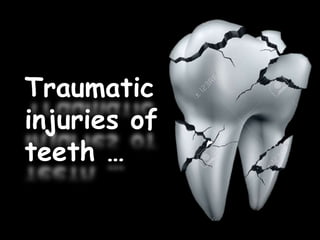
Traumatic injuries of teeth
- 2. General aspects Crown fracture Crown root fracture Root fracture
- 3. Contents Introduction Classification general aspects - history of presenting illness - clinical examination
- 4. - Mechanical stimulation - Thermal tests - Electrometric tests - Laser doppler flowmetry - Radiographic examination Crown fractures Crown root fractures Root fractures
- 5. Endodontic management of traumatized teeth Orthodontic management of traumatized teeth Conclusion References
- 6. Introduction Traumatic dental injuries usually imply wound healing processes in the periodontium, the pulp and sometimes associated soft tissue. The outcome of these determines the final healing of such injuries
- 7. The general response of soft and mineralized tissues to surgical and traumatic injuries is a sensitive process, where even minor changes in the treatment procedure may have an impact upon the rate and quality of healing
- 8. Epidemiology Dental trauma is common in childhood & adolescent Incidence of dental trauma is 31-40% of boys & 16-30% of girls at 5 years of age Incidence of dental trauma is 12-33% of boys & 4-19% of girls at 12 years of age Boys are affected almost twice as often as girls in both the dentitions
- 9. Classification -Classification by Rabinowitch(1956) - Classification by Ellis and Davey(1960) - Classification by WHO 1978 - Andreasen’s modification of WHO classification 1981 - Classification by Garcia Godoy(1981) - modification of Ellis classification by Mc Donald,Avery and Lynch(1983) - Classification by Ulfohn(1985) - Classification by Heithersay and Marde - Classification by Hargreaves and Craig - Currently recommended classification by WHO modified by Andreasen and Andreasen
- 10. I. Classification by Rabinowitch 1956 Injuries to primary teeth - Fracture of enamel, or slightly into dentine - Fractures into dentine - Fractures into the pulp - Fractures of the root - Comminuted fractures - Displaced teeth
- 11. II. Classification by Ellis and Davey 1960 - Class I – simple fracture of crown involving only enamel - Class II- extensive fracture of crown with considerable amount of dentine, with no pulp exposure - Class III- extensive fracture of crown, with dentinal involvement and pulp exposure - Class IV- traumatized tooth becomes non vital with or without loss of crown structure
- 12. - Class V – tooth lost due to trauma - Class VI- fracture of root with or without crown fracture - Class VII – displacement of the tooth without crown or root fracture - Class VIII – fracture of the crown en masse - Class IX- fracture of deciduous teeth
- 13. III. WHO Classification 1978 873.60- enamel fracture 873.61- crown fracture involving enamel, dentine without pulpal involvement 873.62- crown fracture with pulpal involvement 873.63- root fracture 873.64- crown root fracture 873.66- luxation 873.67- intrusion or extrusion 873.68- avulsion 873.69- other injuries like soft tissue lacerations
- 14. IV. WHO classification modified by Andreasen 1981 873.64- uncomplicated crown root fracture without pulp exposure 873.64 – complicated crown root fracture without pulp exposure 873.66- concussion- injury to tooth supporting structure without loosening or displacement of tooth
- 15. 873.66- subluxation – an injury to tooth supporting with abnormal loosening or displacement of tooth 873.66- lateral luxation, displacement of tooth in a direction other than maxillary and accompanied by fracture of alveolar socket
- 16. V. Classification by Garcia and Godoy(1981) Class I- enamel fracture Class II- enamel and dentine fracture Class III- enamel and dentine fracture with pulp exposure Class IV- enamel, dentine and cementum fracture
- 17. Class V- root fracture Class VI – concussion Class VII- luxation Class VIII- extrusion Class IX- avulsion – total displacement
- 18. VI. Modification of Ellis classification by Mc Donald, Avery,Lynch (1983) Class 1: simple fracture of crown involving little or no dentine Class 2: extensive fracture of crown involving considerable dentine but not dental pulp Class 3: extensive fracture of crown with pulp exposure Class 4: loss of entire crown
- 19. VII. Classification by Ulfohn(1985) a. Fracture of enamel b. Fracture of the crown with indirect pulp exposure through the dentine c. Fracture of crown with direct pulp exposure
- 20. VIII. Classification by Heithersay and Marde Class I- fracture line does not extent below the level of attached gingiva Class II- fracture line extends below attached gingiva, not below alveolar craest Class III- fracture line extends below alveolar crest Class IV- fracture line is within coronal third of root but below level of alveolar crest
- 21. Class I: no fracture /fracture of enamel only with or without displacement of tooth Class II: fracture of crown involving enamel, dentine without pulp exposure, with/without tooth displacement Class III: Fracture of crown exposing pulp, with/without displacement of tooth Class IV: fracture of root with/ without coronal fracture, with/without displacement of tooth Class V: total displacement of tooth IX. Classification by Hargreaves and Craig
- 22. X. Classification currently recommended by WHO modified by Andreasen and Andreasen Soft tissues Lacerations – 873.69 Contusion-N 902.0 Abrasions-N 910.00
- 23. Tooth fractures N 873.60- enamel fracture N873.61- crown fractures- uncomplicated (no pulp exposure) N873.62-crown fractures- complicated(with pulp exposure) N873.64-crown root fractures N873.63- root fractures
- 24. Luxation injuries 873.66- tooth concussion 873.66- subluxation 873.66- extrusive luxation 873.66- lateral luxation 873.67- intrusive luxation 873.68- avulsion
- 25. Facial skeletal injuries Alveolar process- maxilla/mandible Body of the maxillary/ mandibular bone Temporomandibular joint
- 26. Classification according to J O Andreasen & F M Andreasen Injuries to hard dental tissues and pulp Injuries to the periodontal tissues Injuries to the supporting bone Injuries to gingiva or oral mucosa
- 27. General aspects in dental injury History Clinical examination Mechanical stimulation Thermal tests Electrometric tests Laser doppler flowmetry Radiographic examination
- 28. History Delirium Time elapsed b/n injury and treatment Place of accident Nature of accident
- 29. Previous treatment Multiple injuries Medical history Cerebral involvement
- 30. Spontaneous pain Reaction to thermal stimuli Pain during mastication
- 31. Clinical examination Extraoral wounds Injury – oral mucosa, gingiva Crown – extent of fracture, pulp exposure, change of colour
- 32. Displacement of teeth Occlusion Mobility of teeth , alveolar fragments Palpation of alveolar process
- 33. Tenderness of teeth Pulp sensibility test
- 34. Mechanical stimualtion Exposed dentine – probe – sensibility Test cavity Exposed pulp – cotton pledeget soaked in saline
- 35. Thermal test Heated gutta percha Ethyl chloride Ice Carbon dioxide snow Dichlor- difluormethane
- 36. Electric pulp test Current is carried ionically through the electrolytes of tooth - Teeth wity temporary crown- dist b/n crown and electrode-1mm or else thermal pulp test with carbon dioxide snow - Stage of eruption - Teeth orthodontic treatment
- 37. Laser doppler flowmetry Laser beam – coronal aspect of pulp Reflected light scattered by RBCs- Doppler frequency shift Scattered light- detected and processed to yield a signal
- 38. Laser doppler flowmetry Laser beam – coronal aspect of pulp Reflected light scattered by RBCs- Doppler frequency shift Scattered light- detected and processed to yield a signal
- 39. - Disadvantages Blood pigment – interferes with laser light transmission Equipment – refinement Expensive
- 40. - Radiographic examination Stage of root formation Injuries affecting root, periodontal structures Root fractures – fracture line runs parallel to central beam
- 41. - Displacement – Lateral, extrusive luxations- widening of PDL space Intrusive – blurred PDL Differing angulations - necessary
- 42. - Steep occlusal exposure – root fractures, lateral luxations with oral displacement of crown Extraoral r/g- direction of dislocation of Intruded primary incisors Bone fractures – intraoral r/g unless fracture is confined to facial/ lingual bony plate
- 43. - Jaw fractures – extraoral r/g – mandatory
- 45. Classification based on anatomic, therapeutic and prognostic considerations 1.Enamel infraction 2.Enamel fracture 3.Enamel dentine fracture 4.Complicated crown fracture * Permanent dentition – 26- 76% of dental injuries
- 47. Etiology - falls - contact sports - automobile accidents - foreign bodies striking the teeth - Ocupational hazard
- 48. Infractions - Cause - Patterns - Visualization - Associated injuries
- 49. Fractures - Single tooth involveemnt - Max incisors - Incisal edge, facial surface - Concomitant luxation injuries - Fracture of unerupted permanent teeth
- 50. Fractures - Single tooth involveemnt - Max incisors - Incisal edge, facial surface - Concomitant luxation injuries - Fracture of unerupted permanent teeth
- 51. - Examination – cleansing the area with water spray - Pulp exposure - Dentine sensitivity - Pulp sensibility test
- 54. Radiographic examination - Size of pulp, root development - Record - Verify hard tissue barrier over exposed pulp
- 55. Pathology Infractions - ground sections : dark lines parallel to enamel rods Fractures - dentine exposure - 20,000- 45,000 tubules
- 56. - Bacterial penentration - Fluid movement . Pulpal blood flow . Neurogenic stimulation - Inflammation of pulp
- 57. Treatment Enamel infraction - No treatment - Multiple lines – seal with unfilled resin
- 58. Enamel dentine fractures - Smoothing fracture lines - Combined with orthodontic extrusion of fractured tooth - Restoration
- 59. - Maintain anatomy and occlusion crown - Reattachment - Temporary crown ?? – risk of leakage
- 62. Treatment strategy - Use of dentine bonding agents : should exposed dentine be lined ? - Ca(OH)2 lining - Microleakage
- 63. - Dentine bonding agent - Pulpal response ?? ** hard setting Ca(OH)2 liner/ GIC + dentine bonding agent - hermetic seal
- 64. Complicated crown fractures/ enamel dentine crown fractures with pulp exposure - Treatment of exposed pulp - Provisional treatment of crown fractures . Stainless steel crowns . Composite resin- moulded using acrylic crowns . Splints
- 65. Definitive management of crown fractures Previously – cast semi permanent restorations • Composite restorations • Reattachment • Full coverage crown • Laminate veneers
- 67. Composite resin - Microfilled /macrofilled /hybrid composites - Maxillomandibular relation - Support for centric, protrusive and protrusive lateral functions
- 68. • Highly polishable hybrids – 0.04microns microparticles, 1-3 micron macroparticles • Heavy filled microfilled composites – 0.04 microns • Semi polishable hybrids- 1-3 microns
- 69. Shade selection – utmost importance Customized composite shade guide
- 70. Pulp protection Dentine exposure – changes in the pulp ? Lining the deepest part of fracture with hard setting Ca(OH)2 followed by resin bonding Splint – prior to application of splint, pulp dentine protection
- 71. Primary teeth • Sharp edges • F varnish
- 72. Preparation Bevel – not placed Chamfer/shoulder Bullet nosed diamond Cervically – 1mm beyond fractured enamel and Depth – half of enamel thickness
- 73. Chamfer – not too shallow Functions - Removes acid resistant enamel - Resin lap joint - Finish line - Prism orientation
- 74. Acid etching 4 important considerations 1. Method 2. Time 3. Concentration 4. Type of acid used
- 75. Bonding resins - Allows clinician –manipulate the composite material - Air inhibited layer – tight bond b/n the composite and the bonding resin
- 76. Insertion of composite material Setting against a matrix – smooth finish Resin crown form Odus Pella and Interberg crown forms
- 77. Points to bear in mind : - Minimum labial finishing after composite polymerizing - Composite excess should not extend > 1mm - Vent holes , air escape
- 78. • Crown matrix adapted • Wooden wedge • Crown form compressed labiolingually
- 79. Polymerising composite material Factors - Time of application - Plane of direction of light source - Distance b/n light and composite - Shade of composite - Nature of filler particle - Temperature
- 80. Caution exercised to avoid “Skin effect” Undercuring
- 81. Finishing Labial margin – thin tapered carbide bur White line Finishing knife, interproximal finishing knives Abrasive strips White stone
- 82. Disc contouring Aluminium oxide disks Superfine disc – hybrid composite Paste polishing Paste and rubber cup Abrasive points and Prisma Gloss
- 83. Occlusion – verified , microfilled composite- cohesive failure is seen Light filled on heavy filled composite resin sandwich technique
- 85. 1) It is a conservative procedure 2) Appears more natural than any other composite. 3). color stability (Busato et al, 1998).
- 86. 4) Total chairside time for re-attachment of incisal edge is less than constructing a composite resin incisal edge(Cavalleri and Zerman, 1995). 5) The method is much more economical.
- 87. [I; Techniques 1.Placement of a circumferential bevel before are attaching the fragment (Simonsen, 1979 and Burke, 1991 andWalker, 1996). 2. Placement of an external chamfer at the fracture line after bonding(Baratieri, 1994). 3. Use of a v-shaped enamel notch.
- 88. 4. Placement of an internal groove ( Walker, 1996 and Baratieri, 1994). 5. Leaving a superficial overcontour of Restorative material over the fracture line (Reis et al, 2001 and Badami et al, 1995). Indian J.Sci.Res. 6(2) : 163-170, 2015 Chaudhary et al
- 89. Adv- restoring fracture with material that abrades the same way as the opposing teeth Minimal chair time Prior to advent of DBA, reattachment was done with acid etching
- 90. Bonding using DBA – bond strength – 50-60 % of intact teeth Remaining dentine thickness – min 0.2mm Gluma Dentine bonding system Most important aspect – retrieval of the fragment
- 91. Treatment strategy Depends on distance b/n pulp and fracture surface If small- fracture surface – covered with hard setting Ca(OH)2, provisional restoration Liner placement on enamel- removal of temporary restoration
- 92. Interim period – fragment kept moist If distance b/n pulp and fracture surface is more – reattachment at the time of injury Temporary restoration – not eugenol Due to complexing action of eugenol with calcium in sound dentine
- 93. ** The purpose of this study was to determine the effects of various drying and wetting storage periods on the fractured fragment and to reach a final conclusion as to which rehydration period is both better and more practical under clinical conditions.
- 95. Results -Compared to a 30-minute period, a 24-hour rehydration of the tooth fragment before treatment seems to salvage enough moisture to result in an increase in reattachment strength. Operative Dentistry, 2012, 37-5, 501-508 F Shirani et al
- 111. Esthetics following reattachment - Discoloration - Light cured composite , double chamfer preparation
- 112. Fragment retention – questionable - Nowadays treated as a method to maintain esthetics until a final treatment option is planned
- 113. Laminate veneers …
- 114. Fracture strength – increased , prep limited to enamel Better esthetic results Good material adaptation Minimal plaque retention Strength and survival- improved
- 115. Patient selection - If composite build up is done – at least 2 weeks should elapse before initiating preparation and impressions for ceramic veneer - Dimensional stability of composite
- 116. Prognosis Permanent teeth Follow up after trauma - Pulp sensibility – lowered imm after injury - 1-8 weeks - Pulp testing done without removing temp restn
- 117. Enamel infractions - Isolated infractions – risk if pulp necrosis less Enamel fractures - Risk of pulp necrosis – 0.2-1.0%
- 118. - Enamel dentine fractures without pulp Involvement - risk- 1-6% - Stage of root development • Constricted apices – greater risk • Extent of periodontal injury
- 119. • Extent and location of fracture horizontal, proximal superficial fractures - >> Deep proximal fractures • Effect of treatment • Time interval
- 123. CROWN ROOT FRACTURES
- 124. A fracture involving enamel, dentine and cementum Complicated/ uncomplicated Etiology – direct trauma
- 125. Clinical presentation Fracture starts incisal to marginal gingiva- oblique course Coronal aspect – held by PDL fibers Slightly displaced fragments
- 126. Single/ multiple fracture line Pain
- 127. Radiographic presentation Imperceptible Fracture line perpendicular to central beam
- 130. Pathology Pulpal inflammation Proliferation of gingiva Osteodentine deposition ?
- 131. Treatment Emergency – stabilization of coronal fragment Definitive treatment – initiated soon Multiple fractures, removal of loose fragments , coverage with glass ionomer cement
- 132. Incomplete fractures of immature permanent teeth- amenable to orthodontic extrusion, Pulp capping, restoration
- 133. Definitive therapy includes 1. Removal of coronal fragment and supragingival restoration 2. Surgical exposure of fracture surface 3. Orthodontic extrusion of apical fragment 4. Surgical extrusion of apical fragment
- 138. Surgical exposure of fracture site
- 142. Surgical extrusion
- 143. Surgical extrusion
- 144. Surgical extrusion
- 145. Surgical extrusion
- 149. Root fractures
- 150. Fractures involving dentine,cementum and pulp and relatively uncommon among dental traumas 0.5- 7 % of injuries affecting permanent dentition Mechanism – frontal impact – compression zones labially and lingually
- 151. Histologic level – injury to periodontal ligament, stretching or laceration of pulp at fracture level Clinical findings Max central incisors, 11-20 years Uncommon in younger individuals
- 152. Associated with alveolar fractures Slightly extruded tooth , lingual direction Site of fracture – tooth mobility Diagnosis entirely based on radiograph
- 153. Radiographic findings Fracture line – at optimal angle for radiographic disclosure Visible only if central beam directed within a maximum range of 15-20 degree of fracture plane If ellipsoid radiolucent line – seen , two additional r/g should be taken
- 154. One with increased angulation – 15 degree Second – negative angulation of 15 degree to original Usually missed, later revealed – due to devpt of haemorrhage or granulation tissue b/n fragments / resorption
- 156. The aim of this study was to investigate the accuracy of cone-beam computed tomography (CBCT) in the diagnosis of vertical root fractures in a tooth with gutta-percha and prefabricated posts The CBCT scans revealed a high accuracy in the diagnosis of vertical root fractures; the accuracy did not decrease in the presence of gutta-percha. .
- 157. The presence of prefabricated posts also had little effect on the accuracy of the system, which was, of course, not statistically significant
- 160. Fracture line – Single line – usual finding Direction varies Apical/mid root fractures – facio oral direction in an incisal direction Cervical fractures – more horizontal
- 161. Fractures at the apical third – detected with steep occlusal exposure Teeth with incomplete root formation – ‘green stick fracture’ Seen as – break in continuity of root canal wall/ root surface of immature root Can also heal by hard tissue formation
- 162. Pathology Root fracture healing Initiated at the site of pulpal and Periodontal involvement- two types of wound healing response Occur independantly
- 163. Healing with calcified tissue Interposition of connective tissue Interposition of bone and connective tissue Interposition of granulation tissue
- 165. Healing with calcified tissue Uniting callus of hard tissue Dentine, osteodentin, cementum Inner part – dentine , outer- cementum First layer- cellular, atubular Later – normal tubular dentin
- 166. Cementum deposition – preceded by resorptive processes Discontinuous layer interspersed with connective tissue derived from PDL Widening of root canal space- internal surface resorption, followed by hard tissue formation Limited peripheral rounding of fracture edges
- 167. Partial pulp canal obliteration confined to apical fragment C/E - Normal mobility - Normal percussion to percussion, decreased response to pulp sensibility testing
- 168. Seen in cases of concussion, subluxation No dislocation of coronal fragment Immature root formation
- 171. Interposition of connective tissue Moderately injured pulp Pulp revascularization >> healing Periodontal ligament cells – dominate healing Connective tissue b/n fragments
- 172. Connective tissue fibres – running // fracture surface New apical foramen – formed at fracture level Peripheral rounding of fracture edges Slight ingrowth of bone PDL space around apical fragment
- 173. r/g Rounding of fracture edges Radiolucent line external, internal surface resorption & pulp canal obliteration
- 175. c/p Teeth – firm/ slightly mobile Weak pain- percussion Sensibility test- normal
- 176. Post resorptive mineralization – ankylosis Follow up
- 177. Interposition of bone and connective tissue b/n fragments Bone into root canal Result of trauma prior to completed alveolar Process, coronal fragment continues to erupt
- 179. r/g Bony bridge b/n fragments with periodontal space around pulp canal obliteration c/p Teeth – firm React normally to pulp test
- 180. Interposition of granulation tissue Histologically- granulation tissue Coronal pulp – necrotic – inflammatory changes , Often, communication b/n gingival crevice & fracture line Apical – contain vital pulp
- 181. r/g Widening of fracture line, rarefaction of alveolar bone c/p Coronal fragment – loose, extruded, sensitive to percussion Fistulae labial mucosa
- 182. Negative pulpal sensibility Fixation of orthodontic bands prior to advent of acid etching – was a cause for pulp necrosis
- 183. Treatment Reduction of displaced coronal fragment and firm immobilization Digital manipulation – repositioning Socket wall fracture ? Radiographic evaluation
- 191. Immobilization – rigid splint (acid etch resin splint)- passively applied 2-3 months – fixation period Immature teeth with incomplete root fractures – heal by hard tissue union If splinted/ not – teeth monitored radiographically and for pulp sensibility
- 192. Proximity of cervical fracture line to the gingival crevice – poor chance of healing Consider – removal of coronal fragment, orthodontic/surgical extrusion Fracture at cervical third of root – conservative management Good oral hygiene- permanent fixation of coronal fragment to adjacent teeth
- 193. If tooth – unsalvagable- extraction With great caution- with little / no sacrifice to labial bone Preserve the marginal socket wall Another option – preservation of apical fragment containing vital pulp
- 194. Primary teeth with root fractures without dislocation – preserved Coronal fragment – usually removed inorder to prevent pulp necrosis Never apical fragment is removed – damage
- 195. Pulpal healing Survival following root fracture >> than luxation injury Periodontal ligament contributes to revascularization Escape of pulpal edema through fracture Minimizing pressure
- 196. Fracture reduces the impact to the apical area Close c/l and r/g follow ups Extrusion of coronal fragment, tenderness to percussion r/g signs of pulp necrosis – seen after 2 months
- 197. Factors contributing to necrosis - Displacement of coronal fragment - Forceful application of splints - Non splinting - Teeth with completed root formation
- 200. Factors affecting type of healing Presence of restorations Presence of marginal periodontitis - Interposition of connective tissue
- 201. Management of pulp necrosis - Apical fragment – vital - Coronal fragment – root filled If fracture line is in cervical third of root, pulp necrotic – intraradicular splinting – uniting fragments and serving as root filling
- 206. Using metal endodontic implants Implant – shifts the fulcrum to more apical position
- 207. Pulp canal obliteration Common finding Partial pulp canal obliteration – fracture region & apical region Can extend into 1-2 mm of coronal fragment Complete obliteration
- 208. Both types – progress at the same rate Well advanced – 9-12 months Approach full density – 1-2 years Obliteration of Apical segment –healing with calcified tissue Apical and coronal – connective tissue
- 209. Other criteria Resorption of bone at the level of fracture Loss of sensibility Loosening of coronal fragment
- 210. Root resorption 60% cases Detected within 1 year after injury Should be differentiated from resorption of bone at the level of root fracture Types External surface resorption
- 211. External inflammatory resorption External replacement resorption Internal surface resorption Internal tunneling resorption
- 213. Endodontic management of traumatized teeth …
- 214. Crown fractures with pulp exposure - Pulp – lacerated, exposed - Healing does not occur - Bacteria - Hemorrage , inflammation
- 215. Crown fractures with pulp exposure - Pulp – lacerated, exposed - Healing does not occur - Bacteria - Hemorrage , inflammation
- 216. Proliferative/destructive changes Treatment goals Preservation of vitality Hard tissue barrier
- 217. • Pulp capping • Pulpotomy • Pulpectomy
- 218. • Factors - Maturity of root formation - Luxation injury - Age - Surgical procedure - Wound dressing
- 219. Pulp capping 24 hrs Small exposure Caoh 1* and 2* contamination
- 221. Pulpotomy Removal of damaged / inflamed tissue – clinically healthy pulp Time Exposure size
- 222. Partial / cervical pulpotomy Treatment Diamond bur, contrangled handpiece Cooling , no pressure 2mm below exposure site
- 223. Hemostasis Caoh , cotton pellet GIC, ZnOE Complications – pulp canal obliteration, pup necrosis
- 224. Pulpectomy Complete removal of pulp Interim caoh dressing
- 225. Clinical evaluation of pulp healing Histological techniques Criteria No c/l symptoms r/g Root devpt- immature
- 226. Sensitivity to electrical stimulation Follow up at 3 yrs Hard tissue barrier – clinical probing
- 227. Root fractures 25 % - pulp necrosis Apical fragment – vital Diagnosis- pulp sensibility, color change Widening of space b/n fragments
- 228. Changes- periradicular bone 3 months after injury Splinting – mandate Internal surface/ tunneling resorption – healing External inflammatory root resorption - necrosis
- 229. Treatment Endodontic treatment Coronal fragment – RCT Both fragments – necrotic – treatment Not treatable – surgical removal
- 230. Orthodontic treatment of root fractured teeth
- 231. Crown , crown root fracture - Extrusion - Heithersay 1973, - Ingber 1976
- 233. Coronal shift of marginal gingiva Stretching & readjusting of periodontal fibres To avoid relapse – fibrotomy Before retention period 3-5mmduring 3-4 weeks Resorption - rare
- 234. Root fractures Depends upon the type of healing that has occurred
- 235. Prevention Children , professional players Mouth guards -
- 236. Conclusion management of traumatic dental injuries can be both demanding and challenging that requires immediate Intervention as they are accompanied by the emotional factors of the patient The successful treatment requires often a multidisciplinary approach involing Different specialities
- 237. References 1.Ingle JI, Bakland LK. Endodontics. 5th ed. Hamilton, Ontario, Canada: BC Decker; 2002:795. 2. Malmgren O, Malmgren B, and Frykholm A. Rapid orthodontic extrusion of crown root and cervical root fractured teeth. Endod Dent Traumatol 1991;7(2):49-54. 3. Ehrmann EH. Restoration of a fractured incisor with exposed pulp using original tooth fragment: report of case. JADA 1989; 118(2):188-185. 4. Morse DR, O’Larnic J, Yesilsoy C. Apexification: review of the literature. Quintessence Int 1990;21(7):589-598. 5. Baratieri LN, Monteiro S Jr, Caldeira de Andrada MA. Tooth fracture reattachment: case reports. Quintessence Int 1990;21(4): 261-270.
- 238. . Deliperi S, Bardwell DN, Coina C. Reconstruction of devital teeth using direct fiber-reinforced composite resins: a case report. J Adhes Dent 2005;7(2):165-171. 7. Villat C, Machtou P, Naulin-Ifi C. Multidisciplinary approach to the immediate esthetic repair and long-term treatment of an oblique crown-root fracture. Dent Traumatol 2004;20(1):56-60. 8. Textbook and color atlas of traumatic dental injuires - andreasen
- 239. Thank you…