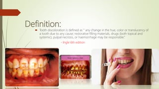
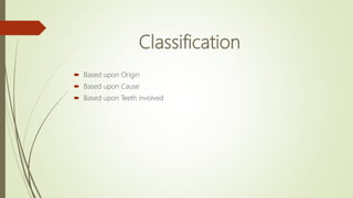
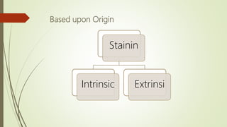
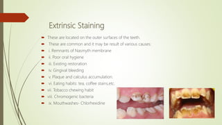
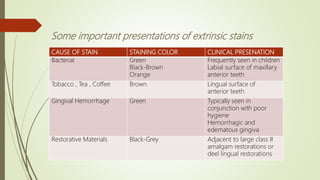
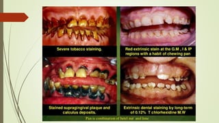
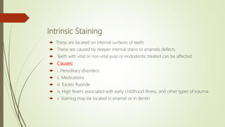
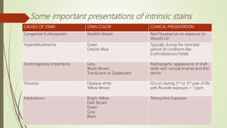
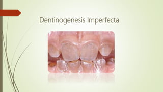
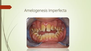
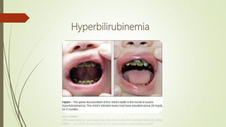
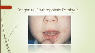
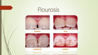
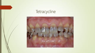
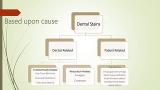
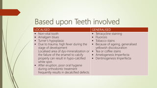
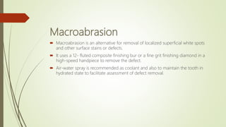
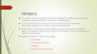
TOOTH STAINING AND
DISCOLORATION
Jabran Anwar
(BDS)
Definition:
 Tooth discoloration is defined as “ any change in the hue, color or translucency of
a tooth due to any cause; restorative filling materials, drugs (both topical and
systemic), pulpal necrosis, or haemorrhage may be responsible.”
- Ingle 6th edition
Classification
 Based upon Origin
 Based upon Cause
 Based upon Teeth involved
Based upon Origin
Stainin
Intrinsic Extrinsi
Extrinsic Staining
 These are located on the outer surfaces of the teeth.
 These are common and it may be result of various causes:
 i. Remnants of Nasmyth membrane
 ii. Poor oral hygiene
 iii. Existing restoration
 iv. Gingival bleeding
 v. Plaque and calculus accumulation.
 vi. Eating habits: tea, coffee stains,etc.
 vii. Tobacco chewing habit
 viii. Chromogenic bacteria
 ix. Mouthwashes- Chlorhexidine
CAUSE OF STAIN STAINING COLOR CLINICAL PRESENATION
Bacterial Green
Black-Brown
Orange
Frequently seen in children
Labial surface of maxillary
anterior teeth
Tobacco , Tea , Coffee Brown Lingual surface of
anterior teeth
Gingival Hemorrhage Green Typically seen in
conjunction with poor
hygiene
Hemorrhagic and
edematous gingiva
Restorative Materials Black-Grey Adjacent to large class II
amalgam restorations or
deel lingual restorations
Some important presentations of extrinsic stains
Intrinsic Staining
 These are located on internal surfaces of teeth
 These are caused by deeper internal stains or enamels defects.
 Teeth with vital or non-vital pulp or endodontic treated can be affected.
 Causes:
 i. Hereditary disorders
 ii. Medications
 iii. Excess fluoride
 iv. High fevers associated with early childhood illness, and other types of trauma.
 v. Staining may be located in enamel or in dentin
CAUSES OF STAIN STAIN COLOR CLINICAL PRESENTATION
Congenital Erythropoietic Reddish-Brown Red Flouresence on exposure to
Wood’s UV
Hyperbilirubinemia Green
Greyish-Blue
Typically during the neonatal
period of conditions like
Erythroblastosis Fetalis
Dentinogenesis Imperfecta Grey
Bluish-Brown
Translucent or Opalescent
Radiographic appearance of shell
teeth with normal enamel and thin
dentin
Flourosis Opaque white
Yellow-Brown
Occurs during 2nd or 3rd year of life
with flouride exposure > 1 ppm
Medications Bright Yellow
Dark Brown
Green
Gray
Black
Tetracycline Exposure
Some important presentations of intrinsic stains
Dentinogenesis Imperfecta
Amelogenesis Imperfecta
Hyperbilirubinemia
Congenital Erythropoietic Porphyria
Flourosis
Tetracycline
HOW TETRACYCLINES CAUSES TOOTH
DISCOLORATION ???
 One of the most obvious and well-documented side effect of tetracycline use is its
incorporation as a fluorescent pigment into tissues that are calcifying at the time of
administration
 It has ability to chelate calcium ions and to be incorporated into teeth cartilage and
bone to form a tetracyline-calcium orthophosphate complex resulting is
discoloration and enamel hypoplasia of both the primary and permanant dentitions
if administered during the period of tooth development
Based upon cause Dental Stains
Dentist Related
Endodontically Related
Pulp Tissue Remnants
Intracanal Medications
Obturating Material
Restoration Related
Amalgam
Composite
Patient Related
Pulp Necrosis
Intrapulpal haemorrhage
Dentin hypercalcification
Tooth formation defects
Developmental defects
related defects
Based upon Teeth involved
LOCALISED GENERALISED
 Non-vital tooth
 Amalgam blues
 Turner’s hypoplasia:
 Due to trauma, high fever during the
stage of development
Localised area of dys-mineralization or
the failure of the enamel to calcify
properly can result in hypo-calcified
white spot.
 After eruption, poor oral hygiene
during orthodontic treatment
frequently results in decalcified defects
 Tetracycline staining
 Fluorosis
 Tobacco stains
 Because of ageing, generalised
yellowish discolouration
 Tea or coffee stains
 Amelogenesis Imperfecta
 Dentinogenesis Imperfecta
Treatment Options
 Scaling
 Microabrasion
 Macroabrasion
 Veneers
 Ceramic Crowns
 Bleaching
Microabrasion
 Microabrasion technique involves the physical removal of tooth structure and
does not remove stains or defects through any bleaching phenomena.
 Fluorosis stains can also be removed by microabrasion if the discoloration is
within the 0.2-0.3 mm removal depth limit.
 Treated area are polished with a fluoride containing paste to restore surface lustre
and enhance re-mineralization.
Macroabrasion
 Macroabrasion is an alternative for removal of localized superficial white spots
and other surface stains or defects.
 It uses a 12- fluted composite finishing bur or a fine grit finishing diamond in a
high-speed handpiece to remove the defect.
 Air-water spray is recommended as coolant and also to maintain the tooth in
hydrated state to facilitate assessment of defect removal.
Veneers
 It is a layer of tooth-coloured material that is applied to a tooth surface to restore
localized or generalised defects and intrinsic discolorations.
 Common indications for veneers are: facial surfaces that are malformed,
discoloured, abraded, or eroded or have faulty restorations.
 Several factors should be evaluated before pursuing full veneers as treatment
option. For example, patient’s age, occlusion, tissue health, position & alignment of
teeth and oral hygiene.
 Based on material, it can be of four types:
a. Composite
b. Processed composite
c. Porcelain
d. Pressed ceramic materials
Bleaching Treatment
 Definition: “ the lightening of the colour of a tooth through the application of a chemical
agent to oxidize the organic pigmentation in the tooth is referred to as bleaching”
Sturdvent 5th edition
 Bleaching is an treatment modality involving an oxidative chemical that alters the light
absorbing and/or light reflecting nature of a material structure ,thereby increasing its
perception of whiteness . -Ingle ,6th edition.
 Bleaching will lighten teeth and the degree of lightening varies with the individual. Teeth
that tend to be yellow in colour are the easiest to lighten and give the best results.
 Darker teeth may need more time to lighten but virtually all cases involving bleaching
can have positive results.
 Whitening offers a conservative, simplified, and economical approach to changing the
color of teeth.
 As a result, tooth whitening has become one of dentistry’s most popular esthetic
treatments.
Bleaching Agents
 The active ingredient in tooth bleaching materials is peroxide compounds.
 Currently a variety of bleaching materials are available.
 the most commonly used peroxide compounds:
a) Hydrogen Peroxide.
b) Sodium Perborate.
c) Carbamide Peroxide.
d) Hydrogen peroxide and carbamide peroxide (extra-coronal).
e) Sodium perborate (intra-coronal bleaching).
Disadvantages
 Normal tooth colour may or may not be restored.
 Bleaching can cause discomfort in children because of their large pulps.
 Sometimes it might irritate the adjacent soft tissue.
 Chances of increased sensitivity are there.
 Extended treatment time may be necessary.
Contra-indications
 Patient selection – Patients with emotional or psychological problem or those with
unrealistic goals do not make good candidates for bleaching.
 Dentinal hypersensitivity – These symptoms may be associated with severe cases of
attrition, erosion, abrasion or abfraction.
 Generalized dental caries and leaking restoration – Use of bleaching agents for such
patients who fall in this category may lead to severe, generalized hypersensitivity.
 Heavily restored teeth – Teeth with visible, tooth colored restorations respond poorly
to bleaching because the composite restorations do not lighten and become more
evident after bleaching
 Teeth with opaque white spots
 Teeth slated for bonded restorations or orthodontic bracketing – Oxygen produced
during bleaching remains in the enamel or dentin oxygen interferes with the bonding
agent and induces bonding failure.
Tooth Staining and Discoloration

Tooth Staining and Discoloration

  • 1.
  • 2.
    Definition:  Tooth discolorationis defined as “ any change in the hue, color or translucency of a tooth due to any cause; restorative filling materials, drugs (both topical and systemic), pulpal necrosis, or haemorrhage may be responsible.” - Ingle 6th edition
  • 3.
    Classification  Based uponOrigin  Based upon Cause  Based upon Teeth involved
  • 4.
  • 5.
    Extrinsic Staining  Theseare located on the outer surfaces of the teeth.  These are common and it may be result of various causes:  i. Remnants of Nasmyth membrane  ii. Poor oral hygiene  iii. Existing restoration  iv. Gingival bleeding  v. Plaque and calculus accumulation.  vi. Eating habits: tea, coffee stains,etc.  vii. Tobacco chewing habit  viii. Chromogenic bacteria  ix. Mouthwashes- Chlorhexidine
  • 6.
    CAUSE OF STAINSTAINING COLOR CLINICAL PRESENATION Bacterial Green Black-Brown Orange Frequently seen in children Labial surface of maxillary anterior teeth Tobacco , Tea , Coffee Brown Lingual surface of anterior teeth Gingival Hemorrhage Green Typically seen in conjunction with poor hygiene Hemorrhagic and edematous gingiva Restorative Materials Black-Grey Adjacent to large class II amalgam restorations or deel lingual restorations Some important presentations of extrinsic stains
  • 8.
    Intrinsic Staining  Theseare located on internal surfaces of teeth  These are caused by deeper internal stains or enamels defects.  Teeth with vital or non-vital pulp or endodontic treated can be affected.  Causes:  i. Hereditary disorders  ii. Medications  iii. Excess fluoride  iv. High fevers associated with early childhood illness, and other types of trauma.  v. Staining may be located in enamel or in dentin
  • 9.
    CAUSES OF STAINSTAIN COLOR CLINICAL PRESENTATION Congenital Erythropoietic Reddish-Brown Red Flouresence on exposure to Wood’s UV Hyperbilirubinemia Green Greyish-Blue Typically during the neonatal period of conditions like Erythroblastosis Fetalis Dentinogenesis Imperfecta Grey Bluish-Brown Translucent or Opalescent Radiographic appearance of shell teeth with normal enamel and thin dentin Flourosis Opaque white Yellow-Brown Occurs during 2nd or 3rd year of life with flouride exposure > 1 ppm Medications Bright Yellow Dark Brown Green Gray Black Tetracycline Exposure Some important presentations of intrinsic stains
  • 10.
  • 11.
  • 12.
  • 13.
  • 14.
  • 15.
  • 16.
    HOW TETRACYCLINES CAUSESTOOTH DISCOLORATION ???
  • 17.
     One ofthe most obvious and well-documented side effect of tetracycline use is its incorporation as a fluorescent pigment into tissues that are calcifying at the time of administration  It has ability to chelate calcium ions and to be incorporated into teeth cartilage and bone to form a tetracyline-calcium orthophosphate complex resulting is discoloration and enamel hypoplasia of both the primary and permanant dentitions if administered during the period of tooth development
  • 18.
    Based upon causeDental Stains Dentist Related Endodontically Related Pulp Tissue Remnants Intracanal Medications Obturating Material Restoration Related Amalgam Composite Patient Related Pulp Necrosis Intrapulpal haemorrhage Dentin hypercalcification Tooth formation defects Developmental defects related defects
  • 19.
    Based upon Teethinvolved LOCALISED GENERALISED  Non-vital tooth  Amalgam blues  Turner’s hypoplasia:  Due to trauma, high fever during the stage of development Localised area of dys-mineralization or the failure of the enamel to calcify properly can result in hypo-calcified white spot.  After eruption, poor oral hygiene during orthodontic treatment frequently results in decalcified defects  Tetracycline staining  Fluorosis  Tobacco stains  Because of ageing, generalised yellowish discolouration  Tea or coffee stains  Amelogenesis Imperfecta  Dentinogenesis Imperfecta
  • 20.
    Treatment Options  Scaling Microabrasion  Macroabrasion  Veneers  Ceramic Crowns  Bleaching
  • 21.
    Microabrasion  Microabrasion techniqueinvolves the physical removal of tooth structure and does not remove stains or defects through any bleaching phenomena.  Fluorosis stains can also be removed by microabrasion if the discoloration is within the 0.2-0.3 mm removal depth limit.  Treated area are polished with a fluoride containing paste to restore surface lustre and enhance re-mineralization.
  • 22.
    Macroabrasion  Macroabrasion isan alternative for removal of localized superficial white spots and other surface stains or defects.  It uses a 12- fluted composite finishing bur or a fine grit finishing diamond in a high-speed handpiece to remove the defect.  Air-water spray is recommended as coolant and also to maintain the tooth in hydrated state to facilitate assessment of defect removal.
  • 23.
    Veneers  It isa layer of tooth-coloured material that is applied to a tooth surface to restore localized or generalised defects and intrinsic discolorations.  Common indications for veneers are: facial surfaces that are malformed, discoloured, abraded, or eroded or have faulty restorations.  Several factors should be evaluated before pursuing full veneers as treatment option. For example, patient’s age, occlusion, tissue health, position & alignment of teeth and oral hygiene.  Based on material, it can be of four types: a. Composite b. Processed composite c. Porcelain d. Pressed ceramic materials
  • 24.
    Bleaching Treatment  Definition:“ the lightening of the colour of a tooth through the application of a chemical agent to oxidize the organic pigmentation in the tooth is referred to as bleaching” Sturdvent 5th edition  Bleaching is an treatment modality involving an oxidative chemical that alters the light absorbing and/or light reflecting nature of a material structure ,thereby increasing its perception of whiteness . -Ingle ,6th edition.  Bleaching will lighten teeth and the degree of lightening varies with the individual. Teeth that tend to be yellow in colour are the easiest to lighten and give the best results.  Darker teeth may need more time to lighten but virtually all cases involving bleaching can have positive results.  Whitening offers a conservative, simplified, and economical approach to changing the color of teeth.  As a result, tooth whitening has become one of dentistry’s most popular esthetic treatments.
  • 25.
    Bleaching Agents  Theactive ingredient in tooth bleaching materials is peroxide compounds.  Currently a variety of bleaching materials are available.  the most commonly used peroxide compounds: a) Hydrogen Peroxide. b) Sodium Perborate. c) Carbamide Peroxide. d) Hydrogen peroxide and carbamide peroxide (extra-coronal). e) Sodium perborate (intra-coronal bleaching).
  • 26.
    Disadvantages  Normal toothcolour may or may not be restored.  Bleaching can cause discomfort in children because of their large pulps.  Sometimes it might irritate the adjacent soft tissue.  Chances of increased sensitivity are there.  Extended treatment time may be necessary.
  • 27.
    Contra-indications  Patient selection– Patients with emotional or psychological problem or those with unrealistic goals do not make good candidates for bleaching.  Dentinal hypersensitivity – These symptoms may be associated with severe cases of attrition, erosion, abrasion or abfraction.  Generalized dental caries and leaking restoration – Use of bleaching agents for such patients who fall in this category may lead to severe, generalized hypersensitivity.  Heavily restored teeth – Teeth with visible, tooth colored restorations respond poorly to bleaching because the composite restorations do not lighten and become more evident after bleaching  Teeth with opaque white spots  Teeth slated for bonded restorations or orthodontic bracketing – Oxygen produced during bleaching remains in the enamel or dentin oxygen interferes with the bonding agent and induces bonding failure.