Nose anatomy and physiology
•Download as PPTX, PDF•
8 likes•3,754 views

This ppt contains anatomy and physiology of Nose and Paranasal sinuses, tried to explain all the structures with the help of CT scans.

Recommended
More Related Content
What's hot
What's hot (20)
Similar to Nose anatomy and physiology
Similar to Nose anatomy and physiology (20)
More from Shraddha Joshi
More from Shraddha Joshi (20)
Recently uploaded
Recently uploaded (20)
Nose anatomy and physiology
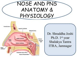
- 1. NOSE AND PNS ANATOMY & PHYSIOLOGY Dr. Shraddha Joshi Ph.D. 1st year Shalakya Tantra ITRA, Jamnagar
- 2. External Nose Skeletal frame work: Bony (upper 1/3rd) and Cartilaginous (Lower 2/3rd). Root of Nose : Continues with forehead. Base of Nose: Triangular in shape. 2 openings- Nostrils (separated by columnella).
- 3. Bony Part: Supports upper part of external nose. Consist of: Nasal Bone. Nasal Spine of Frontal bone. Frontal process of Maxilla.
- 4. Cartilaginous Part: Supports lower part of external nose. Consist of: 1.Paired upper lateral cartilage. 2.Paired lower lateral cartilage. 3.Alar Cartilage. 4.Septal Cartilage.
- 5. Upper lateral cartilages: Intra nasally lower free border is seen as Limen vestibuli/ Nasal Valve/Limen nasi.
- 6. Alar cartilages: Forms contour and nasal tip. Naso- Septal cartilage: Nasal lateral cartilages and septal cartilage.
- 7. Nasal Musculature Procerus Nasalis (transverse and alar parts) Levator labii superioris alaeque nasi Anterior and posterior dilator nares Depressor septi. Nasal Skin Over Nasal Bones and upper lateral cartilages: freely movable. Over alar cartilages: Adherent Contains sebaceous glands: (Hypertrophy- Rhinophyma)
- 8. Internal Nose Nasal cavities: • Divided into 2 parts: Vestibule (Skin lined) Nasal cavity proper (Mucosa lined). • Vestibule: Sebaceous gland, hair follicles and vibrissae. • Nasal valve: Narrowest area of nose and regulates airflow and resistance on inspiration.
- 9. Cottle’s test Used in nasal obstruction due to abnormality of nasal valve. Intended to help focus on the where in the nasal airway congestion is centered. The possibilities are several: congestion can be caused by enlarged nasal turbinates, collapsed nasal valves, a damaged, deviated septum, from nose tip to bridge), or a sinus condition that affects the nasal passages.
- 11. Dangerous area of face
- 12. Dangerous area of Nose Infection may spread into meninges along the pia and arachnoid sheath of olfactory nerves. This area is also connected to superior sagittal sinus and cavernous sinus by venous channels.
- 13. The nose forms the gateway of the respiratory system and serves the following important functions. 1. Respiratory passage: Normally, breathing takes place through the nose. • The inspired air passes upwards in a narrow stream medial to the middle turbinate and then downwards and backwards in the form of an arc, and thus respiratory air currents are restricted to the central part of the nasal chambers. • Any anatomical or pathological obstructive lesion in this region is important, as this disturbs the air flow. Physiology
- 14. 2. Filtration: The nose serves as an effective filter for the inspired air. • Vibrissae (nasal hair) in the nasal vestibule arrest large particulate matter of the inspired air. • The fine particulate matter and bacteria are deposited on the mucus blanket which covers the nasal mucosa. • The mucus contains various enzymes like lysozymes having antibacterial properties. • The mucus with the particulate matter is carried by the ciliary movements posteriorly to the oropharynx, to be swallowed.
- 15. Acute infective condition of the root of the hair follicle or sebaceous gland in the nasal vestibule. Caused by Staphylococcus aureus. Predisposing factors-Trauma as in nose picking Pulling of hair are the usual. C/F- Very painful and these tissues are very tender. Localised redness with swelling of the nasal vestibule and adjacent columella. Dangerous condition as the infection can spread to adjacent tissues of face and upper lip causing cellulitis of the face. Tt.- Local heat, antibiotic, analgesics. Squeezing or incising the furuncle should be avoided as this can lead to dissemination of the infection. Recurrent boils- Either due to frequent trauma like in nose picking or suggest an underlying debilitating disease like diabetes. Furunculosis of Nose
- 17. VESTIBULITIS Diffuse infection of the skin of the anterior nares. Causes: Frequent trauma as in nose picking. This produces traumatic ulceration and crusting, thus giving a foothold to the infection. Types: Acute and Chronic. Persistent nasal discharge leads to excoriation and infection of the skin of the nasal vestibule. T/t: Avoid predisposing factors. Cleaning with hydrogen peroxide. Local ointment
- 18. Erysipelas Inflammation of skin and subcutaneous tissue of the nose. Streptococcal infection. C/F: Bright red, Shiny and well defined lesion in the midface. Resembles butterfly shape. Lesion: Tender and may be associated with lymphadenopathy. Millian’s sign is positive. Millians sign to differentiate erysipelas from cellulitis. Pinna has no deeper dermis and subcutaneous tissue so there cannot be cellulitis.
- 19. Examination of External Nasal Framework Skin: Inflammation, scar, sinus, discoloration, vesicles, fistula, swelling. • Inflammation: Furunculosis. • Scar: Surgery, Trauma • Discoloration: Erysipelas, Sarcoidosis, Lupus Perino. • Vesicles: Herpes simplex and Zoster. • Swelling: Dermoid, Glioma, Encephalocele, Mucocele, Osteoma, Keloid. **Dermoid- in midline of root of the nose and fluctuant, trans- illumination test negative. **Encephalocele and Mningiocele- Frustenberg sign positive.
- 20. Dermoid
- 23. Darier’s Line Thickening of skin of tip of the nose. Fistula BCC- Common site- Ala nasi. • Slightly raised nodule, break down to form crust and edges are raised and rolled, but not elevated so high. SCC • Elevation of skin, which later forms a nodule that breaks to form an ulcer, which is everted, with raised edges, indurated base. Anaesthesia of Malar area.(in orbital fracture, fracture of osteomyelitis of maxillary sinus, malignancy of maxillary sinus). Broadening of nose
- 24. Darier’s line Fistula of nose SCC BCC
- 25. Congenital: Hump, Saddle Nose, Columnellar retraction or destruction, Bifid nose, Supra tip depression, crooked nose. Acquired: • Hump nose Polly beaked nose • Saddle nose Pig nose deformity • Crooked nose Knock knee deformity • Deviated nose Step ladder deformity • Inter-canthal distance increased Hump nose Saddle nose
- 26. Polly beak nose Pig nose deformity Crooked nose
- 27. Palpation Skin mobility: • Decreased- Scar, malignancy, Surgery. • Increased thickness- abscess, cyst, rhinophyma,. • Fluctuation- Abscess and cyst. • Pain, tenderness, rise in temp.- inflammation. • Crepitation over nasal bone- fracture. Columella examination Look for: • Furuncle • Fissure • Crusting • Ulceration
- 28. Vestibule examination Look for: • Follicles- Folliculitis • Pus pointing- Furunculosis • Oedema- Vestibulitis • Scar- Trauma, Sx, Malignancy • Crust- Eczema, Vestibulitis, Rhinitis sicca • Stenosis- Infection, Rhinoscleroma, Post surgical, Synechia
- 29. Nasal Septum Osteo-cartilaginous partition between two halves of nasal cavity. Columella- Small anterior skin lined area. Membranous part.
- 30. Blood Supply
- 31. Venous drainage Nerve Supply Facial Vein Sphenopalatine vein Internal nasal branch of anterior ethmoidal nerve. Nasopalatine branch of maxillary and olfactory nerve
- 32. Nerve Supply
- 33. Bleeding point (in little’s area) DNS Spur Ulcer (due to trauma, Irritant) Perforation (look for site, size, if associated with crust) Swelling ( Septal abscess, Septal Haematoma, Foreign body) Growth (look for site, extent, bleed on touch) Examination of Nasal Septum
- 34. Deviated Nasal Septum Compensatory hypertrophy Cottle’s Classification: Simple DNS, Obstructed DNS, Impacted DNS.
- 36. Anterior Dislocation Septal deviation and perforation
- 37. Etiology: • Trauma • Developmental errors. C/F: OSMHEAD • Nasal Obstruction • Sinusitis • Middle Ear Infection • Headache • Epistaxis • Anosmia • External Deformity
- 38. Nasal Spur
- 39. Caudal dislocation Jacobson’s organ
- 40. Perforation Size- • Small- Upto 1 cm in diameter • Medium- 1-2 cm in diameter • Large- Over 2 cm in diameter
- 41. Septal abscess Septal Haematoma
- 42. Fractures of Nasal septum Etiology: • Trauma- from front, side or below. • Fracture- vertically, horizontally, buckle on itself or crushed into pieces. Clinical features: • Profuse epistaxis (with mucosal tears) • Septal Haematoma (with intact mucosa) Jarjaway fracture Chevallate fracture
- 44. Jarjaway- when front blow. Chevallet- in blow from below. Haematoma- Drain Dislocation and fracture- Repositioned. Nasal Packing
- 45. Nasa Sharira नासा घ्राणेन्द्रिय स्थानं | पान्द्थिवास्तु गरधो गरधेन्द्रियं | Synonyms- नस्य, कुलय, गरधवह, गरध, घ्राण, नान्द्सका, घोण नासा प्रमाण- ‘ स्वाङ्गुली प्रमाणेन चतुरङ्गुला नान्द्सका’ (Ch.Vi.) नासापुट प्रमाण- ‘न्द्िभागेत्यन्द्ि अङ्गुलस्य न्द्िभागेन सह अङ्गुल न्द्िभागा ङ्गुलं’ (Dalhan) Nasaputa is 1 1/3 anguli as inner breadth and 2 anguli as external breadth.
- 46. Features of Nasa suggesting longevity in a baby/ Nasa Sampata: • Riju- Regular straight bridge of nose. • Mahoshvasa- No any obstruction and allows free breath. • (Ch. Sha.) नाताग्र – Downward tip of nose. • (Va. Sha.) उरनताग्र – Tip of nose is upwards. Nasa Asthi/ Bones: • Taruna Asthi (Charaka- Ghonasthi). • Number- 03 Nasa Sandhi: • Number- 02
- 47. Nasa Peshi: • Number- 02 Nasagata Sira: • 24 Sira (V- 06, P-06, K-06, R-06) • Avedhya Sira- 04 (Venesection is contraindicated) • Siravedhana is indicated in case of anosmia and other diseases of nose. Nasagata Dhamni: • Number-02 • For perception of smell.
- 48. Bahirmukha Strotasa: • 2 Nasaputa/ Nasa Randhra. • Pranavaha Strotasa commences from Bahya Nasa Randhra. Marma: घ्राण मागं उभयतः स्रोतोमागि प्रन्द्तबद्धे अभ्यरतरतः फ़णे, ति गरधाज्ञानं |(Su.Sha.) फ़णावुभयतो घ्राण मागं श्रोि पथानुगो | (Va. Sha.) • Sira Marma, • Vaikalyakara Marma
- 49. Shringataka Marma:- • Palm sized • Near Talu, where confluence of nourishing dhamnis/ strotas of nose, ear, eye and tongue are situated. • Number- 04 • Sira Marma • Sadhya Pranahara Marma
- 50. घ्राणे भृशं िाह समन्द्रवते तु न्द्वन्द्नःसरेि धूमं इवेहः वायुः | (Su.Utt.) रक्ते न नासा िग्धेव बह्यरतः स्पशिनासहा | भवेि धूमो उच्च्शच्च्शवास ....... (Va. Utt.) Nasa Dipta Severe burning sensation Pitta and Rakta Dosha is vitiated. Feeling as if nose id ignited both from inside and outside Severe Hyperasthesis
- 51. Nasa Paka घ्राण आन्द्श्रते न्द्पत्तमरुन्द्रि कुयाित अन्द्स्मन न्द्वकारे बलवांश च पाकं | ........... न्द्वक्लेि न्द्वकोथश्च अन्द्प यि .........|| (Su.Utt.) पचेत् नासापुटे न्द्पत्तं त्वक् मांसं िाह शुलवत |(Va. Utt.) Severe suppurative changes in tvaka and mamsa Pitta and Rakta Dosha is vitiated. Multiple boils and ulcerations With burning sensation and pain Necrosis Excessive moistening
- 52. Nasal Cavity Proper Roof Floor Lateral wall Medial wall
- 53. Lateral wall
- 54. Inferior turbinate: Separate bone. NLD opening- Inf. Meatus. Hasner’s valve.
- 55. Middle turbinate: Part of ethmoid bone. Attached to lateral wall by basal lamella. Attachment is in S shape manner. • Ant. 3rd lies in sagittal plane and attached to lateral edge of cribiform plate. • Middle 3rd- frontal plane- lamina papyracea. • Post. 3rd- Horizontally- forms roof of middle meatus- attached to lamina papyracea & medial wall of maxillary sinus.
- 61. Structures of Middle meatus Uncinate process Bulla Ethmoidalis Antrum of middle meatus Agger Nasi cells Uncinate process: • Hiatus semilunaris. • Attachments • Infundibulum
- 62. Lamina Papyracea Middle turbinate Skull base
- 63. Bulla Ethmoidalis: • Ethmoidal cell • Situated behind uncinate process. • May extend superiorly to skull base. • Posteriorly to fuse with ground lamella. • Supra bullar or retro bullar recess.
- 66. Concha bullosa
- 67. Haller cells
- 68. Superior Turbinate Ethmo-turbinal Situated post. and superior to Middle turbinate. May get pneumatized. Important landmark to identify ostium of sphenoid sinus which lies medial to it.
- 72. Onodi cells
- 75. They are air conditioning cavities in certain bones of skull. Anterior group sinuses: They opens into middle meatus. Maxillary Frontal Ethmoidal Posterior group sinuses: Posterior ethmoidal sinuses- Opens into superior meatus Sphenoid sinus- Opens into SER.
- 76. Maxillary Sinus Largest of all Sinuses. Pyramidal in shape. Base- towards lateral wall of nose. Apex- laterally into zygomatic process of maxilla. Capacity- 15 ml (in adult)
- 78. Anterior wall: Facia surface of maxilla. Soft tissues of cheek. Posterior wall: Infra temporal fossa Pterygo-palatine fossa Roof: Floor of orbit. Traversed by infra-orbital nerve and vessles Boundaries:
- 80. Medial wall: Middle and inferior meatuses Uncinate process Floor: Alveolar and palatine processes of maxilla. Roots of 2nd pre molar and 1st molar teeth. **Oro-antral fistula can result from extraction of these teeth. ** Dental infection can also be the cause of maxillary sinusitis.
- 82. Frontal Sinus Between inner and outer tables of frontal bone. Above to the supra orbital margin. Varies in shape and size. 2 frontal sinuses are often asymmetric. May be very large extending into orbital plate in the roof of orbit. May be deficient. Anterior wall: Skin over forehead. Interior wall: To orbit and its contents. Posterior wall: Meninges and frontal lobe of brain.
- 83. Frontal Recess Drainage: through its ostium into the frontal recess. Ostium and frontal recess forms- hour glass structure. Situation: In anterior part of middle meatus. Medially- Middle turbinate Laterally-Lamina Papyracea Anteriorly- Agger Nasi cells Posteriorly- Bulla Ethmoidalis
- 85. Ethmoidal Sinus Thin walled air cavities. Number: 3 to 18. Occupy space between upper 3rd of lateral nasal wall and medial wall of orbit. Anterior Ethmoidal group and Posterior Ethmoidal group divided by basal lamina. Anterior Ethmoidal group- opens into middle meatus. Posterior Ethmoidal group- opens into superior meatus. Roof: Medial extension of orbital plate of frontal bone, which shows depression on its undersurface- Fovea ethmoidalis. Lateral wall: Lamina payracaea
- 88. Anterior group cells: Agger nasi cells- Ethmoidal Bulla- forms posterior boundary of the hiatus semilunaris. Supra orbital cells Fronto ethmoidal cells Haller cells Posterior group of cells- Sphenoethmoidal cell (Onodi cells)- Extends along the lamina papyracea, lateral or superior to the sphenoid.
- 89. Agger nasi cells Supra orbital cells
- 90. Haller cells Onodi cells
- 91. Sphenoid Sinus
- 92. Occupies the body of sphenoid. 2 sinus, separated by a thin bony septum. Ostium of sphenoid sinus is situated high up in the anterior wall and opens into sphenoethmoidal recess. Sphenoethmoidal recess: Medial to superior or supreme turbinate. Slit like, oval or round.
- 94. Mucus Membrane of PNS: Lined by mucus membrane which is continuous with that of nasal cavity. Thinner and less vascular. Ciliated columnar epithelium. Goblet cells present. Cilia are more marked near the ostia.
- 97. Functions of Paranasal Sinuses: Air conditioning of the inspired area. Provide resonance of voice. Act as thermal insulator. Lighten the skull bones. Provide extended surface for olfaction. Provide local immunologic defence. Act as buffers against trauma and this protects brain against injury.
- 98. Ventilation of sinuses: During inspiration Negative pressure in nose. (i.e. -6mm to -200 mmH2O) During Expiration Positive pressure in nose. Sets up eddies Ventilates the sinus **Sinuses are filled during expiration and emptied during inspiration.
- 99. Mucus clearance: Maxillary Sinus: Through natural ostium. Frontal Sinus: Mucus travels to inter-frontal septum (Along the roof, along the floor) and then to natural ostium. One just above the ostium and 2nd in frontal recess, mucus recycles through the sinus. Anti-clockwise in Right frontal sinus. Clock wise in Left frontal sinus. Sphenoid sinus: Towards the ostium into the spheno-ethmoidal recess.
- 101. Ethmoidal sinus: Anterior group of sinuses: Joins that from the frontal and maxillary sinuses and travels towards Eustachian tube, from there to torus tubaris and into the naso-pharynx. Posterior ethmoidal sinuses: superior and supreme meatus, Joins the secretion of sphenoid sinus in SER , passes above and behind the torus tubaris into nasopharynx. ** Infected discharge from the anterior group of sinuses, passes behind posterior pillars and causes hypertrophy of lateral pharyngeal band. **Infected discharge from Posterior group of sinus spreads over the posterior pharyngeal wall.
- 103. Functions of Nose: Respiration Air-conditioning of inspired air Protection of lower airway. Olfaction Vocal resonance Nasal reflex functions
- 104. Natural Pathway Choanal atresia- Asphyxia- Death During Expiration- Entire current is not expelled. Friction offered at limen nasi- coverts it into eddies – ventilates the sinuses through the ostia. Breathing:
- 106. Under autonomic nervous system- Nasal mucosa undergoes rhythmic cyclic congestion and decongestion- Controls air flow. Cycle lasts 2-12 hours. Nasal Cycle:
- 107. Air-conditioner for lungs. Adjusts temperature and humidity of inspired air. Filter and Purify the inspired air. Filtration and Purification: Front of nose: upto 3 micro m Nasal mucus traps: 0.5 to 3.0 micro m Passes to lower tract: smaller than 0.5 micro m Temperature control: Large surface area of nasal mucosa. Highly vascular with cavernous venous spaces or sinusoids which control blood flow. Radiator mechanism. Air Conditioning:
- 108. Humidification: Process goes on simultaneously with temperature control. Air dry in winter and moistened in summer. Nasal mucosa adjusts the relative humidity upto 75% of inspired air. Water is provided by: Mucus and serous secreting glands. 1000 ml of water is evaporated in 24 hrs.
- 109. Mucociliary Mechanism: Mucous blanket. Moves like conveyer belt.- at the speed of 5-10mm/ min. Complete sheet of mucus is cleared in 10-20 min. Turbinates doubles the surface area 600-700 ml of nasal secretions are produced in 24 hrs. Protection of lower airways:
- 110. Beating of Cilia: Effective stoke Recovery stroke ***Immotile cilia syndrome- Rhinosinusitis and bronchoectasis. Enzymes and Immunoglobulins: Muramidase IgA and IgE Interferon Sneezing: Helps noxious substances to wash out. pH is constantly 7.
- 111. Vocal resonance: Nasal consonants (M/N/ NG)- sound passes through nasopharyngeal isthmus and emitted through nose. Nose blocked- M/N/ NG- B/D/G respectively. Nasal Reflexes: Smell- reflex secretion of saliva and gastric juice. Nasal obstruction- Pulmonary resistance increased. Nasal Packing- Lowering of PO2 Pulmonary HTN- in adenoids and tonsilar hypertrophy.