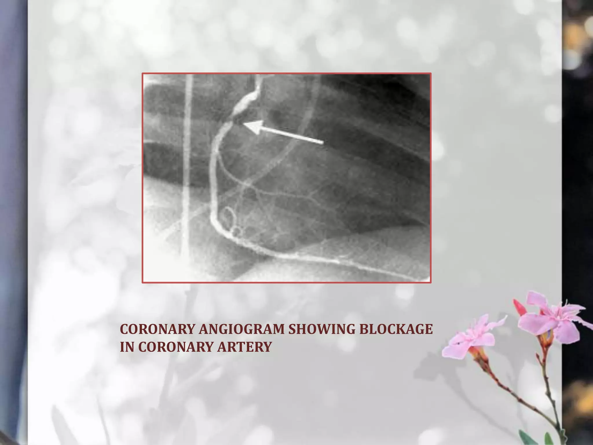
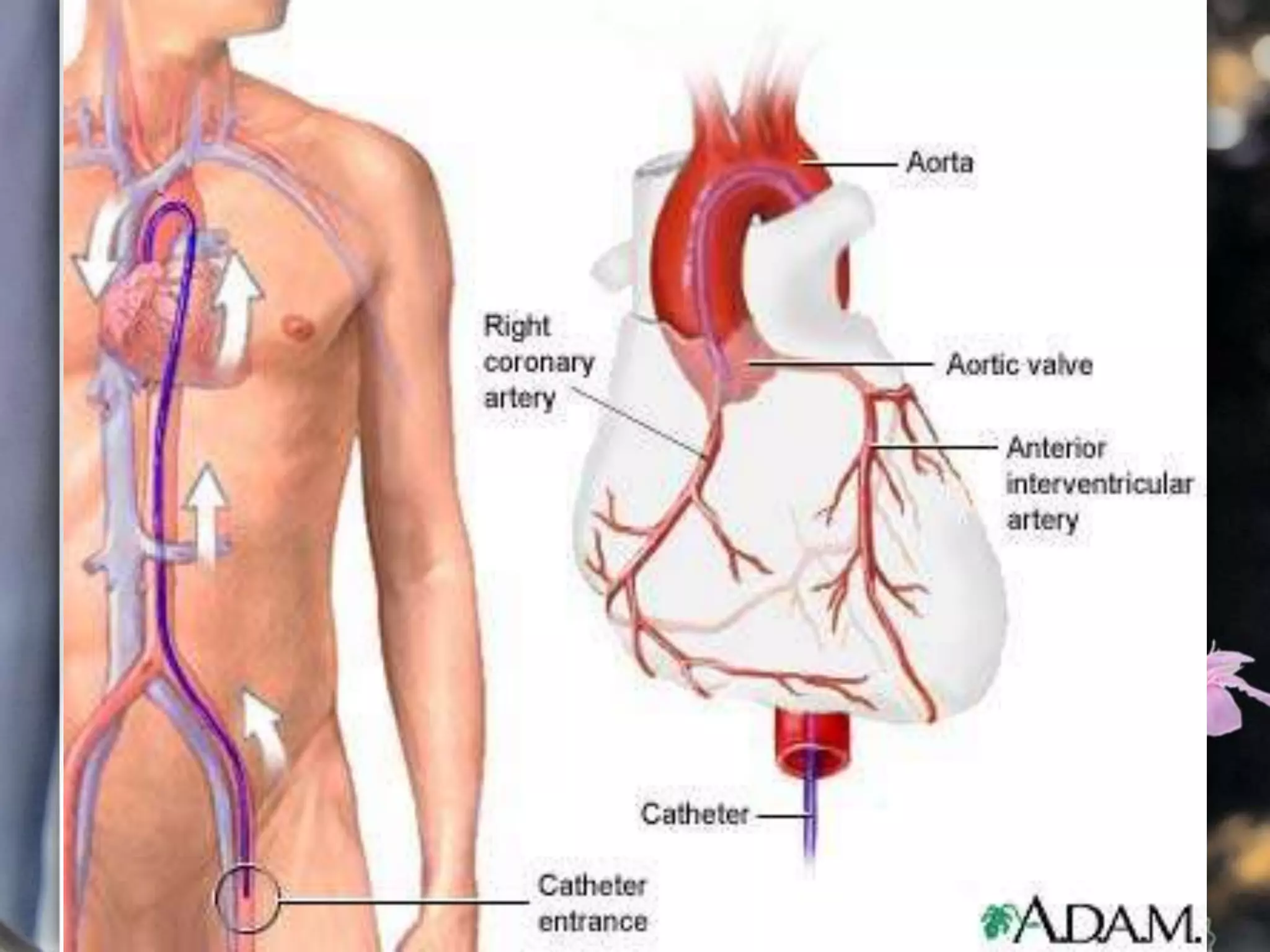
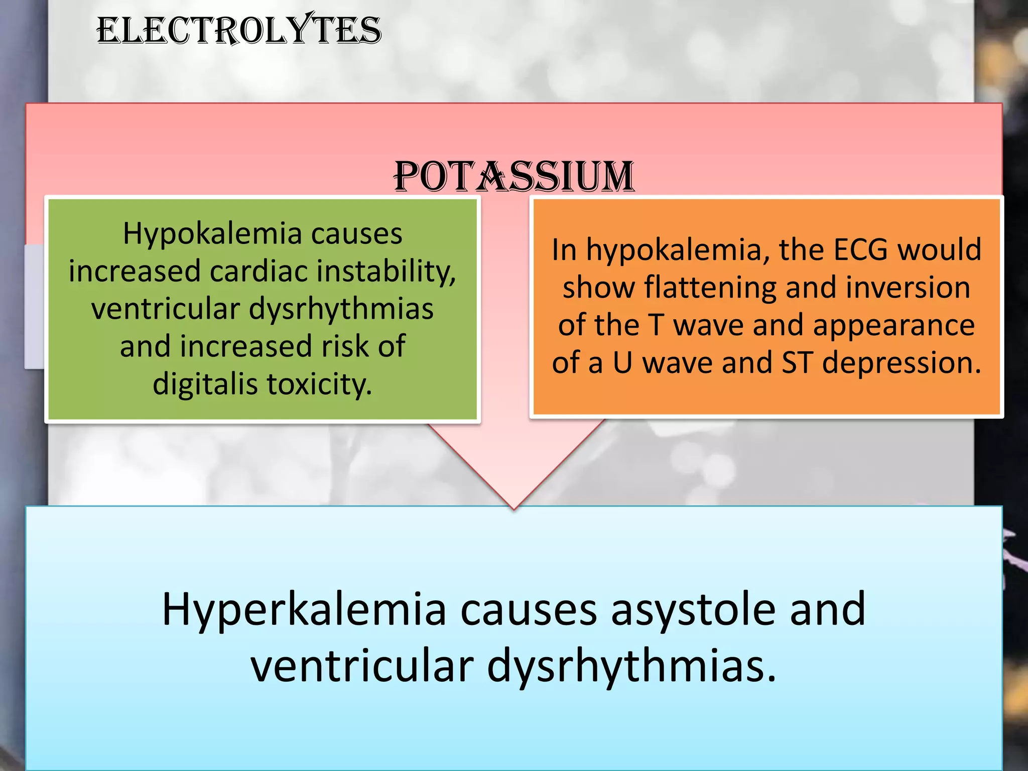
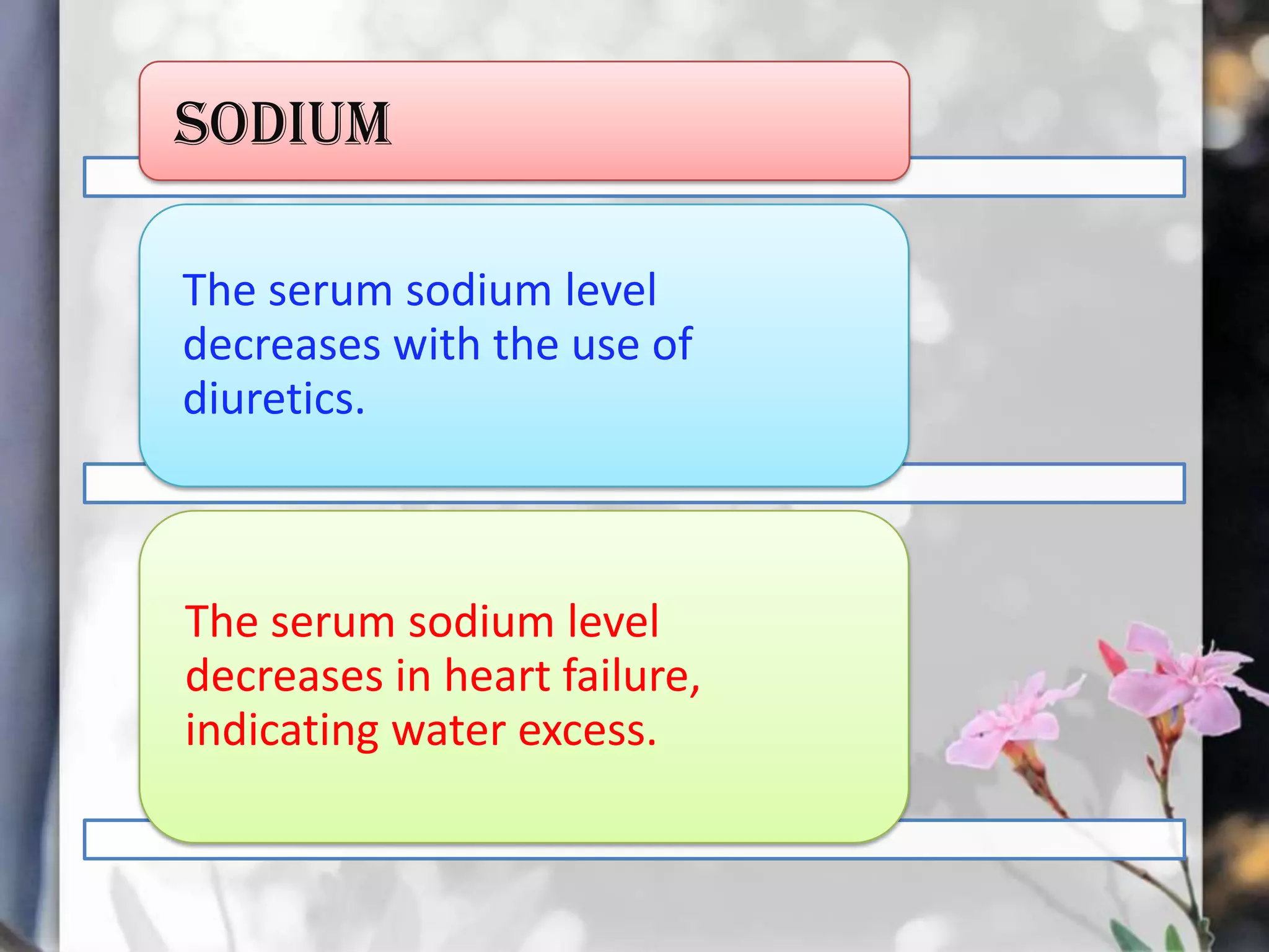
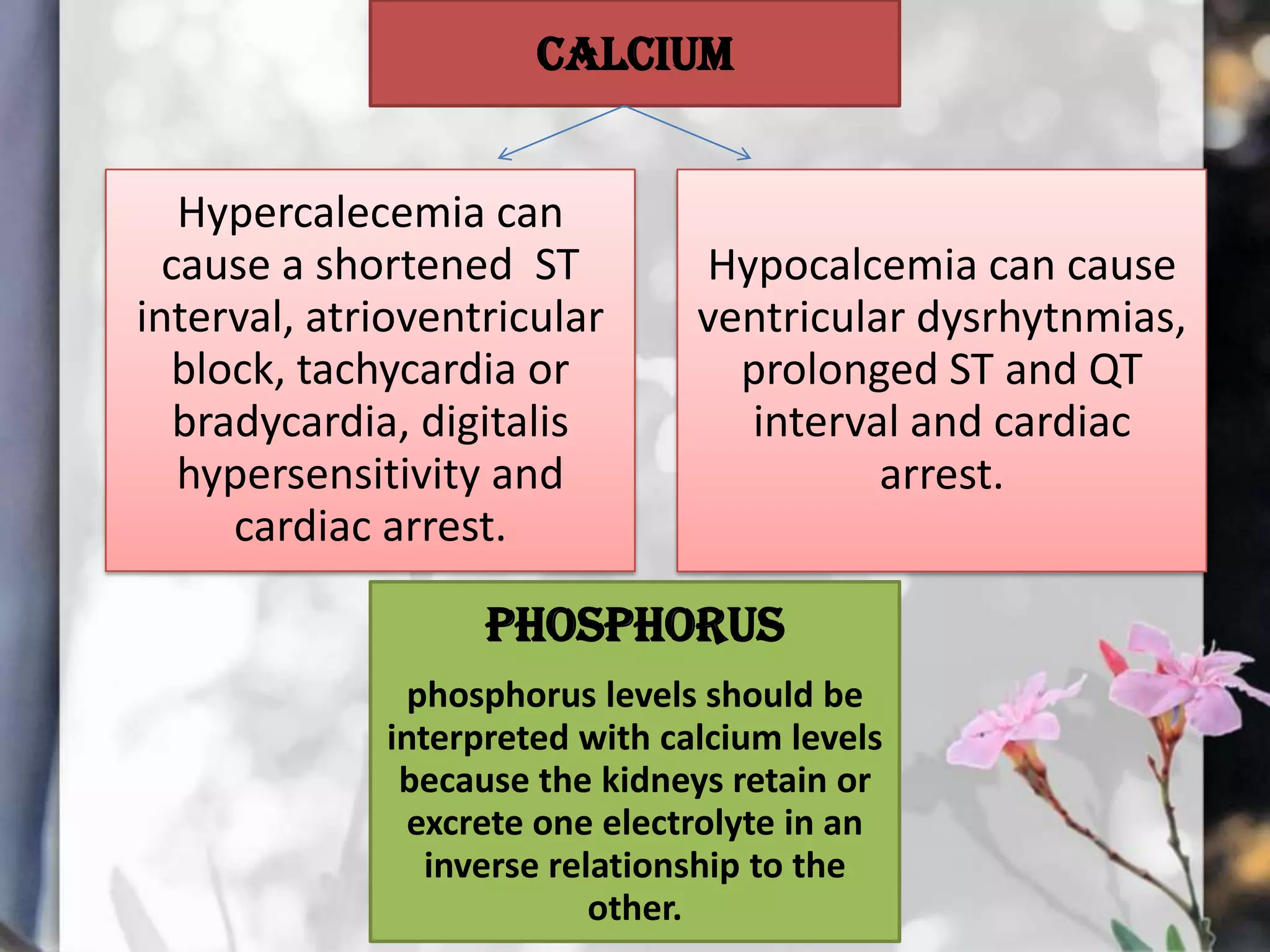
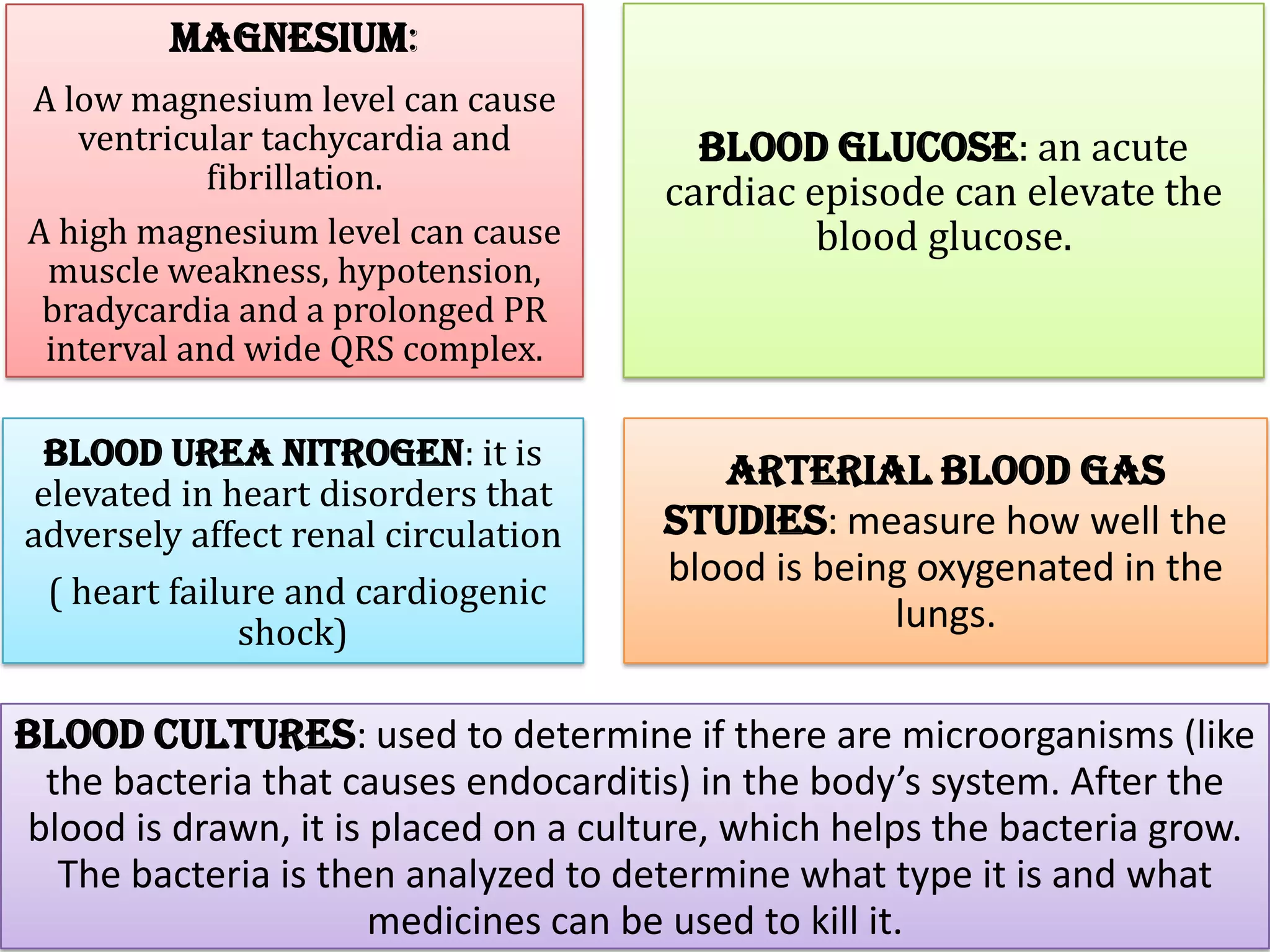
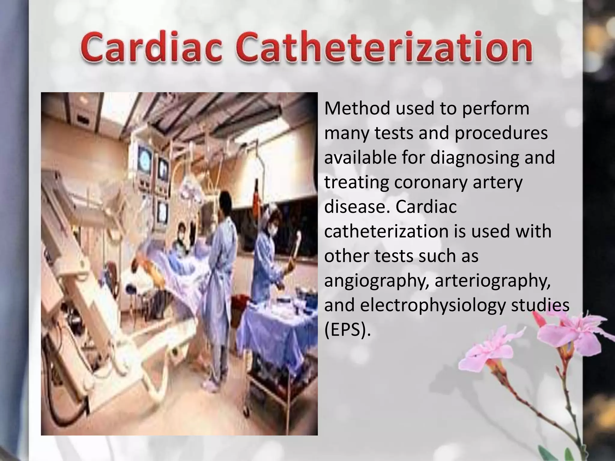
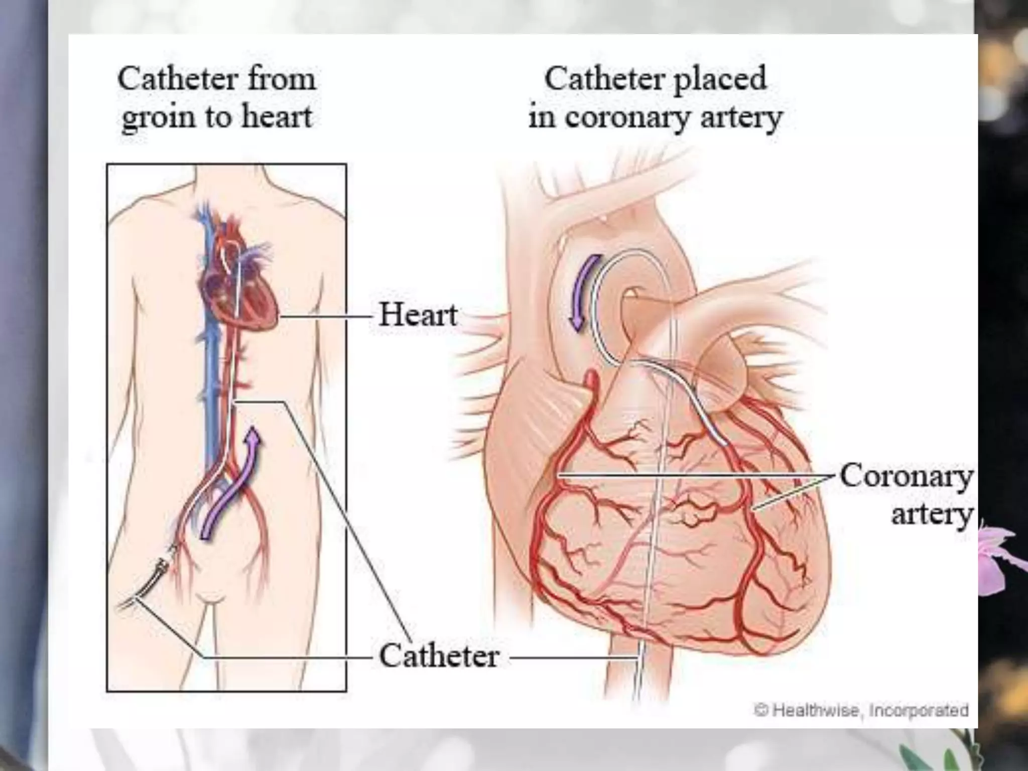
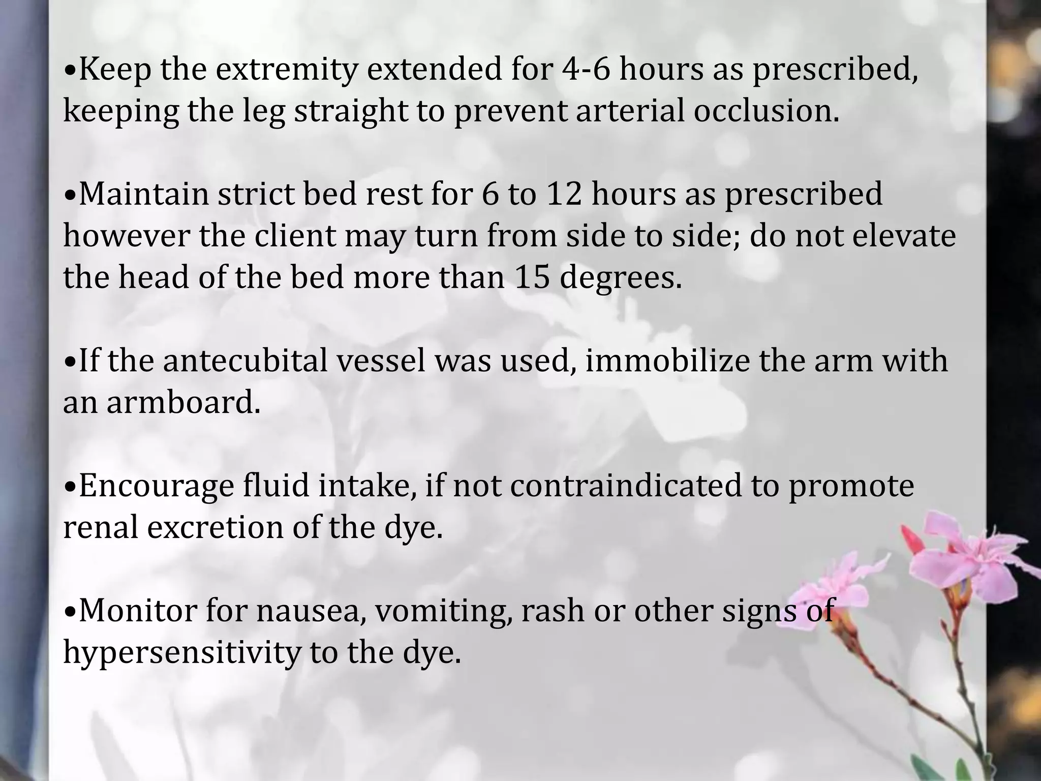
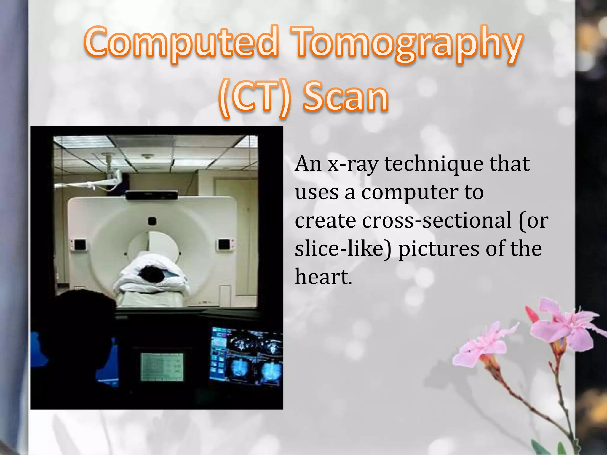
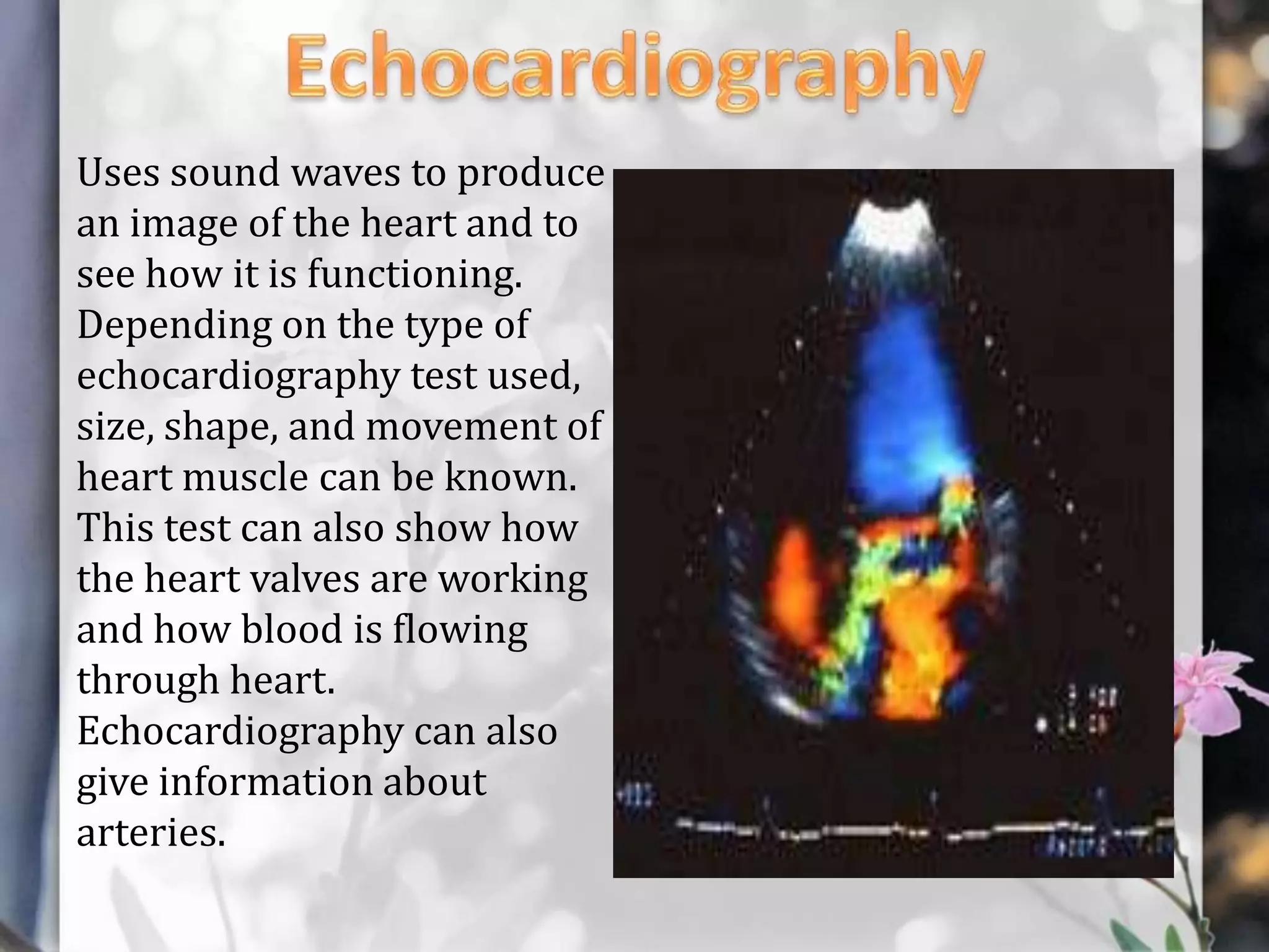
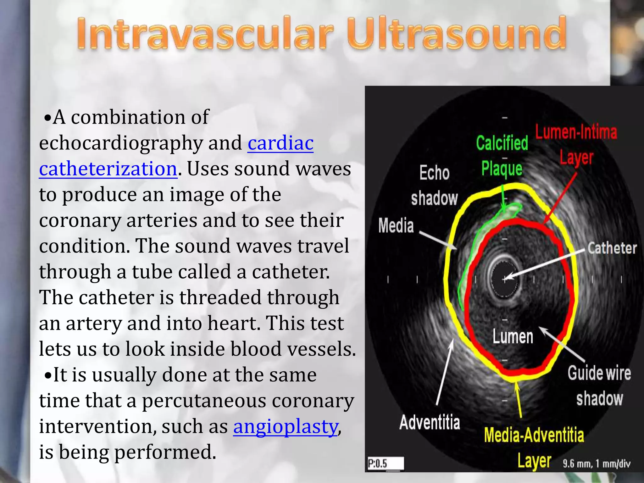
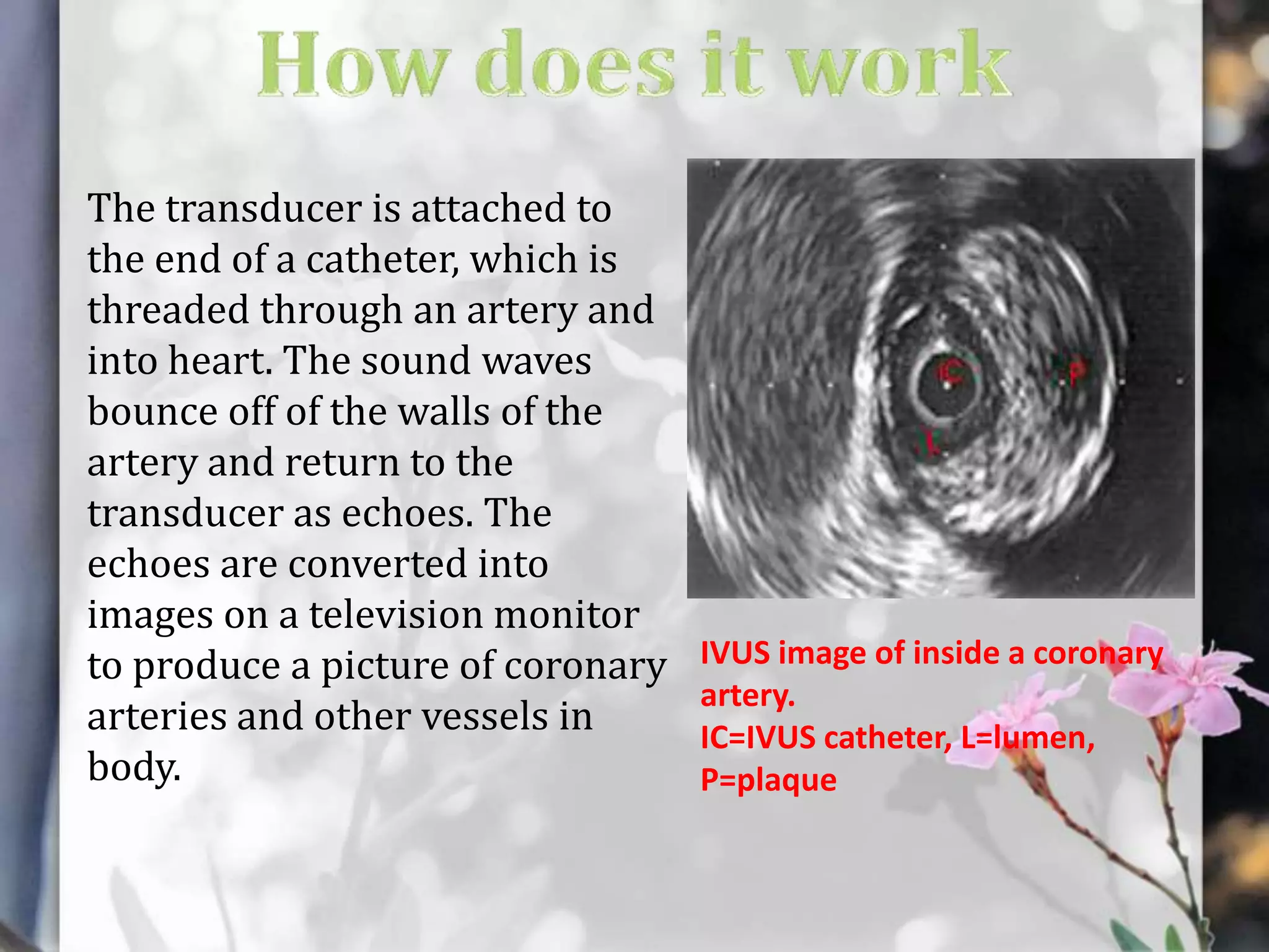
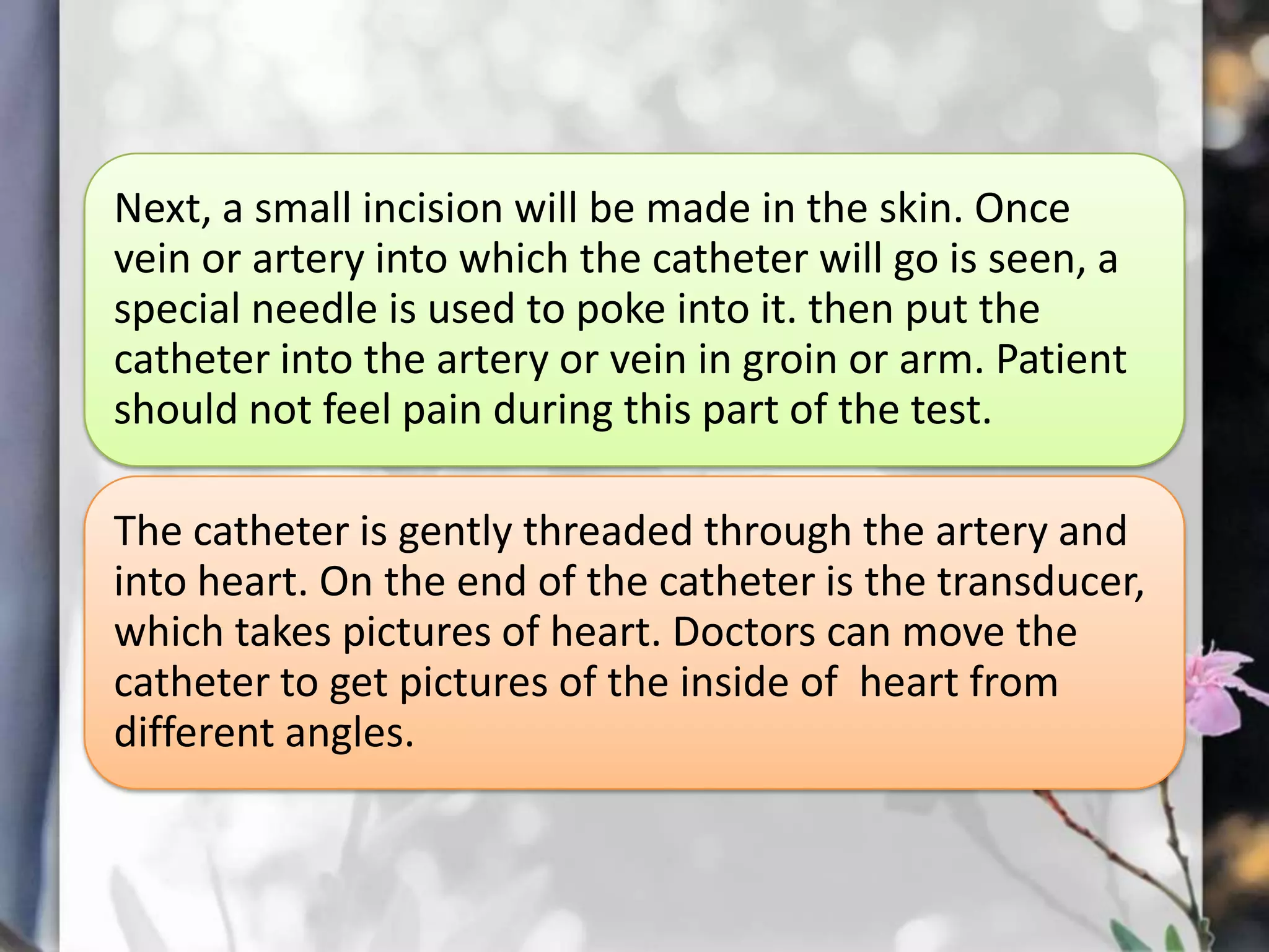
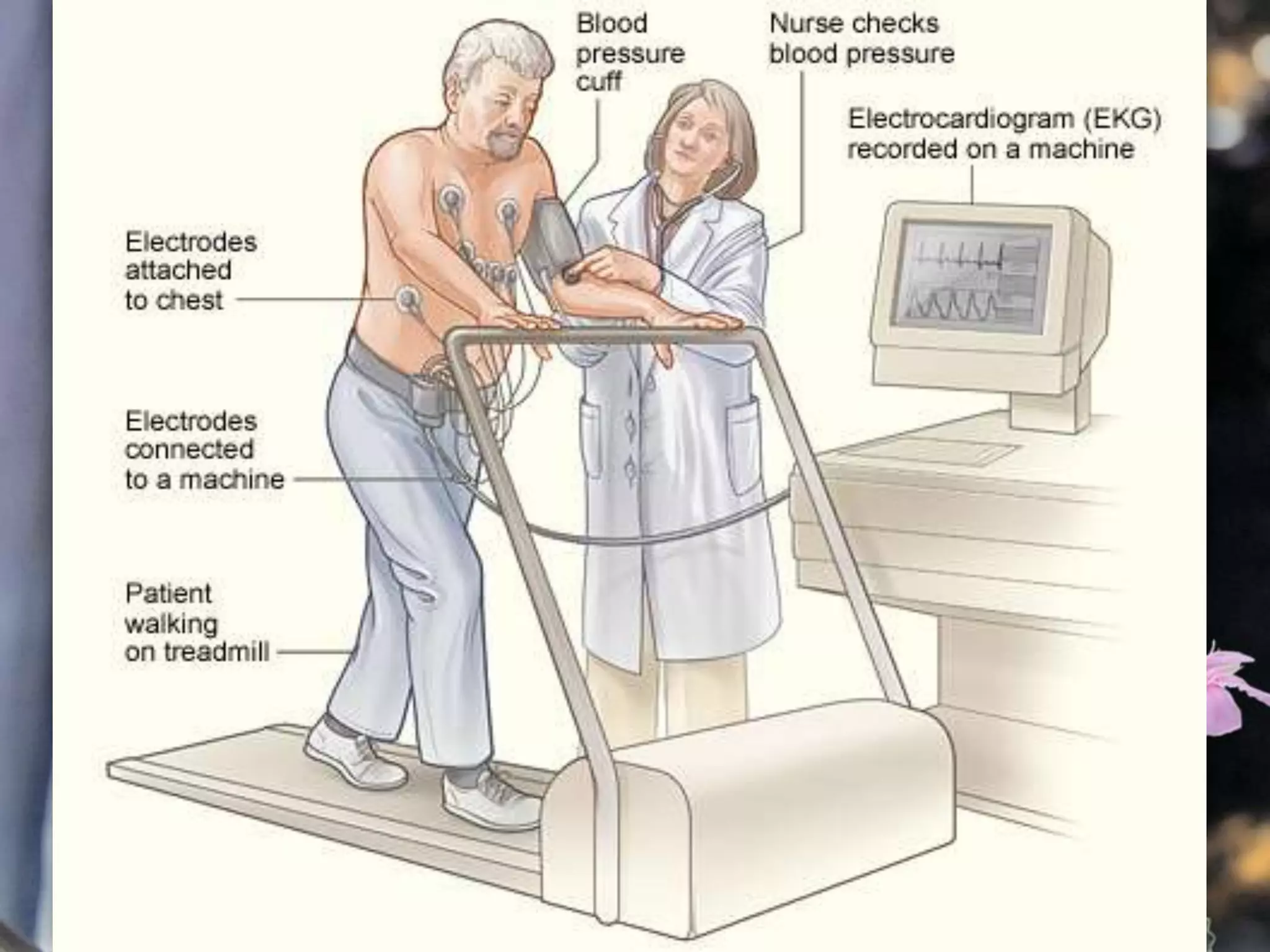
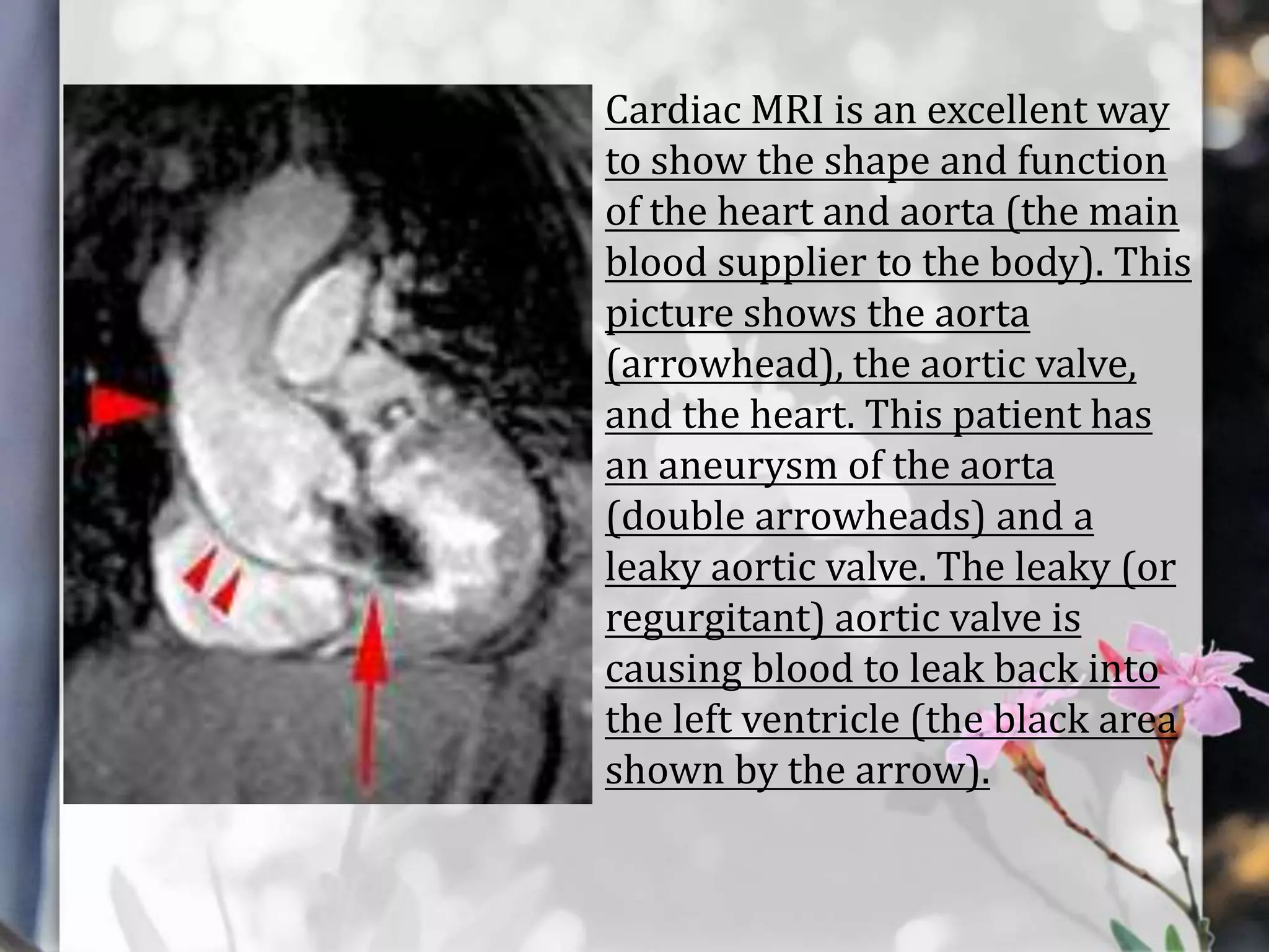
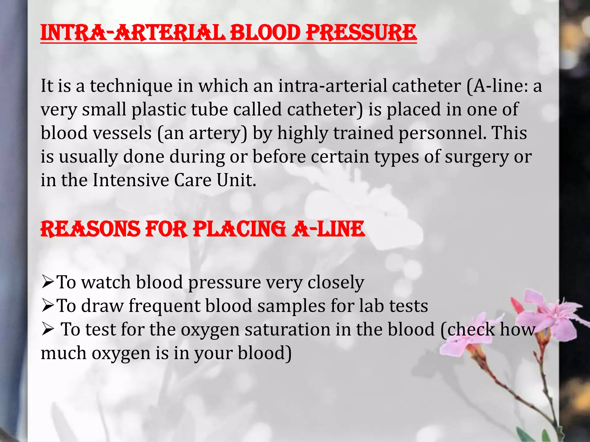
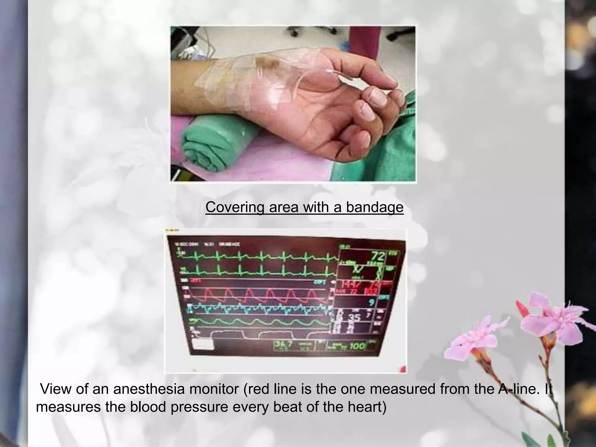
This document describes various cardiac diagnostic tests including imaging tests and blood tests. It provides details on procedures like chest x-rays, echocardiograms, cardiac catheterization, CT scans, and blood tests like cardiac enzymes and electrolyte levels. Precise positioning of patients and monitoring of vital signs during and after procedures is emphasized to obtain clear images and ensure patient safety.
indications, uses and types of cardiac catheterization, about intra cardiac pressure, about angiography and its technique, digital substraction angiography and its technique.
Hemodynamic monitoring- Hemodynamic monitoring refers to the measurement of pressure, flow and oxygenation within the cardiovascular system. Hemodynamic monitoring is amandatory process in all the critical care units to assess the patients progress. This presentation is aimed to create an insight on Hemodynamic monitoring.
Cardiac catheterization (kath-uh-tur-ih-ZAY-shun) is a procedure used to diagnose and treat cardiovascular conditions. During cardiac catheterization, a long thin tube called a catheter is inserted in an artery or vein in your groin, neck or arm and threaded through your blood vessels to your heart.
Using this catheter, doctors can then do diagnostic tests as part of a cardiac catheterization. Some heart disease treatments, such as coronary angioplasty, also are done using cardiac catheterization.
Usually, you'll be awake during cardiac catheterization, but given medications to help you relax. Recovery time for a cardiac catheterization is quick, and there's a low risk of complications.
Cardiovascular assessment and diagnostic proceduresANILKUMAR BR
Cardiovascular disease is the leading killer for both men and women among all racial and ethnic groups in the world wide.
According to the Centers for Disease Control (CDC) studies among coronary heart disease (CAD) patients, 90% of patients have had prior exposure to at least one heart disease risk factor that contributed to their disease.
learn how to obtain an ECG, anyone can do it:
This presentation aims to show the clinical process of obtaining an ECG and features some tips and suggestions to troubleshoot and improve the quality of the tracing.
Please note that you're welcome to use any slides as long as you reference my post when you do so to maintain the integrity of authorship
If interested in detailed answers, please email: aamirdash@yahoo.com
Thanks, Ahmad
invasive non invasive procedures.pdf for bsc nursing studentsshanmukhadevi
Chest X-ray:
The chest X-ray is a noninvasive tool used to visualize internal structures, such as the heart, lungs, soft tissues, and bones.
Most chest X-rays are taken while the patient is inhaling so that the lungs are fully expanded.
Several types of chest X-rays can be used to assess heart size, contour, and position; other types reveal cardiac and pericardial calcification as well as physiologic alterations in pulmonary circulation.
Tyde Pavlinik shares a brand new, breakthrough presentation about the major cardiology advancements in history. Enjoy the presentation and please share!
I am a Neurosurgeon with advanced training in Interventional vascular Neurosurgery(FINR) from Zurich, Switzerland, and FMINS-Fellowship in minimally invasive and Endoscopic Neurosurgery from Germany.
I am presently working in Columbia asia hospitals, Bangalore.
My areas of interest are Vascular Neurosurgery, Stroke specialist, interventional neuroradiology, Endoscopic and minimally invasive Neurosurgery, Endoscopic spine surgery.
this document includes all the essential headings under which physical examination of the patient is done. it helps in collecting the objective information from the patient
format of case study : a Nursing point of view. it includes all the headings or points about which the information regarding the patient needs to be collected and helps to write a detailed case study
Flu Vaccine Alert in Bangalore Karnatakaaddon Scans
As flu season approaches, health officials in Bangalore, Karnataka, are urging residents to get their flu vaccinations. The seasonal flu, while common, can lead to severe health complications, particularly for vulnerable populations such as young children, the elderly, and those with underlying health conditions.
Dr. Vidisha Kumari, a leading epidemiologist in Bangalore, emphasizes the importance of getting vaccinated. "The flu vaccine is our best defense against the influenza virus. It not only protects individuals but also helps prevent the spread of the virus in our communities," he says.
This year, the flu season is expected to coincide with a potential increase in other respiratory illnesses. The Karnataka Health Department has launched an awareness campaign highlighting the significance of flu vaccinations. They have set up multiple vaccination centers across Bangalore, making it convenient for residents to receive their shots.
To encourage widespread vaccination, the government is also collaborating with local schools, workplaces, and community centers to facilitate vaccination drives. Special attention is being given to ensuring that the vaccine is accessible to all, including marginalized communities who may have limited access to healthcare.
Residents are reminded that the flu vaccine is safe and effective. Common side effects are mild and may include soreness at the injection site, mild fever, or muscle aches. These side effects are generally short-lived and far less severe than the flu itself.
Healthcare providers are also stressing the importance of continuing COVID-19 precautions. Wearing masks, practicing good hand hygiene, and maintaining social distancing are still crucial, especially in crowded places.
Protect yourself and your loved ones by getting vaccinated. Together, we can help keep Bangalore healthy and safe this flu season. For more information on vaccination centers and schedules, residents can visit the Karnataka Health Department’s official website or follow their social media pages.
Stay informed, stay safe, and get your flu shot today!
Knee anatomy and clinical tests 2024.pdfvimalpl1234
This includes all relevant anatomy and clinical tests compiled from standard textbooks, Campbell,netter etc..It is comprehensive and best suited for orthopaedicians and orthopaedic residents.
Lung Cancer: Artificial Intelligence, Synergetics, Complex System Analysis, S...Oleg Kshivets
RESULTS: Overall life span (LS) was 2252.1±1742.5 days and cumulative 5-year survival (5YS) reached 73.2%, 10 years – 64.8%, 20 years – 42.5%. 513 LCP lived more than 5 years (LS=3124.6±1525.6 days), 148 LCP – more than 10 years (LS=5054.4±1504.1 days).199 LCP died because of LC (LS=562.7±374.5 days). 5YS of LCP after bi/lobectomies was significantly superior in comparison with LCP after pneumonectomies (78.1% vs.63.7%, P=0.00001 by log-rank test). AT significantly improved 5YS (66.3% vs. 34.8%) (P=0.00000 by log-rank test) only for LCP with N1-2. Cox modeling displayed that 5YS of LCP significantly depended on: phase transition (PT) early-invasive LC in terms of synergetics, PT N0—N12, cell ratio factors (ratio between cancer cells- CC and blood cells subpopulations), G1-3, histology, glucose, AT, blood cell circuit, prothrombin index, heparin tolerance, recalcification time (P=0.000-0.038). Neural networks, genetic algorithm selection and bootstrap simulation revealed relationships between 5YS and PT early-invasive LC (rank=1), PT N0—N12 (rank=2), thrombocytes/CC (3), erythrocytes/CC (4), eosinophils/CC (5), healthy cells/CC (6), lymphocytes/CC (7), segmented neutrophils/CC (8), stick neutrophils/CC (9), monocytes/CC (10); leucocytes/CC (11). Correct prediction of 5YS was 100% by neural networks computing (area under ROC curve=1.0; error=0.0).
CONCLUSIONS: 5YS of LCP after radical procedures significantly depended on: 1) PT early-invasive cancer; 2) PT N0--N12; 3) cell ratio factors; 4) blood cell circuit; 5) biochemical factors; 6) hemostasis system; 7) AT; 8) LC characteristics; 9) LC cell dynamics; 10) surgery type: lobectomy/pneumonectomy; 11) anthropometric data. Optimal diagnosis and treatment strategies for LC are: 1) screening and early detection of LC; 2) availability of experienced thoracic surgeons because of complexity of radical procedures; 3) aggressive en block surgery and adequate lymph node dissection for completeness; 4) precise prediction; 5) adjuvant chemoimmunoradiotherapy for LCP with unfavorable prognosis.
263778731218 Abortion Clinic /Pills In Harare ,sisternakatoto
263778731218 Abortion Clinic /Pills In Harare ,ABORTION WOMEN’S CLINIC +27730423979 IN women clinic we believe that every woman should be able to make choices in her pregnancy. Our job is to provide compassionate care, safety,affordable and confidential services. That’s why we have won the trust from all generations of women all over the world. we use non surgical method(Abortion pills) to terminate…Dr.LISA +27730423979women Clinic is committed to providing the highest quality of obstetrical and gynecological care to women of all ages. Our dedicated staff aim to treat each patient and her health concerns with compassion and respect.Our dedicated group ABORTION WOMEN’S CLINIC +27730423979 IN women clinic we believe that every woman should be able to make choices in her pregnancy. Our job is to provide compassionate care, safety,affordable and confidential services. That’s why we have won the trust from all generations of women all over the world. we use non surgical method(Abortion pills) to terminate…Dr.LISA +27730423979women Clinic is committed to providing the highest quality of obstetrical and gynecological care to women of all ages. Our dedicated staff aim to treat each patient and her health concerns with compassion and respect.Our dedicated group of receptionists, nurses, and physicians have worked together as a teamof receptionists, nurses, and physicians have worked together as a team wwww.lisywomensclinic.co.za/
Basavarajeeyam is an important text for ayurvedic physician belonging to andhra pradehs. It is a popular compendium in various parts of our country as well as in andhra pradesh. The content of the text was presented in sanskrit and telugu language (Bilingual). One of the most famous book in ayurvedic pharmaceutics and therapeutics. This book contains 25 chapters called as prakaranas. Many rasaoushadis were explained, pioneer of dhatu druti, nadi pareeksha, mutra pareeksha etc. Belongs to the period of 15-16 century. New diseases like upadamsha, phiranga rogas are explained.
- Video recording of this lecture in English language: https://youtu.be/lK81BzxMqdo
- Video recording of this lecture in Arabic language: https://youtu.be/Ve4P0COk9OI
- Link to download the book free: https://nephrotube.blogspot.com/p/nephrotube-nephrology-books.html
- Link to NephroTube website: www.NephroTube.com
- Link to NephroTube social media accounts: https://nephrotube.blogspot.com/p/join-nephrotube-on-social-media.html
HOT NEW PRODUCT! BIG SALES FAST SHIPPING NOW FROM CHINA!! EU KU DB BK substit...GL Anaacs
Contact us if you are interested:
Email / Skype : kefaya1771@gmail.com
Threema: PXHY5PDH
New BATCH Ku !!! MUCH IN DEMAND FAST SALE EVERY BATCH HAPPY GOOD EFFECT BIG BATCH !
Contact me on Threema or skype to start big business!!
Hot-sale products:
NEW HOT EUTYLONE WHITE CRYSTAL!!
5cl-adba precursor (semi finished )
5cl-adba raw materials
ADBB precursor (semi finished )
ADBB raw materials
APVP powder
5fadb/4f-adb
Jwh018 / Jwh210
Eutylone crystal
Protonitazene (hydrochloride) CAS: 119276-01-6
Flubrotizolam CAS: 57801-95-3
Metonitazene CAS: 14680-51-4
Payment terms: Western Union,MoneyGram,Bitcoin or USDT.
Deliver Time: Usually 7-15days
Shipping method: FedEx, TNT, DHL,UPS etc.Our deliveries are 100% safe, fast, reliable and discreet.
Samples will be sent for your evaluation!If you are interested in, please contact me, let's talk details.
We specializes in exporting high quality Research chemical, medical intermediate, Pharmaceutical chemicals and so on. Products are exported to USA, Canada, France, Korea, Japan,Russia, Southeast Asia and other countries.
TEST BANK for Operations Management, 14th Edition by William J. Stevenson, Ve...kevinkariuki227
TEST BANK for Operations Management, 14th Edition by William J. Stevenson, Verified Chapters 1 - 19, Complete Newest Version.pdf
Title: Sense of Taste
Presenter: Dr. Faiza, Assistant Professor of Physiology
Qualifications:
MBBS (Best Graduate, AIMC Lahore)
FCPS Physiology
ICMT, CHPE, DHPE (STMU)
MPH (GC University, Faisalabad)
MBA (Virtual University of Pakistan)
Learning Objectives:
Describe the structure and function of taste buds.
Describe the relationship between the taste threshold and taste index of common substances.
Explain the chemical basis and signal transduction of taste perception for each type of primary taste sensation.
Recognize different abnormalities of taste perception and their causes.
Key Topics:
Significance of Taste Sensation:
Differentiation between pleasant and harmful food
Influence on behavior
Selection of food based on metabolic needs
Receptors of Taste:
Taste buds on the tongue
Influence of sense of smell, texture of food, and pain stimulation (e.g., by pepper)
Primary and Secondary Taste Sensations:
Primary taste sensations: Sweet, Sour, Salty, Bitter, Umami
Chemical basis and signal transduction mechanisms for each taste
Taste Threshold and Index:
Taste threshold values for Sweet (sucrose), Salty (NaCl), Sour (HCl), and Bitter (Quinine)
Taste index relationship: Inversely proportional to taste threshold
Taste Blindness:
Inability to taste certain substances, particularly thiourea compounds
Example: Phenylthiocarbamide
Structure and Function of Taste Buds:
Composition: Epithelial cells, Sustentacular/Supporting cells, Taste cells, Basal cells
Features: Taste pores, Taste hairs/microvilli, and Taste nerve fibers
Location of Taste Buds:
Found in papillae of the tongue (Fungiform, Circumvallate, Foliate)
Also present on the palate, tonsillar pillars, epiglottis, and proximal esophagus
Mechanism of Taste Stimulation:
Interaction of taste substances with receptors on microvilli
Signal transduction pathways for Umami, Sweet, Bitter, Sour, and Salty tastes
Taste Sensitivity and Adaptation:
Decrease in sensitivity with age
Rapid adaptation of taste sensation
Role of Saliva in Taste:
Dissolution of tastants to reach receptors
Washing away the stimulus
Taste Preferences and Aversions:
Mechanisms behind taste preference and aversion
Influence of receptors and neural pathways
Impact of Sensory Nerve Damage:
Degeneration of taste buds if the sensory nerve fiber is cut
Abnormalities of Taste Detection:
Conditions: Ageusia, Hypogeusia, Dysgeusia (parageusia)
Causes: Nerve damage, neurological disorders, infections, poor oral hygiene, adverse drug effects, deficiencies, aging, tobacco use, altered neurotransmitter levels
Neurotransmitters and Taste Threshold:
Effects of serotonin (5-HT) and norepinephrine (NE) on taste sensitivity
Supertasters:
25% of the population with heightened sensitivity to taste, especially bitterness
Increased number of fungiform papillae
NVBDCP.pptx Nation vector borne disease control programSapna Thakur
NVBDCP was launched in 2003-2004 . Vector-Borne Disease: Disease that results from an infection transmitted to humans and other animals by blood-feeding arthropods, such as mosquitoes, ticks, and fleas. Examples of vector-borne diseases include Dengue fever, West Nile Virus, Lyme disease, and malaria.
Ozempic: Preoperative Management of Patients on GLP-1 Receptor Agonists Saeid Safari
Preoperative Management of Patients on GLP-1 Receptor Agonists like Ozempic and Semiglutide
ASA GUIDELINE
NYSORA Guideline
2 Case Reports of Gastric Ultrasound
5. Chest X-RAY
•A radiology test that involves exposing the chest briefly to
radiation to produce an image of the chest and the internal
organs of the chest.
•An X-ray film is positioned against the body opposite the
camera, which sends out a very small dose of a radiation
beam.
• As the radiation penetrates the body, it is absorbed in
varying amounts by different body tissues depending on the
tissue's composition of air, water, blood, bone, or muscle.
6. Procedure:Patient is asked to wear X-ray gown, and
extra metallic objects such as jewellery are
removed from the chest and/or neck areas.
Patients may be asked to take a deep
breath and hold it during the chest X-ray in
order to inflate the lungs to their
maximum
.
The radiology technologist or technician is
a trained, certified assistant to the
radiologist who will help the patient
during the X-ray and actually perform the
X-ray testprocedure.
7. POSITIONING A PATIENT FOR PA FRONTAL CHEST
RADIOGRAPH. THE X-RAY TUBE IS BEHIND THE PATIENT
AND THE X-RAY FILM IS CLOSE TO HIS ANTERIOR CHEST.
8. TYPICAL CARDIAC CARE UNIT (CCU) PATIENT POSITIONED FOR
AN AP CHEST RADIOGRAPH. NOTE THAT, AS OFTEN OCCURS IN
THE CCU, THE PATIENT’S CHEST IS NOT PERFECTLY
PERPENDICULAR TO X-RAY TUBE. THIS CAUSES THE HEART TO
APPEAR LARGE AND INDISTINCT. THIS IS CALLED A LORDOTIC
POSITION.
9.
10. an x-ray technique
where dye is injected
into the chambers of
your heart or the
arteries that lead to
your heart (the
coronary arteries).
13. PREPROCEDURE
Informed
consent is
taken
from the
patient.
Assess for
allergies to
seafood,
iodine or
radiopque
dyes.
Ask the patient
to not to eat or
drink anything
after midnight
before test. If
patient is having
diabetes, doctor
should be
consulted about
the food and
insulin intake.
Talk to doctor
about any
medicines
that patient is
taking,
because he or
she may want
the patient to
stop taking
them before
the test.
14. DURING PROCEDURE
• The patient is carried to the lab. Patient will lie on an
examination table, which is usually near an x-ray
camera.
• Small metal disks called electrodes will be placed on
his chest. These electrodes have wires called leads,
which hook up to an electrocardiogram machine. This
machine will monitor heart rhythm during the test.
To prevent infection, patient will be cleansed
around the area of leg where the catheter will
be inserted.
A needle with a tube connected to it will be put
in patient’s arm. This is called an intravenous
line or IV. Patient will be given a mild sedative
through the IV to relax throughout the test.
15. CONT…….
•The patient will be given an anesthetic medicine with a
needle to numb the area around where the catheter will be
inserted. The patient may feel mild discomfort.
•Next, a small incision will be made in the skin. Once doctors
see the artery into which the catheter will go, a special
needle is used to poke into it. Then the catheter is put into
the artery in leg.
•The catheter is gently threaded through the artery and into
heart. If we want to measure blood pressure in the left
ventricle (the heart's main pumping chamber), then position
the catheter there and pump some dye into heart.
16. CONT……
•If we want to see blood flow through coronary arteries,
then position the catheter at the opening of each of the
arteries and pump the dye into them. The dye will let us
see whether the patient have blockages in a main artery
or its branches. This information is recorded on a
television monitor.
•then, the catheter and IV will be removed. Firm
pressure will be applied to the site where the catheter
was inserted to stop any bleeding. The patient will also
be bandaged.
17. Post procedure:•The patient will be moved to another room where patient
will need to rest for a few hours. Patient may feel a little
sleepy until the sedative has worn off. Tell the patient to lie
still and not bend knee too much. Nurses will watch to see
that heart rate and blood pressure are normal. After this
time of rest, the patient will be able to go home.
•Angiography is a very safe test. The dye used for the test is
harmless, and by drinking lots of liquids after the test, which
can help rid body of the dye.
18. CBC- one of the most common blood tests. It is usually
done as part of a routine checkup and can help detect a
number of blood disorders, such as anemia, infections,
clotting problems, blood cancers, and immune system
problems.
It also measures the hemoglobin (iron) levels in blood and
hematocrit, which is how much space red blood cells take up
in blood. An elevated hematocrit can result from vascular
volume depletion. Decreases in hematocrit and hemoglobin
indicates anemia.
19. • Mean corpuscular volume, which is a measure of the average size of
red blood cells. Specific blood tests can be performed to see if there
is a problem with heart or blood vessels
• Blood coagulation factors: an increase in coagulation factors can
occur during and after MI which places the client at greater risk for
thrombophlebitis and extension of clots in coronary arteries.
• Serum lipids: the lipid profile measures serum cholesterol,
triglycerides and lipoprotein levels. It is also used to assess the risk of
developing coronary artery disease. The desirable range for serum
cholesterol is less than 200mg/dl, with LDL cholesterol less than
130mg/dl and HDL cholesterol at 30-70mg/dl.
20. ELECTROLYTES
POTASSIUM
Hypokalemia causes
increased cardiac instability,
ventricular dysrhythmias
and increased risk of
digitalis toxicity.
In hypokalemia, the ECG would
show flattening and inversion
of the T wave and appearance
of a U wave and ST depression.
Hyperkalemia causes asystole and
ventricular dysrhythmias.
21. SODIUM
The serum sodium level
decreases with the use of
diuretics.
The serum sodium level
decreases in heart failure,
indicating water excess.
22. Calcium
Hypercalecemia can
cause a shortened ST
interval, atrioventricular
block, tachycardia or
bradycardia, digitalis
hypersensitivity and
cardiac arrest.
Hypocalcemia can cause
ventricular dysrhytnmias,
prolonged ST and QT
interval and cardiac
arrest.
Phosphorus
phosphorus levels should be
interpreted with calcium levels
because the kidneys retain or
excrete one electrolyte in an
inverse relationship to the
other.
23. Magnesium:
A low magnesium level can cause
ventricular tachycardia and
fibrillation.
A high magnesium level can cause
muscle weakness, hypotension,
bradycardia and a prolonged PR
interval and wide QRS complex.
Blood urea nitrogen: it is
elevated in heart disorders that
adversely affect renal circulation
( heart failure and cardiogenic
shock)
Blood glucose: an acute
cardiac episode can elevate the
blood glucose.
Arterial blood gas
studies: measure how well the
blood is being oxygenated in the
lungs.
Blood cultures: used to determine if there are microorganisms (like
the bacteria that causes endocarditis) in the body’s system. After the
blood is drawn, it is placed on a culture, which helps the bacteria grow.
The bacteria is then analyzed to determine what type it is and what
medicines can be used to kill it.
24. CARDIAC ENZYMES
•CK-MB( creatine kinase, myocardial muscles)Certain
enzymes will be present if the heart muscle (myocardium)
has been damaged by a heart attack, because damaged heart
cells release these enzymes into the blood. The most
common cardiac enzyme that is released is creatine kinase.
• An elevation occurs within 4-6 hours and peaks 18-24
hours following an acute ischemic attack. Normal value is 05% of total; total CK is 26-174 units/L.
25. Lactate dehydrogenase (LDH), elevations in LDH occur 24
hours following MI and peak in 48-72 hours. When serum
concentration of LDH1 is higher than LDH2 the pattern is
indicated as “flipped”, signifying myocardial necrosis. Normal
value in conventional units is 140 to 280 international units.
Troponin I has a high
Myoglobin is an oxygen
affinity for myocardial
binding protein found in
injury, it rises within 3
cardiac and skeletal
hours and persists upto 7
muscle. Level rises within
days. Normal values are
1 hour after cell death,
low, with troponin T
peaks in 4-6hours and
normally ranging from 0returns to normal within
0.2ng/ml and troponin I
24-36 hours.
being less than 0.6ng/ml.
26. Method used to perform
many tests and procedures
available for diagnosing and
treating coronary artery
disease. Cardiac
catheterization is used with
other tests such as
angiography, arteriography,
and electrophysiology studies
(EPS).
27.
28. INTERVENTIONS (PREPROCEDURE)
• First of all informed consent is taken and patient is assessed for allergies to
seafood, iodine or radiaopaque dyes.
• Patients are usually told not to eat or drink anything after midnight the night
before the test to prevent vomiting and aspiration during the procedure.
• If the patient is taking some medicines to stop taking them before the test.
This is especially important if patient is taking blood-thinning medicines or
anti-platelet medicines. patient will most likely have blood tests, an
electrocardiogram, and a chest x-ray taken before the procedure.
• If the patient is having diabetes, physician is consulted about food and insulin
intake, because not eating can affect blood sugar levels.
• Document the client’s height and weight because these data will be needed to
determine the amount of dye administered.
29. • Document baseline vital signs and note the quality and presence
of peripheral pulses for postprocedure comparison.
• Inform the client that a local anesthetic will be administered
before catheter insertion.
• Inform the client that he/she may feel fatigued because of the
need to lie still on a hard table for upto 2 hours.
• Inform the client that he may have a fluttering feeling as the
catheter passes through the heart, a flushed warm feeling
when dye is injected, a desire to cough and palpitations
caused by heart irritability.
• Prepare the insertion site by shaving and cleaning with an
antiseptic solution if prescribed.
• Administer preprocedure medications if prescribed.
• Insert the IV line if prescribed.
30. POST PROCEDURE
The patient will be moved to another room where he will
need to rest for a few hours. He may feel a little sleepy until
the sedative has worn off. Ask the patient to lie still and not
bend knee too much.
Monitor vital signs and cardiac rhythm for dysrhythmias at
least every 30 minutes for 2 hours initially.
Assess for chest pain and if dysrhythmias or chest pain
occurs notify the physician.
Monitor peripheral pulses and the colour , warmth and
sensation of the extremity distal to insertion site atleast
every 30 minutes for 2 hours initially.
31. •Notify the physician if the client complains of numbness and
tingling, if extremity becomes cool, pale, cyanotic or if loss of
the peripheral pulses occurs.
•Monitor the pressure dressing for bleeding or hematoma
formation.
•Apply sandbag or compression device to the insertion site
to provide additional pressure if required
•Monitor for bleeding and if bleeding occurs apply pressure
immediately and notify the physician.
•Monitor for hematoma and if hematoma develops notify the
physician.
32. •Keep the extremity extended for 4-6 hours as prescribed,
keeping the leg straight to prevent arterial occlusion.
•Maintain strict bed rest for 6 to 12 hours as prescribed
however the client may turn from side to side; do not elevate
the head of the bed more than 15 degrees.
•If the antecubital vessel was used, immobilize the arm with
an armboard.
•Encourage fluid intake, if not contraindicated to promote
renal excretion of the dye.
•Monitor for nausea, vomiting, rash or other signs of
hypersensitivity to the dye.
33. An x-ray technique that
uses a computer to
create cross-sectional (or
slice-like) pictures of the
heart.
34.
35. How does it work?
The CT scanner is a large X-ray machine that has a short,
open-ended tube in the middle (like a very short tunnel). The
patient lies on a scanning table, which slides through the
middle of the CT scanner. The CT scanner takes many x-ray
pictures of thin slices of heart. A computer then puts these
images together to make one detailed picture. In some cases,
a contrast dye is injected into the bloodstream to help
physician get a clearer picture.
36. Interventions
•If a contrast dye is not going to be used during CT scan, ask
the patient not to eat for about 2 hours before the test. If a
contrast dye is going to be used, ask patient not to eat for
about 4 hours before the test. The contrast dye may cause hot
flushing in some patients.
•Patient will be asked to undress and put on a hospital gown.
Then, patient is asked to lie down on a table, which will be
slowly moved through the hollow center of the CT scanner.
The patient is asked to lie still and to hold breath briefly as
each picture is taken.
37. •After the test, patient may go about normal activities. Some
people find that they have a bad reaction to the contrast dye,
but this is rare. If this happens, patient will be treated at the
hospital after test.
•CT scanning is a safe test. Although exposure to radiation is
small, pregnant female should not have a CT scan.
38. A CT scan does not get a moving picture of the heart. Instead, CT scans
are used to see if part of the heart has calcified. This patient has had a
heart attack that scarred the heart's main pumping chamber (the left
ventricle). The red arrows point to the scarred part of the heart that has
since formed a thick, calcified wall at the tip of the left ventricle
39. A CT scan gets a number of images that can be looked at one by one. New
computer technology now lets technicians stack the images on top of each
other to get a 3-D image that can be rotated and viewed from any angle.
The red arrows in the images above point to a large aneurysm of the
abdominal aorta seen from the front (left picture) and the side (right
picture).
40. Faster type of CT scanning,
which takes an x-ray of the
heart in about one-tenth of
a second. Ordinary CT
scanning can take anywhere
from 1 to 10 seconds. EBCT
takes pictures so quickly
that it can avoid blurred
pictures caused by the
beating of the heart, a
problem with a regular CT
scan
41. Used routinely to measure
the amount of calcium in the
coronary arteries—similar
to EBCT—but are also now
able to take images of the
coronary arteries that are
nearly comparable to those
taken during a cardiac
catheterization.
42. The two MDCT views above show that the
left coronary artery (red arrows) and its
side branches are normal.
43. This 3D MDCT view is looking down on top of
the left ventricle. It shows a normal coronary
artery (black arrow) and side branches.
44. Uses sound waves to produce
an image of the heart and to
see how it is functioning.
Depending on the type of
echocardiography test used,
size, shape, and movement of
heart muscle can be known.
This test can also show how
the heart valves are working
and how blood is flowing
through heart.
Echocardiography can also
give information about
arteries.
45. •One-dimensional or M-mode echocardiography is one beam
of ultrasound directed toward the heart. Doctors most often
use M-mode echocardiography to see just the left side (or
main pumping chamber) of heart.
•Two-dimensional echocardiography produces a broader
moving picture of heart. Two-dimensional echocardiography
is one of the most important diagnostic tools.
•Doppler echocardiography measures blood flowing through
the arteries and shows the pattern of flow through the heart.
46. INTERVENTIONS
1.
2
3
• No special preparation is needed before an
echocardiogram.
• During the test, patient lies on an examination table.
A technician will place small metal disks called
electrodes on chest. These electrodes have wires
called leads, which hook up to an electrocardiogram
machine. This machine will monitor heart rhythm
during the test.
• Next, the technician will put a thick gel on patient’s
chest. The gel may feel cold, but it does not harm
skin. Then, the technician will use the transducer to
send and receive the sound waves.
47. 4.
5.
• The transducer will be placed directly on the left
side of chest, above heart. The technician will
press firmly as he or she moves the transducer
across chest. Patient may be asked to breathe in
or out or to briefly hold breath during the test.
But, for most of the test, patient will lie still.
• An echocardiogram may take up to 45 minutes
to perform. Patient should not have any pain
or discomfort during the test
48. •A combination of
echocardiography and cardiac
catheterization. Uses sound waves
to produce an image of the
coronary arteries and to see their
condition. The sound waves travel
through a tube called a catheter.
The catheter is threaded through
an artery and into heart. This test
lets us to look inside blood vessels.
•It is usually done at the same
time that a percutaneous coronary
intervention, such as angioplasty,
is being performed.
49. The transducer is attached to
the end of a catheter, which is
threaded through an artery and
into heart. The sound waves
bounce off of the walls of the
artery and return to the
transducer as echoes. The
echoes are converted into
images on a television monitor
to produce a picture of coronary
arteries and other vessels in
body.
IVUS image of inside a coronary
artery.
IC=IVUS catheter, L=lumen,
P=plaque
50. Ask the patient - Do not eat or drink anything after
midnight, the night before test. Doctor is consulted about
any medicines that patient is taking, because some
medicines are to be stopped taking them before the test.
Patient will most likely have blood tests, an
electrocardiogram, and a chest x-ray taken before the
procedure.
Once patient is in the catheterization laboratory (also called
the cath lab), television monitors, heart monitors, and
blood pressure machines are there to monitoring. Patient
will lie on an examination table, which is usually near an xray camera.
51. Electrodes will be placed on patient’s chest. These electrodes
have wires called leads, which hook up to an electrocardiogram
machine. This machine will monitor heart rhythm during the test.
To prevent infection, patient will be shaved and cleansed around
the area of groin or arm where the catheter will be inserted.
A needle with a tube connected to it will be put in arm. This is
called an intravenous line or IV. Patient will get a mild sedative
through the IV to relax throughout the test. Patient will be given
an anesthetic medicine with a needle to numb the area around
where the catheter will be inserted. Patient may feel mild
discomfort
52. Next, a small incision will be made in the skin. Once
vein or artery into which the catheter will go is seen, a
special needle is used to poke into it. then put the
catheter into the artery or vein in groin or arm. Patient
should not feel pain during this part of the test.
The catheter is gently threaded through the artery and
into heart. On the end of the catheter is the transducer,
which takes pictures of heart. Doctors can move the
catheter to get pictures of the inside of heart from
different angles.
53. After having enough pictures, the catheter and IV line will be
removed. Patient will also be disconnected from the
electrocardiogram machine. Firm pressure will be applied to
the site where the catheter was inserted to stop any bleeding.
Patient will also be bandaged.
Patient will be moved to another room where he will need to
rest for 5 or 6 hours. Patient may feel a little sleepy until the
sedative has worn off. Patient will be told to lie still. If the
catheter was inserted in groin, try not to bend knee. If the
catheter was inserted in arm, try not to bend elbow. Nurse
will watch you to see that heart rate and blood pressure are
normal. After this time of rest, patient can go home.
54. Uses sound waves to produce an
image of the heart and to see
how it is functioning. Stress
echocardiography (or stress
echo) lets to see the wall motion
of the heart's pumping
chambers before and after
exercise. The test can show if
certain areas of the heart
muscle are not getting enough
oxygen-rich blood.
55.
56. After heart has reached a certain rate during exercise on a
treadmill or stationary bike, patient is asked to lie down on
an examination table. Next, the technician will put a thick gel
on chest. The gel may feel cold, but it does not harm skin.
Then the technician will use the transducer to send and
receive the sound waves.
The transducer will be placed directly on chest, above
heart. The technician will press firmly as he or she moves the
transducer across chest. Patient is asked to breathe in or out
or to hold breath briefly during the test. For most of the test,
patient will lie still.
Most stress echo tests take about 45 minutes, but a
dobutamine stress echo takes longer.
57. TEE uses sound waves to produce an image of the heart
and to see how it is functioning. But unlike standard
echocardiography, the sound waves travel through a tube-like
device put in the mouth and passed down the throat into the
esophagus.
Shows the detailed size, shape, and movement of heart
muscle, and the condition of aorta, which is the main blood
vessel supplying blood to body. It can also show how the
heart valves are working and how blood is flowing through
heart. TEE also gives doctors information about arteries.
Doctors use this test when they cannot get a clear picture of
heart using standard echocardiography.
58.
59. How does it work?
TEE uses high-frequency sound waves (also called
ultrasound) that can provide a moving picture of heart. The
test is like standard echocardiography except that the
pictures of the heart come from inside the esophagus rather
than through the chest wall. The sound waves are sent
through the body with a device called a transducer, which is
attached to a tube and put down esophagus. The sound
waves bounce off of the heart and return to the transducer
as echoes. The echoes are converted into images on a
television monitor to produce a picture of heart and aorta.
60. Patient need to stop eating for at least 4 hours
before the test. Consult the doctor about any
medicines that patient is taking, because some
medicines need to stopped before the test.
The test will begin with the technician spraying
throat with an anesthetic, which will make it feel
numb. Then, patient will lie down on the
examination table. Small metal disks called
electrodes will be placed on chest. These
electrodes have wires called leads, which hook up
to an electrocardiogram machine. This machine will
monitor heart rhythm during the test.
Then, a small flexible tube called a probe will be put
down throat. The technician will ask patient to
swallow, and the probe will be gently moved down
throat. This is often the most uncomfortable part of
the test. Once the probe is in place, patient should
not feel any pain.
61. On the end of the probe is the transducer,
which takes pictures of heart. The
technician can also move the probe to get
pictures of heart from different angles.
After the technician has enough pictures,
the probe and IV line will be removed.
Patient will also be disconnected from the
electrocardiogram machine. Patient may
feel a little sleepy until the sedative has
worn off, and you will be watched to see
that your heart rate and blood pressure are
normal.
If the patient is having a sore throat or
trouble swallowing after the procedure.
These side effects usually go away after a
day or so.
62. An electrocardiogram (ECG
or EKG) test.
(ECG or EKG) is a routine test that is used to look at
the electrical activity of the heartbeat. An
electrocardiogram tells a lot about heart and how it
is working. This test can help us learn more about
heart rhythm, the size and function of the chambers
of heart, and heart muscle. A healthy person's
electrocardiogram has a certain pattern. When there
are changes in that pattern, then there is a problem
with heart. For example, during a heart attack, the
EKG machine records the changing pattern of the
heart's electrical activity.
63. How does it work?
For heart to beat, an electrical impulse is sent from the
sinoatrial (SA) node, which is located in heart. The SA
node helps heart keep a steady pace. An
electrocardiogram can trace the path of electrical energy
that is sent from the SA node and through heart. This
helps us to know whether patient have a problem that
might cause heart to beat irregularly.
Small metal disks called electrodes are placed on skin.
The electrodes are used to pick up the electrical
impulses of the heart. The impulses are recorded, giving
doctors a record of heart's electrical activity.
64.
65. Electrocardiogram basics
•An electrocardiogram reflects the electrical activity of
cardiac cells and records electrical activity at the speed of
25mm/sec.
•An electrocardiogram strip consists of horizontal squares
representing seconds and vertical squares representing
voltage.
•Each small square represents 0.04 second.
•Each large square represents 0.20 second.
•The P-wave represents atrial depolarization.
•The PR interval represents the time it takes an impluse to
travel from the atria through the AV node, bundle of his and
bundle branches to the purkinje fibres.
66. •Normal PR interval duration ranges from 0.12 to .20 second.
•The PR interval is measured from the beginning of the P wave
to end of the PR segment.
•The QRS complex represents ventricular depolarization.
•Normal QRS complex duration ranges from 0.04 to 0.1
second.
•The Q wave appears as the first negative deflection in the QRS
complex and reflects initial ventricular septal depolarization
•The R wave is the first positive deflection in the QRS complex.
•The S wave appears as the second negative deflection in the
QRS complex.
67. •The J point marks the end of the QRS complex
and beginning of the ST segment.
•The QRS duration is measured from the end of
the PR segment to the J point.
•The ST segment represents part of the
ventricular repolarization.
•The T wave represents ventricular
repolarization and ventricular diastole.
•The U wave may follow the T wave.
68. •A prominent U wave may indicate an
electrolyte abnormality such as hypokalemia.
•The QT interval represents ventricular
refractory time or the total time required for
ventricular depolarization and repolarization.
•The QT interval is measured from the
beginning of the QRS complex to the end of the
T wave.
•The QT interval normally lasts 0.32 to 0.4
second but varies with the client’s heart rate,
age and sex.
69. INTERVENTIONS
No special preparation is needed before an
electrocardiogram.
During the test, patient will lie on an examination
table. A technician will clean the areas on body where
the electrodes will be placed, usually chest, back,
wrists, and ankles. The electrodes have wires called
leads, which hook up to the electrocardiogram
machine.
Once the electrodes are in place, patient will be asked
to lie down. The technician will enter some
information into the electrocardiogram machine and
then tell patient to lie still for about a minute while
the machine takes its readings. The test is completely
safe and painless.
70. Phonocardiography
The graphic recording of heart sounds and murmurs. The
trem also includes pulse tracing ( carotid, apex and venous
pulse).
It involves picking up, through highly sensitive microphone,
sonic vibrations from the heart which are then converted into
electrical energy and fed into a galvanometer, where they are
recorded on paper.
71. •The procedure is most useful when there is
evidence of heart murmurs or unusual heart sounds,
such as gallops, that are difficult to discern by the
human ear. Most recordings are made through an
externally applied microphone but intracardiac
recordings, made through a phonocatheter, are
possible.
•Phonocardiography is a promising non-invasive
diagnostic tool for the assessment of aortic stenosis
(AS), and time-frequency representation is a
potential tool to extract information from the
phonocardiogram (PCG) signal.
72. VECTORCARDIOGRAPHY
Vectorcardiography is a method of recording the magnitude
and direction of the electrical forces that are generated by
the heart by means of a continuous series of vectors that
form curving lines around a central point.
Vectorcardiography was developed by E. Frank in the early
30's. Since the human body is a three dimesional structure,
the basic idea is to construct 3 orthogonal leads containing
all the electric information. The three leads are represented
by right-left axis (X), head-to-feet axis (Y) and front-back
(anteroposterior) axis (Z).
73. ELECTROPHYSIOLOGY STUDIES
Electrophysiology studies, or EPS, use cardiac
catheterization techniques to study patients
who have irregular heartbeats (called
arrhythmias). EPS shows how the heart
reacts to controlled electrical signals. These
signals can help doctors find out where in the
heart the arrhythmia starts and what
medicines will work to stop it. EPS can also
helps know what other catheter techniques
could be used to stop the arrhythmia.
74. How does it work?
EPS uses electrical signals to find out what kind of arrhythmia
patient have and what can be done to prevent or control it.
Doctors will perform a cardiac catheterization procedure in
which a long, thin tube (called a catheter) will be put into an
artery in leg and threaded into heart. This catheter can be
used to send the electrical signals into heart.
Stimulating the heart will cause an arrhythmia, so record
where in the heart it started. In some cases, patient might be
given a medicine to cause an arrhythmia. Certain medicines
can also be given through the catheter to see which ones will
stop the arrhythmia.
75. INTERVENTIONS
Do not eat or drink anything after
midnight the night before test
Electrodes will be placed on chest. These
electrodes have wires called leads, which
hook up to an electrocardiogram machine.
This machine will monitor heart rhythm
during the test.
To prevent infection, patient will be shaved
and cleansed around the area of leg where
the catheter will be inserted.
76. patient will be given a mild
sedative through the IV to
relax throughout the test.
Patient will be given an
anesthetic medicine with a
needle to numb the area around
where the catheter will be
inserted. Patient may feel mild
discomfort. Next, a small
incision will be made in the skin.
Once the artery into which the
catheter will go is seen, a special
needle is used to poke into it.
Then the catheter is inserted
into the artery in leg.
77. The catheter is gently threaded through the artery and into
heart. Once the catheter is in place, doctors will give heart
small electrical impulses to make it beat at different speeds.
patient will be able to feel his heartbeat changing speeds, and
this may cause some mild discomfort. After the information is
gathered, the catheter and IV will be removed. Firm pressure
will be applied to the site where the catheter was inserted to
stop any bleeding. Patient will also be bandaged.
78. Patient will be moved to another room
where he will need to rest for a few hours.
Patient may feel a little sleepy until the
sedative has worn off. Ask the patient to lie
still and not bend knee too much. Nurses
will watch you to see that heart rate and
blood pressure are normal. After this time
of rest, patient will be able to go home
79. EXERCISE STRESS TEST
A common test that is used to
diagnose coronary artery disease.
The test helps to see how the
heart performs during exercise. It
may also be called exercise
tolerance tests, stress tests,
exercise EKGs, or treadmill tests.
An exercise stress test may also
use echocardiography (called a
stress echocardiogram) or
radioisotope dyes that are
injected into the bloodstream
(called nuclear stress tests).
80. GATED POOL BLOOD SCAN
Its a test using radioisotope dye
that shows how blood pools in
heart during rest, exercise, or both.
The test can tell how well the heart
is pumping blood and if it is
working harder to make up for one
or more blocked arteries. This test
is also very useful for finding
"ejection fraction," which is the
percentage of blood that is
pumped out of heart's lower
chambers with each heartbeat.
This test is also called multi-unit
gated analysis or MUGA.
81. How does it work?
Gated blood pool scanning makes use of a radioactive
substance that is injected into bloodstream. The
radioactive substance "tags" or "labels" the red blood cells
in blood. This substance is safe and will not harm blood or
organs. A gamma-ray camera is used to take pictures of
heart as the "tagged" red blood cells circulate.
82. INTERVENTIONS
No special preparation is needed before
patient have a resting gated blood pool scan.
Ask the patient not to eat or drink anything
(Patient may only have water) after midnight the
night before the test
The electrodes have wires called leads, which are
attached to a nuclear imaging computer. Then the
technician will give 2 injections to the patient: the
first injection prepares the red blood cells, and the
second is used to "label" the red blood cells. The
technician will ask the patient to lie down on a
small examination table, which has a special
camera around it. Next, the technician will take a
number of pictures of heart with the gamma-ray
camera.
83. If doctor orders an exercise gated blood
pool test, patient will be moved to a
different examination table. When patient
lie down, there will be pedals at the end
of the bed. Patient is asked to put his feet
in the pedals and, while still lying down,
begin to pedal as if riding a bicycle. Using
the gamma-ray camera, the technician
will take a number of pictures of heart.
After the test, patient may feel tired,
but patient will be allowed to resume
his normal activities as soon as
patient is done with the test. The
harmless radioactive substance will
leave the body within 2 or 3 days.
Women who are pregnant or
breastfeeding should not have a gated
blood pool scan.
84. Holter
monitoring
Gives a constant reading of
heart rate and rhythm over a
24-hour period (or longer).
The Holter monitor can record
heart rate and rhythm when
patient feels chest pain or
symptoms of an irregular
heartbeat (called arrhythmia).
85. How does it work?
The Holter monitor is a recording device. The monitor has a
strap that patient wear over shoulder or around his waist.
The Holter monitor is battery-powered and holds a regularsized cassette tape, much like one patient would use in an
audio tape player. The monitor has 5 to 7 wires called leads.
The leads attach to metal disks called electrodes, which
patient wear on his chest. These electrodes are very
sensitive, and they can pick up the electrical impulses of the
heart. The impulses are recorded by the Holter monitor and
give a 24-hour record of patient’s heart's electrical activity.
86. Holter monitoring is a
painless test. patient needs
to go into doctor's office to
be fitted for the monitor. It
is a good idea to bathe
before pateint go to the
doctor's office, because
once patient is fitted with
the Holter monitor, he
cannot get it wet in the
shower or bathtub.
A nurse will clean the areas with
alcohol and then place the
electrodes on patient’s chest.
For men, the nurse may have to
shave some small areas of chest.
The electrodes stick to the skin
with a gel. Sometimes, an
electrode and lead wire will be
taped to chest to prevent from
moving around.
87. Patient will wearHolter
monitor for at least 12 to 24
hours. While patient is
wearing the monitor, he will
be asked to keep a log of his
daily activities: what he did
and at what time. This will
help the to figure out what
patient was doing during the
times that there were
abnormal readings.
After 24 hours patient goes
back to doctor's office to
have the electrodes
removed. This may cause
some discomfort, similar
to having a bandage pulled
off.
88. CARDIAC RESONANCE IMAGING
MRI is a scan that lets us see
inside the body without having
to perform surgery. The test is
painless, and uses no radiation.
Cardiac MRI is a test that gives
a detailed picture of the heart,
including the chambers and
valves, without patients having
to undergo cardiac
catheterization.
89. How does it work?
The MRI machine looks like a long, narrow tube. When
patient is placed inside of the tube, he is surrounded by a
magnetic field. The human body is made up of different
elements, most of which are also magnetic. The magnetic
field surrounding body reacts with the magnetic elements
within body to transmit a faint radio signal.
90. This cardiac MRI picture
shows the 4 chambers of
the heart using what is
called a "bright blood"
technique. Blood within the
heart shows up as white,
and the heart muscle shows
up as dark gray. The 4
chambers of the heart are
the left ventricle (LV), the
right ventricle (RV), the left
atrium (LA), and the right
atrium (RA).
91. INTERVENTIONS
No special preparation is needed
before an MRI.
The MRI machine will surround the
patient during the test, and some
people may feel closed in or
claustrophobic. patient will have to lie
still, and he may be asked to hold his
breath briefly while the technician
takes pictures of his heart.
An MRI is a completely painless test, and
because the MRI machine uses
magnetism, patient is not exposed to any
radiation. MRI cannot be done if patient
have a pacemaker.
92. MAGNETIC RESONANCE
ANGIOGRAPHY (MRA)
When an MRI machine is used to study the blood vessels
leading to the brain, heart, kidneys, and legs, it is called
magnetic resonance angiography (MRA). MRA uses the same
technology as MRI, but technicians use special settings on the
machine to detect and diagnose blood vessel diseases. MRA
can usually gives very clear images of the blood vessels
without exposing the patient to radiation. In some cases, a
harmless dye may be used to make the images even clearer.
The MRA dye highlights the blood vessels, making them
stand out from the tissues around them.
93. Cardiac MRI is an excellent way
to show the shape and function
of the heart and aorta (the main
blood supplier to the body). This
picture shows the aorta
(arrowhead), the aortic valve,
and the heart. This patient has
an aneurysm of the aorta
(double arrowheads) and a
leaky aortic valve. The leaky (or
regurgitant) aortic valve is
causing blood to leak back into
the left ventricle (the black area
shown by the arrow).
94. NUCLEAR (THALLIUM) STRESS
TEST
Used to see the heart while resting and shortly
after exercise. The test can give information
about the size of the heart's chambers, how
well the heart is pumping blood, and whether
the heart has any damaged or dead muscle.
Nuclear stress tests can also give information
about arteries and whether they might be
narrowed or blocked because of coronary
artery disease.
95. Images taken at rest (left) and during exercise (right) from
a nuclear stress test. The dark area shows a location
where blood flow is abnormal.
96. HOW DOES IT WORKS….?????
The results of the nuclear stress test can show if the heart
is not working properly while resting, exercising, or both. If
the test shows that blood flow is normal while resting but
not normal while exercising, then it is evident that blood
flow to heart is not adequate during times of stress. The
heart normally pumps more blood during times of physical
exertion.
If the test results are not normal during both parts of the
test (rest and exercise), part of heart is permanently
deprived of blood or is scarred. If radioactive substance in
one part of heart is not seen, it probably means that
section of heart muscle has died, either because of a
previous heart attack or because the coronary arteries
supplying blood to that area of the heart are blocked.
97. INTERVENTIONS
Just like the exercise stress test, patient will
have small metal disks called electrodes
placed on chest and back. The electrodes are
attached to wires called leads, which are
attached to an electrocardiogram machine.
then patient walks walk on a treadmill.
After getting the information which is
required from the exercise part of the test,
patient is asked to step off of the treadmill
and go into another room. patient will be
given an injection of a radioactive
substance, and he will be asked to lie on an
examination table, which has a gamma-ray
camera above it. The camera is used to take
pictures of heart. The camera can pick up
traces of the radioactive substance in body
and then send a picture to a television
monitor.
98. After this part of the test is over, patient
can leave the testing area for 3 or 4 hours.
Patient is asked not to exercise or drink or
eat anything with caffeine, such as coffee,
tea, sodas, or chocolate. When patient
returns, another injection of the
radioactive substance is given . patient will
be asked to lie down on the examination
table, and the gamma-ray camera will take
pictures of heart while patient is resting.
This will give your an idea of how heart
works during both exercise and rest.
After the test is over, patient may
eat, drink, and go back to his
normal activities right away.
99. POSITRON EMISSION TOMOGRAPHY
Positron emission tomography (PET) is a scan
that uses information about the energy of
certain elements in body to show whether
parts of the heart muscle are alive and
working. A PET scan can also show if heart is
getting enough blood to keep the muscle
healthy. A PET scan is very accurate because
it actually shows heart at work.
100. How does it work?
PET scanning uses a radioactive substance, which is injected
into bloodstream. This radioactive substance goes to areas
inside body, where tissue either is damaged or not working
properly. These areas usually have increased or decreased
"metabolic" activity. The PET scan machine then has
hundreds of radiation detectors that can find this
radioactive substance in body. The PET scanner measures
this radioactivity throughout body and uses computers to
create pictures of heart or other body tissues.
101. INTERVENTIONS
No special preparation is needed
before a PET scan. If patient have
diabetes, blood sugar levels will be
monitored during the test, because
the test results are not always
accurate in patients with diabetes.
The PET scanner is a large
machine that has a short, openended tube in the middle (like a
very short tunnel). The patient
lies on a scanning table, which
slides through the middle of the
PET scanner.
102. Patient will be asked to remove all clothes above waist. A technician
will put a ring of detectors around chest. patient will then lie down
on a table, which will be moved inside of the PET scan machine.
Doctors will take a picture of heart before the radioactive substance
is injected into bloodstream. This takes about 15 to 30 minutes.
patient will need to keep his arms above his head during this part of
the test. Next, a radioactive substance will be injected with a needle.
patient will need to wait about 45 minutes for the substance to move
through his bloodstream and into heart. Patient will be asked to hold
his arms above his head as doctor takes another picture of heart.
103. Central venous pressure
(CVP)
Describes the pressure of blood in the thoracic vena cava,
near the right atrium of the heart. CVP reflects the amount of
blood returning to the heart and the ability of the heart to
pump the blood into the arterial system.
It is a good approximation of right atrial pressure, which is a
major determinant of right ventricular end diastolic volume.
Normal CVP can be measured from two points of reference:
•Sternum: 0–5 cm H2O
•Midaxillary line: 5–10 cm H2O
104. CONT……...
Normal values are 2–6 mmHg
Nursing Alert: Don’t rely on CVP alone, use them in
conjunction with other assessment data. Report abnormal
findings to the doctor.
Equipment: Venous pressure tray, cutdown tray, infusion
solution and infusion set, 3-way- or 4-way stopcock (a
pressure transducer may be used), IV pole attached to bed,
arms board, adhesive tape, ECG monitor, carpenter’s level
(for establishing zero point)
105. ACTIONS:
1.Assemble equipment according to manufacturer’s
directions.
2.Explain that the procedure is similar to an IV and that the
patient may move in bed as desired after passage of the CVP
catheter.
3.Place the patient in a position of comfort. This is the
baseline used for subsequent readings.
4.Attached manometer to the IV pole. The zero point of the
manometer should be on a level with the patient’s right
atrium.
106. 5. Mark the midaxillary line on the patient with an indelible
pencil.
6.The CVP catheter is connected to a 3-way stopcock that
communicates to an open IV and to a manometer.
7.Start the IV flow and fill the manometer 10 cm above
anticipated reading (or until the level of 20cm, HOH is
reached). Turn the stopcock and fill the rubbing with fluid.
107. 8.The CVP site is surgically cleansed. The physician,
introduces the CVP catheter percutaneously or by direct
venous cutdown and threaded through an antecubital,
subclavian, or internal or external jugular vein into the
superior vena cava just before it enters the right atrium.
9.When the catheter enters the thorax an inspiratory fall and
expiratory rise in venous pressure are observed.
108. 10.The patient may be monitored by ECG during catheter
insertion.
11.The catheter may be sutured and taped in place. A sterile
dressing is applied.
12.The infusion is adjusted to flow into the patient’s vein by
a slow continuous drip.
109. TO MEASURE CVP
1.Place the patient in the identified position and confirm zero point.
Intravascular pressures are measured to the atmospheric pressure
at the middle of the right atrium; this is the zero point or external
reference point.
2.Position the zero point of the manometer at the level of the right
atrium.
3.Turn the stopcock so that the IV solution flows into the
manometer filling to about the 20-25cm level. Then turn the
stopcock so that the solution in manometer flows into the patient.
4.Observe the fall in the height of the column of fluid in the
manometer. Record the level at which the solution stabilizes or
stops moving downward. This is the central venous pressure.
Record CVP and the position of the patient.
110. 5.The CVP my range from 5-12cm. HOH.
6. Assess patient’s clinical condition. Frequent changes in
measurements (interpreted within the context of the clinical
situation) will serve as a guide to detect whether the heart
can handle its fluid load and whether hypovolemia or
hypervolemia is present.
7.Turn the stopcock again to allow IV solution to flow from
solution bottle into the patient’s veins.
111. Pulmonary capillary wedge
pressure
PCWP (also called the pulmonary wedge pressure or PWP,
or pulmonary artery occlusion pressure or PAOP), is the
pressure measured in by wedging a pulmonary catheter
with a deflated balloon into a small pulmonary arterial
branch.
Physiologically, distinctions can be drawn among
pulmonary venous pressure, pulmonary artery pressure,
pulmonary capillary wedge pressure, and left atrial
pressure, but not all of these can be measured in a clinical
context.
112. How is it measured?
The PCWP is measured by inserting a balloontipped, multi-lumen catheter (Swan-Ganz catheter)
into a peripheral vein, then advancing the catheter
into the right atrium, right ventricle, pulmonary
artery, and then into a branch of the pulmonary
artery. Just behind the tip of the catheter is a small
balloon that can be inflated with air (~1 cc). The
catheter has one opening (port) at the tip (distal to
the balloon) and a second port several centimeters
proximal to the balloon. These ports are connected
to pressure transducers
113. When properly positioned in a branch of the
pulmonary artery, the distal port measures pulmonary
artery pressure (~ 25/10 mmHg) and the proximal port
measures right atrial pressure (~ 0-3 mmHg). The
balloon is then inflated, which occludes the branch of
the pulmonary artery. When this occurs, the pressure
in the distal port rapidly falls, and after several
seconds, reaches a stable lower value that is very
similar to left atrial pressure (LAP, normally about 8-10
mmHg). The balloon is then deflated.
114. Why is it measured?
It is important to measure PCWP to diagnose the severity of
left ventricular failure and to quantify the degree of mitral
valve stenosis. Both of these conditions elevate LAP and
therefore PCWP. These pressures are normally 8-10 mmHg.
Aortic valve stenosis and regurgitation, and mitral
regurgitation also elevate LAP. When these pressures are
above 20 mmHg, pulmonary edema is likely to be present,
which is a life-threatening condition.
115. Intra-arterial blood pressure
It is a technique in which an intra-arterial catheter (A-line: a
very small plastic tube called catheter) is placed in one of
blood vessels (an artery) by highly trained personnel. This
is usually done during or before certain types of surgery or
in the Intensive Care Unit.
Reasons for Placing A-Line
To watch blood pressure very closely
To draw frequent blood samples for lab tests
To test for the oxygen saturation in the blood (check how
much oxygen is in your blood)
116. Method of Placement of A-Line
This is usually placed on the inner side of the wrist. It could
also be placed in the artery on the inner side of the elbow,
the groin or the foot.
The equipment used to place an A-line
117. The area is decided mainly upon how well your pulse is felt.
That area of the skin cleaned well with a disinfecting solution
and alcohol.
Preparation and cleaning of skin
118. Then, the pulse is felt. With a small needle the skin is
numbed with local anesthesia. Then, using a needle with a
plastic catheter the skin is entered.
Entering the skin
119. Once inside the artery, the plastic catheter is advanced
further in and the needle is removed.
Removal of needle
121. Covering area with a bandage
View of an anesthesia monitor (red line is the one measured from the A-line. It
measures the blood pressure every beat of the heart)