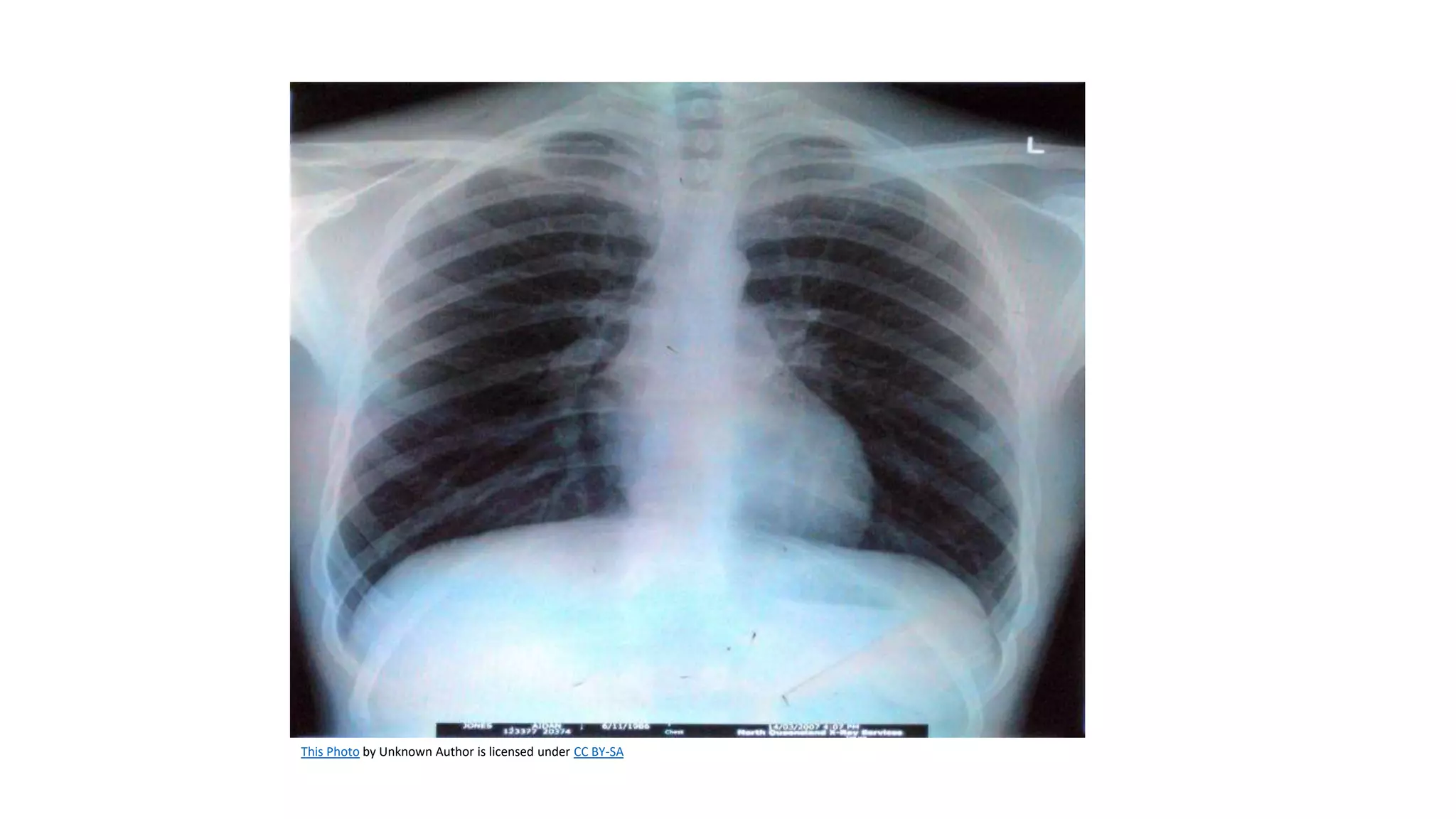
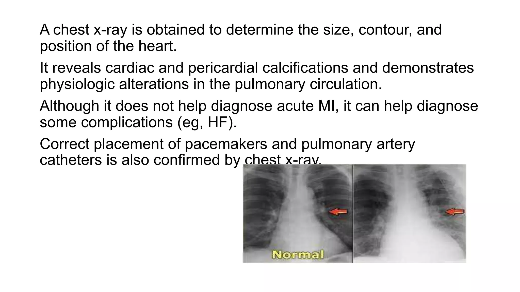
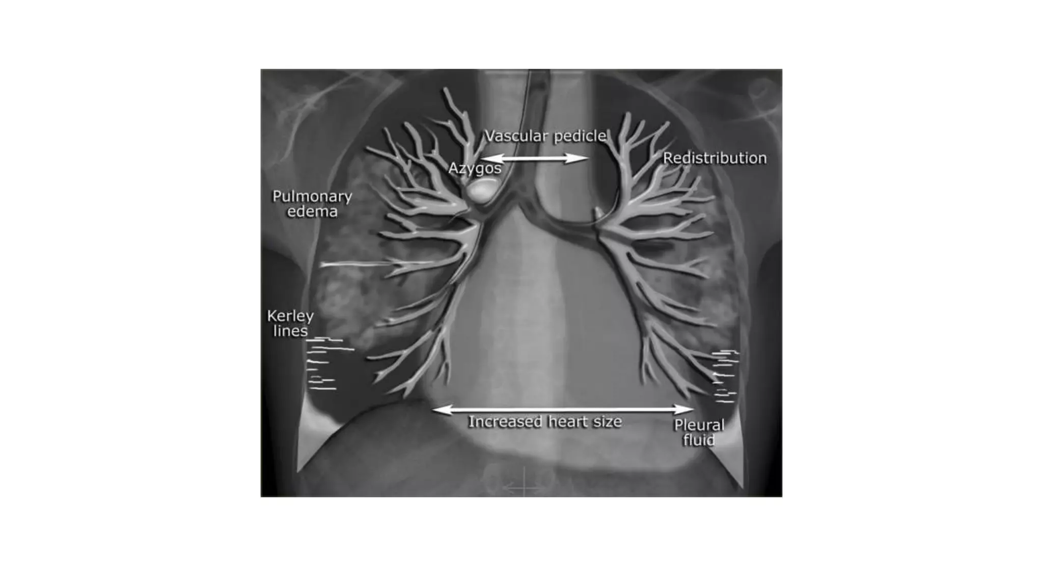
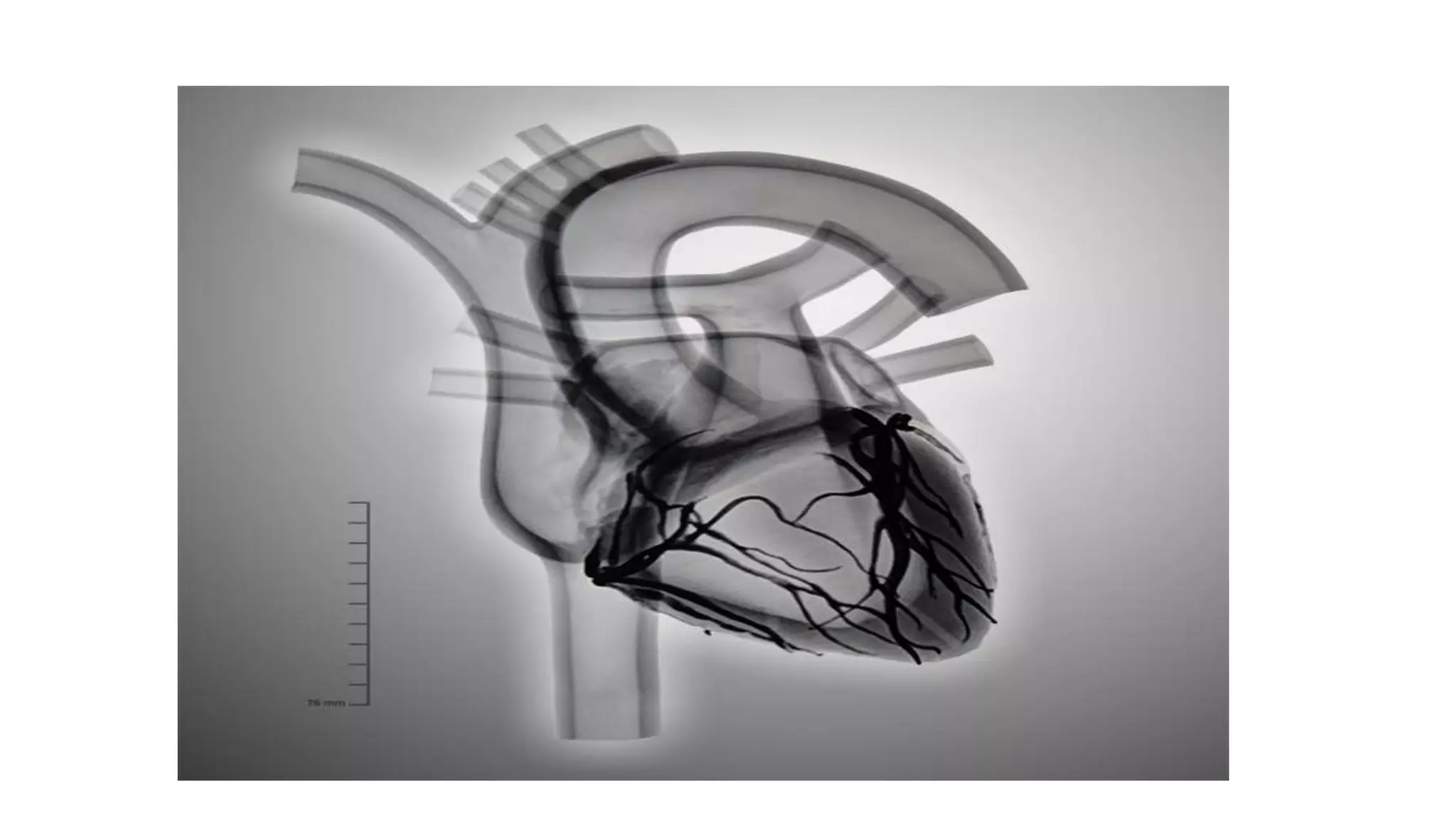
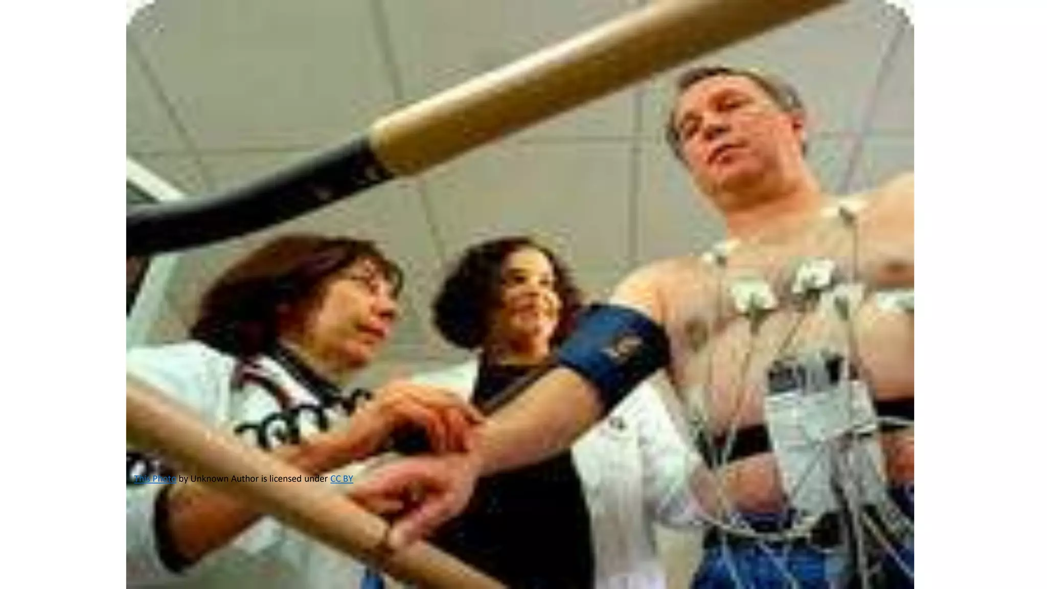
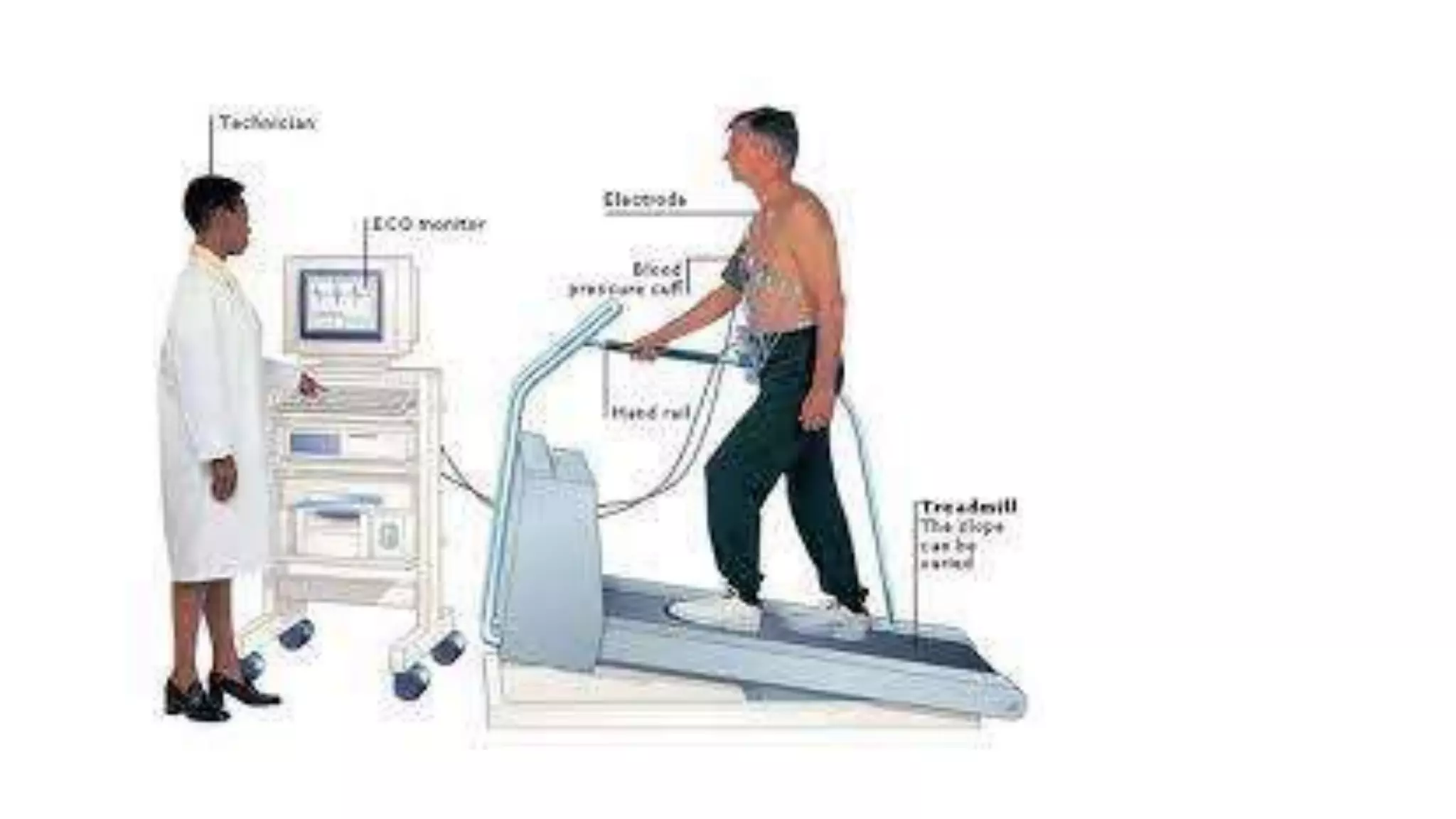
This Photo by Unknown Author is licensed under CC BY-SA
A chest x-ray is obtained to determine the size, contour, and
position of the heart.
It reveals cardiac and pericardial calcifications and demonstrates
physiologic alterations in the pulmonary circulation.
Although it does not help diagnose acute MI, it can help diagnose
some complications (eg, HF).
Correct placement of pacemakers and pulmonary artery
catheters is also confirmed by chest x-ray.
• Fluoroscopy is an x-ray imaging technique that allows
visualization of the heart on a screen.
• It shows cardiac and vascular pulsations and unusual cardiac
contours.
• During a fluoroscopy procedure, an X-ray beam is passed
through the body. The image is transmitted to a monitor so the
movement of a body part or of an instrument or contrast agent
("X-ray dye") through the body can be seen in detail.
This Photo by Unknown Author is licensed under CC BY
The stress test helps determine the following:
(1) presence of CAD,
(2) cause of chest pain
(3) functional capacity of the heart after an MI or heart surgery,
(4) effectiveness of antianginal or antiarrhythmic medications,
(5) dysrhythmias that occur during physical exercise, and
(6) specific goals for a physical fitness program.
Contraindications
severe aortic stenosis, acute myocarditis or pericarditis, severe
hypertension, suspected left main CAD, HF, and unstable angina
• Procedure
• During an exercise stress test, the patient walks on a
tread- mill pedals a stationary bicycle, or uses an arm
crank. Exercise intensity progresses according to
established protocols.
• The goal is to increase the heart rate to the “target heart
rate,” which is 80% to 90% of the maximum predicted
heart rate based on the patient’s age and gender.
• During the test, the following are monitored:
• two or more ECG leads for heart rate, rhythm, and
ischemic changes; BP; skin temperature; physical
appearance; perceived exertion; and symptoms,
including chest pain, dyspnea, dizziness, leg cramping,
and fatigue.
• The test is terminated when the target heart rate is achieved or
when the patient experiences chest pain, extreme fatigue, a
decrease in BP or pulse rate, serious dysrhythmias or ST-
segment changes on the ECG, or other complications.
• When significant ECG abnormalities occur during the stress
test (ST-segment depressions or elevations), the test result is
reported as positive and further diagnostic testing such as a
cardiac catheterization is required.
•Pharmacologic Stress Testing
• Procedure
Two vasodilating agents, dipyridamole (Persantine) and
adenosine (Adenocard) dobutamine administered IV, are
used to mimic the effects of exercise by maximally
dilating the coronary arteries.
• Nursing Interventions
• In preparation for the pharmacologic stress test, the
patient is instructed not to eat or drink anything for at
least 4 hours before the test.(caffeine)
• Patients taking aminophylline, theophylline, or
dipyridamole are instructed to stop taking these
medications for 24 to 48 hours before the test (if
tolerated).
• The patient is informed about the transient sensations
that may occur during infusion of the vasodilating agent,
such as flushing or nausea.
Echo cardiography
Traditional Echocardiography
• Echocardiography is a non invasive ultrasound test that is used
to measure the ejection fraction and examine the size, shape,
and motion of cardiac structures.
• It is particularly useful for diagnosing pericardial effusions;
determining chamber size and the etiology of heart murmurs;
evaluating the function of heart valves, including prosthetic
heart valves; and evaluating ventricular wall motion.
• Echocardiography involves transmission of high-frequency
sound waves into the heart through the chest wall and recording
of the return signals.
• The ultrasound is generated by a handheld transducer applied
to the front of the chest.
• The transducer picks up the echoes, converts them to electrical
impulses, and transmits them for display on an oscilloscope and
recording on a videotape.
• An ECG is recorded simultaneously to assist with interpreting
the echocardiogram.
• Myocardial ischemia from decreased perfusion during stress
causes abnormalities in ventricular wall motion and is easily
detected by echocardiography.
Before echocardiography, the nurse informs the patient about the
test, explaining that it is painless. Echocardiographic monitoring
is performed while a transducer that emits sound waves is moved
over the surface of the chest wall. Gel applied to the skin helps
transmit the sound waves. Periodically, the patient is asked to
turn onto the left side or hold a breath. The test takes about 30 to
45 minutes
TRANS ESOPHAGEAL ECHO
A probe with an ultrasound transducer at the tip is swallowed
while the physician controls angle and depth.
As it passes down the esophagus, it sends back clear images of
heart size, wall motion, valvular abnormalities, endocarditis
vegetation, and possible source of thrombi without interference
from lungs or chest ribs.
A contrast medium may be injected IV for evaluating direction
of blood flow if an atrial or ventricular septal defect is
suspected.
Doppler ultrasound and colorflow imaging can also be used
concurrently.
• Instruct patient to be NPO for at least 6 hr before test.
Remove dentures.
• IV sedation is administered and throat locally anesthetized.
• A bite block is placed in the mouth.
• Monitor vital signs and oxygen saturation levels and perform
suctioning as needed during procedure.
• Patient may not eat or drink until gag reflex returns.
• Sore throat is temporary.
• Radionuclide Imaging
• Radionuclide imaging studies involve the use of radioisotopes to
noninvasively evaluate coronary artery perfusion, to detect
myocardial ischemia and infarction, and to assess left ventricular
function.
• Thallium 201 (Tl201) and technetium 99m (Tc99m) are two
radioisotopes used in cardiac nuclear medicine When these
radioisotopes are injected into the bloodstream, the energy emitted
can be detected by a gamma scintillation camera positioned over the
body.
• Planar imaging, used with thallium, provides a one-dimensional view
of the heart from three locations.
• Single photon emission computed tomography (SPECT) provides
three- dimensional images. With SPECT, the patient is positioned
supine with arms raised above the head, while the camera moves
around the patient’s chest in a 180- to 360-degree arc to more
precisely identify the areas of decreased myocardial perfusion.
• MYOCARDIAL PERFUSION IMAGING
• Test of Ventricular Function and Wall Motion
Equilibrium radionuclide angiocardiography (ERNA)
multiple-gated acquisition (MUGA) scanning
• Non invasive technique that uses a conventional scintillation
camera interfaced with a computer to record images of the
heart during several hundred heartbeats. The computer
processes the data and allows for sequential viewing of the
functioning heart. The sequential images are analyzed to
evaluate left ventricular function, wall motion, and ejection
fraction.
• MUGA scanning can also be used to assess the differences in
left ventricular function during rest and exercise.
non invasive and invasive cardiac monitoring.pptx

non invasive and invasive cardiac monitoring.pptx

  • 2.
    This Photo byUnknown Author is licensed under CC BY-SA
  • 3.
    A chest x-rayis obtained to determine the size, contour, and position of the heart. It reveals cardiac and pericardial calcifications and demonstrates physiologic alterations in the pulmonary circulation. Although it does not help diagnose acute MI, it can help diagnose some complications (eg, HF). Correct placement of pacemakers and pulmonary artery catheters is also confirmed by chest x-ray.
  • 5.
    • Fluoroscopy isan x-ray imaging technique that allows visualization of the heart on a screen. • It shows cardiac and vascular pulsations and unusual cardiac contours. • During a fluoroscopy procedure, an X-ray beam is passed through the body. The image is transmitted to a monitor so the movement of a body part or of an instrument or contrast agent ("X-ray dye") through the body can be seen in detail.
  • 7.
    This Photo byUnknown Author is licensed under CC BY
  • 9.
    The stress testhelps determine the following: (1) presence of CAD, (2) cause of chest pain (3) functional capacity of the heart after an MI or heart surgery, (4) effectiveness of antianginal or antiarrhythmic medications, (5) dysrhythmias that occur during physical exercise, and (6) specific goals for a physical fitness program. Contraindications severe aortic stenosis, acute myocarditis or pericarditis, severe hypertension, suspected left main CAD, HF, and unstable angina
  • 10.
    • Procedure • Duringan exercise stress test, the patient walks on a tread- mill pedals a stationary bicycle, or uses an arm crank. Exercise intensity progresses according to established protocols. • The goal is to increase the heart rate to the “target heart rate,” which is 80% to 90% of the maximum predicted heart rate based on the patient’s age and gender. • During the test, the following are monitored: • two or more ECG leads for heart rate, rhythm, and ischemic changes; BP; skin temperature; physical appearance; perceived exertion; and symptoms, including chest pain, dyspnea, dizziness, leg cramping, and fatigue.
  • 11.
    • The testis terminated when the target heart rate is achieved or when the patient experiences chest pain, extreme fatigue, a decrease in BP or pulse rate, serious dysrhythmias or ST- segment changes on the ECG, or other complications. • When significant ECG abnormalities occur during the stress test (ST-segment depressions or elevations), the test result is reported as positive and further diagnostic testing such as a cardiac catheterization is required.
  • 12.
    •Pharmacologic Stress Testing •Procedure Two vasodilating agents, dipyridamole (Persantine) and adenosine (Adenocard) dobutamine administered IV, are used to mimic the effects of exercise by maximally dilating the coronary arteries. • Nursing Interventions • In preparation for the pharmacologic stress test, the patient is instructed not to eat or drink anything for at least 4 hours before the test.(caffeine)
  • 13.
    • Patients takingaminophylline, theophylline, or dipyridamole are instructed to stop taking these medications for 24 to 48 hours before the test (if tolerated). • The patient is informed about the transient sensations that may occur during infusion of the vasodilating agent, such as flushing or nausea.
  • 14.
  • 15.
    Traditional Echocardiography • Echocardiographyis a non invasive ultrasound test that is used to measure the ejection fraction and examine the size, shape, and motion of cardiac structures. • It is particularly useful for diagnosing pericardial effusions; determining chamber size and the etiology of heart murmurs; evaluating the function of heart valves, including prosthetic heart valves; and evaluating ventricular wall motion.
  • 16.
    • Echocardiography involvestransmission of high-frequency sound waves into the heart through the chest wall and recording of the return signals. • The ultrasound is generated by a handheld transducer applied to the front of the chest. • The transducer picks up the echoes, converts them to electrical impulses, and transmits them for display on an oscilloscope and recording on a videotape. • An ECG is recorded simultaneously to assist with interpreting the echocardiogram. • Myocardial ischemia from decreased perfusion during stress causes abnormalities in ventricular wall motion and is easily detected by echocardiography.
  • 17.
    Before echocardiography, thenurse informs the patient about the test, explaining that it is painless. Echocardiographic monitoring is performed while a transducer that emits sound waves is moved over the surface of the chest wall. Gel applied to the skin helps transmit the sound waves. Periodically, the patient is asked to turn onto the left side or hold a breath. The test takes about 30 to 45 minutes
  • 18.
  • 20.
    A probe withan ultrasound transducer at the tip is swallowed while the physician controls angle and depth. As it passes down the esophagus, it sends back clear images of heart size, wall motion, valvular abnormalities, endocarditis vegetation, and possible source of thrombi without interference from lungs or chest ribs. A contrast medium may be injected IV for evaluating direction of blood flow if an atrial or ventricular septal defect is suspected. Doppler ultrasound and colorflow imaging can also be used concurrently.
  • 21.
    • Instruct patientto be NPO for at least 6 hr before test. Remove dentures. • IV sedation is administered and throat locally anesthetized. • A bite block is placed in the mouth. • Monitor vital signs and oxygen saturation levels and perform suctioning as needed during procedure. • Patient may not eat or drink until gag reflex returns. • Sore throat is temporary.
  • 22.
    • Radionuclide Imaging •Radionuclide imaging studies involve the use of radioisotopes to noninvasively evaluate coronary artery perfusion, to detect myocardial ischemia and infarction, and to assess left ventricular function. • Thallium 201 (Tl201) and technetium 99m (Tc99m) are two radioisotopes used in cardiac nuclear medicine When these radioisotopes are injected into the bloodstream, the energy emitted can be detected by a gamma scintillation camera positioned over the body. • Planar imaging, used with thallium, provides a one-dimensional view of the heart from three locations. • Single photon emission computed tomography (SPECT) provides three- dimensional images. With SPECT, the patient is positioned supine with arms raised above the head, while the camera moves around the patient’s chest in a 180- to 360-degree arc to more precisely identify the areas of decreased myocardial perfusion.
  • 24.
    • MYOCARDIAL PERFUSIONIMAGING • Test of Ventricular Function and Wall Motion Equilibrium radionuclide angiocardiography (ERNA) multiple-gated acquisition (MUGA) scanning • Non invasive technique that uses a conventional scintillation camera interfaced with a computer to record images of the heart during several hundred heartbeats. The computer processes the data and allows for sequential viewing of the functioning heart. The sequential images are analyzed to evaluate left ventricular function, wall motion, and ejection fraction. • MUGA scanning can also be used to assess the differences in left ventricular function during rest and exercise.