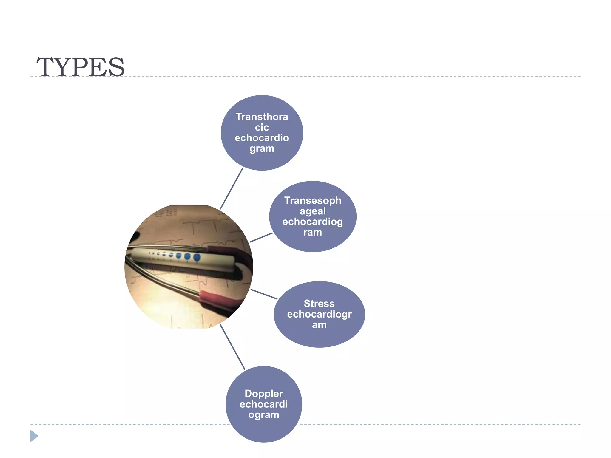
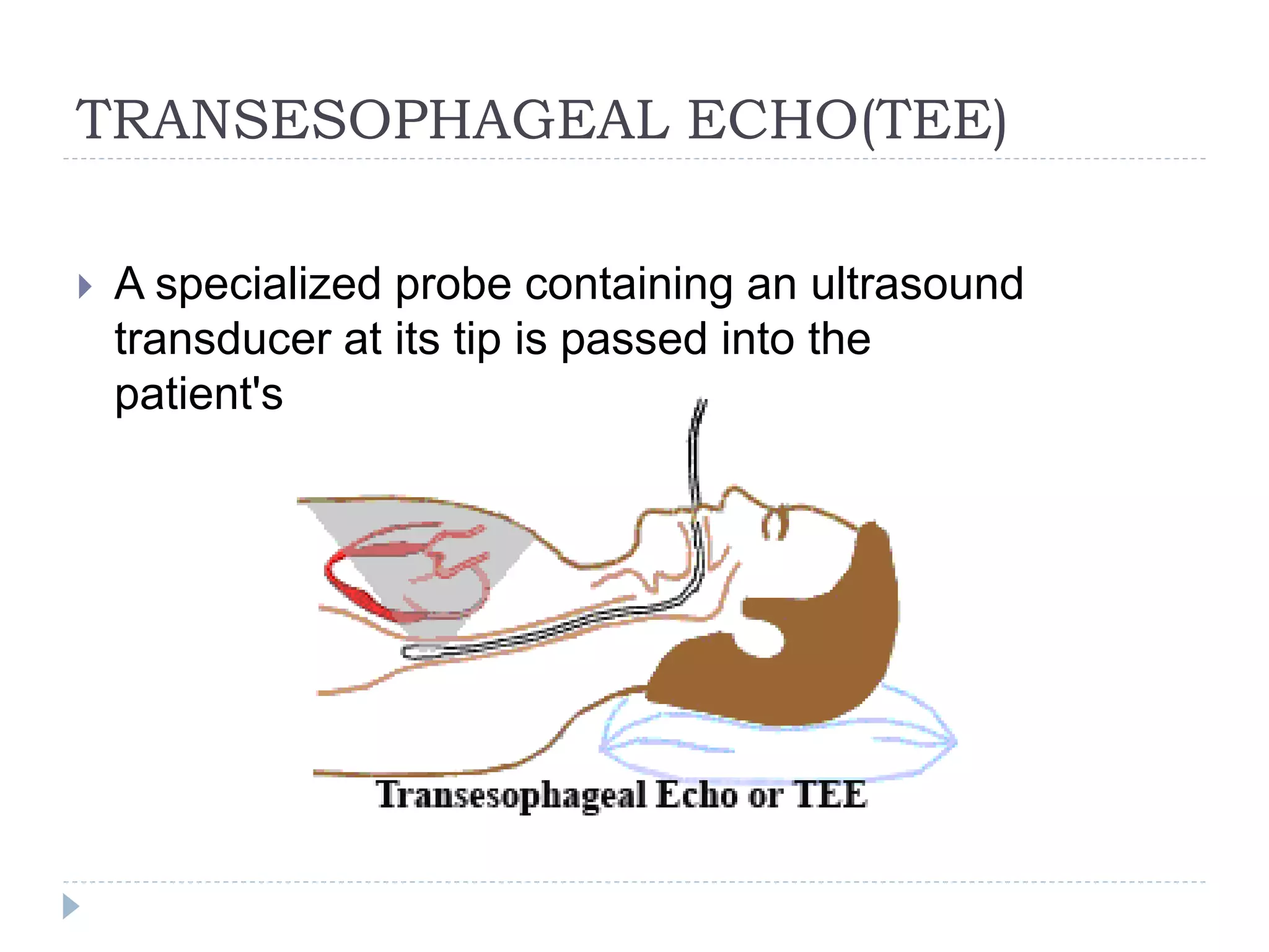
Echocardiography uses ultrasound to create images of the heart. The main types are transthoracic and transesophageal echocardiograms. Transthoracic echocardiograms image the heart non-invasively from the chest, while transesophageal echocardiograms pass an ultrasound probe down the esophagus for clearer images. Doppler echocardiography assesses blood flow velocity and direction. Echocardiography is used to evaluate heart structures and function, detect abnormalities, and help diagnose conditions like heart valve problems, heart muscle diseases, and blood clots.
CARDIAC TAMPONADE ( Cardiac emergency) • Cardiac Tamponade is a life threatening complication caused by excessive accumulation of fluid in the pericardium. Or • Compression of all cardiac chambers due to excessive accumulation of pericardial fluid leading to compromised cardiac out put.
commonly used for medical students, and helpful to use this ppt to study for them, and also a common man can understand very easily what is coarctation of aorta.
A transesophageal echocardiogram (TEE) uses echocardiography to assess the structure and function of the heart. During the procedure, a transducer (like a microphone) sends out ultrasonic sound waves. When the transducer is placed at certain locations and angles, the ultrasonic sound waves move through the skin and other body tissues to the heart tissues, where the waves bounce or "echo" off of the heart structures. The transducer picks up the reflected waves and sends them to a computer. The computer displays the echoes as images of the heart walls and valves.
A traditional echocardiogram is done by putting the transducer on the surface of the chest. This is called a transthoracic echocardiogram. A transesophageal echocardiogram is done by inserting a probe with a transducer down the esophagus. This provides a clearer image of the heart because the sound waves do not have to pass through skin, muscle, or bone tissue. The TEE probe is much closer to the heart since the esophagus and heart are right next to each other.
Transesophaheal echo cardiography, the basic views. It is a diagnostic procedure to visualize the heart and have a better understanding of the structure and functions of the heart
A transesophageal echocardiogram, or TEE, is an alternative way to perform an echocardiogram. A specialized probe containing an ultrasound transducer at its tip is passed into the patient's esophagus. This allows image and Doppler evaluation which can be recorded. It has several advantages and some disadvantages compared with a transthoracic echocardiogram.
These simplified slides by Dr. Sidra Arshad present an overview of the non-respiratory functions of the respiratory tract.
Learning objectives:
1. Enlist the non-respiratory functions of the respiratory tract
2. Briefly explain how these functions are carried out
3. Discuss the significance of dead space
4. Differentiate between minute ventilation and alveolar ventilation
5. Describe the cough and sneeze reflexes
Study Resources:
1. Chapter 39, Guyton and Hall Textbook of Medical Physiology, 14th edition
2. Chapter 34, Ganong’s Review of Medical Physiology, 26th edition
3. Chapter 17, Human Physiology by Lauralee Sherwood, 9th edition
4. Non-respiratory functions of the lungs https://academic.oup.com/bjaed/article/13/3/98/278874
Recomendações da OMS sobre cuidados maternos e neonatais para uma experiência pós-natal positiva.
Em consonância com os ODS – Objetivos do Desenvolvimento Sustentável e a Estratégia Global para a Saúde das Mulheres, Crianças e Adolescentes, e aplicando uma abordagem baseada nos direitos humanos, os esforços de cuidados pós-natais devem expandir-se para além da cobertura e da simples sobrevivência, de modo a incluir cuidados de qualidade.
Estas diretrizes visam melhorar a qualidade dos cuidados pós-natais essenciais e de rotina prestados às mulheres e aos recém-nascidos, com o objetivo final de melhorar a saúde e o bem-estar materno e neonatal.
Uma “experiência pós-natal positiva” é um resultado importante para todas as mulheres que dão à luz e para os seus recém-nascidos, estabelecendo as bases para a melhoria da saúde e do bem-estar a curto e longo prazo. Uma experiência pós-natal positiva é definida como aquela em que as mulheres, pessoas que gestam, os recém-nascidos, os casais, os pais, os cuidadores e as famílias recebem informação consistente, garantia e apoio de profissionais de saúde motivados; e onde um sistema de saúde flexível e com recursos reconheça as necessidades das mulheres e dos bebês e respeite o seu contexto cultural.
Estas diretrizes consolidadas apresentam algumas recomendações novas e já bem fundamentadas sobre cuidados pós-natais de rotina para mulheres e neonatos que recebem cuidados no pós-parto em unidades de saúde ou na comunidade, independentemente dos recursos disponíveis.
É fornecido um conjunto abrangente de recomendações para cuidados durante o período puerperal, com ênfase nos cuidados essenciais que todas as mulheres e recém-nascidos devem receber, e com a devida atenção à qualidade dos cuidados; isto é, a entrega e a experiência do cuidado recebido. Estas diretrizes atualizam e ampliam as recomendações da OMS de 2014 sobre cuidados pós-natais da mãe e do recém-nascido e complementam as atuais diretrizes da OMS sobre a gestão de complicações pós-natais.
O estabelecimento da amamentação e o manejo das principais intercorrências é contemplada.
Recomendamos muito.
Vamos discutir essas recomendações no nosso curso de pós-graduação em Aleitamento no Instituto Ciclos.
Esta publicação só está disponível em inglês até o momento.
Prof. Marcus Renato de Carvalho
www.agostodourado.com
263778731218 Abortion Clinic /Pills In Harare ,sisternakatoto
263778731218 Abortion Clinic /Pills In Harare ,ABORTION WOMEN’S CLINIC +27730423979 IN women clinic we believe that every woman should be able to make choices in her pregnancy. Our job is to provide compassionate care, safety,affordable and confidential services. That’s why we have won the trust from all generations of women all over the world. we use non surgical method(Abortion pills) to terminate…Dr.LISA +27730423979women Clinic is committed to providing the highest quality of obstetrical and gynecological care to women of all ages. Our dedicated staff aim to treat each patient and her health concerns with compassion and respect.Our dedicated group ABORTION WOMEN’S CLINIC +27730423979 IN women clinic we believe that every woman should be able to make choices in her pregnancy. Our job is to provide compassionate care, safety,affordable and confidential services. That’s why we have won the trust from all generations of women all over the world. we use non surgical method(Abortion pills) to terminate…Dr.LISA +27730423979women Clinic is committed to providing the highest quality of obstetrical and gynecological care to women of all ages. Our dedicated staff aim to treat each patient and her health concerns with compassion and respect.Our dedicated group of receptionists, nurses, and physicians have worked together as a teamof receptionists, nurses, and physicians have worked together as a team wwww.lisywomensclinic.co.za/
Basavarajeeyam is a Sreshta Sangraha grantha (Compiled book ), written by Neelkanta kotturu Basavaraja Virachita. It contains 25 Prakaranas, First 24 Chapters related to Rogas& 25th to Rasadravyas.
Lung Cancer: Artificial Intelligence, Synergetics, Complex System Analysis, S...Oleg Kshivets
RESULTS: Overall life span (LS) was 2252.1±1742.5 days and cumulative 5-year survival (5YS) reached 73.2%, 10 years – 64.8%, 20 years – 42.5%. 513 LCP lived more than 5 years (LS=3124.6±1525.6 days), 148 LCP – more than 10 years (LS=5054.4±1504.1 days).199 LCP died because of LC (LS=562.7±374.5 days). 5YS of LCP after bi/lobectomies was significantly superior in comparison with LCP after pneumonectomies (78.1% vs.63.7%, P=0.00001 by log-rank test). AT significantly improved 5YS (66.3% vs. 34.8%) (P=0.00000 by log-rank test) only for LCP with N1-2. Cox modeling displayed that 5YS of LCP significantly depended on: phase transition (PT) early-invasive LC in terms of synergetics, PT N0—N12, cell ratio factors (ratio between cancer cells- CC and blood cells subpopulations), G1-3, histology, glucose, AT, blood cell circuit, prothrombin index, heparin tolerance, recalcification time (P=0.000-0.038). Neural networks, genetic algorithm selection and bootstrap simulation revealed relationships between 5YS and PT early-invasive LC (rank=1), PT N0—N12 (rank=2), thrombocytes/CC (3), erythrocytes/CC (4), eosinophils/CC (5), healthy cells/CC (6), lymphocytes/CC (7), segmented neutrophils/CC (8), stick neutrophils/CC (9), monocytes/CC (10); leucocytes/CC (11). Correct prediction of 5YS was 100% by neural networks computing (area under ROC curve=1.0; error=0.0).
CONCLUSIONS: 5YS of LCP after radical procedures significantly depended on: 1) PT early-invasive cancer; 2) PT N0--N12; 3) cell ratio factors; 4) blood cell circuit; 5) biochemical factors; 6) hemostasis system; 7) AT; 8) LC characteristics; 9) LC cell dynamics; 10) surgery type: lobectomy/pneumonectomy; 11) anthropometric data. Optimal diagnosis and treatment strategies for LC are: 1) screening and early detection of LC; 2) availability of experienced thoracic surgeons because of complexity of radical procedures; 3) aggressive en block surgery and adequate lymph node dissection for completeness; 4) precise prediction; 5) adjuvant chemoimmunoradiotherapy for LCP with unfavorable prognosis.
CDSCO and Phamacovigilance {Regulatory body in India}NEHA GUPTA
The Central Drugs Standard Control Organization (CDSCO) is India's national regulatory body for pharmaceuticals and medical devices. Operating under the Directorate General of Health Services, Ministry of Health & Family Welfare, Government of India, the CDSCO is responsible for approving new drugs, conducting clinical trials, setting standards for drugs, controlling the quality of imported drugs, and coordinating the activities of State Drug Control Organizations by providing expert advice.
Pharmacovigilance, on the other hand, is the science and activities related to the detection, assessment, understanding, and prevention of adverse effects or any other drug-related problems. The primary aim of pharmacovigilance is to ensure the safety and efficacy of medicines, thereby protecting public health.
In India, pharmacovigilance activities are monitored by the Pharmacovigilance Programme of India (PvPI), which works closely with CDSCO to collect, analyze, and act upon data regarding adverse drug reactions (ADRs). Together, they play a critical role in ensuring that the benefits of drugs outweigh their risks, maintaining high standards of patient safety, and promoting the rational use of medicines.
Adv. biopharm. APPLICATION OF PHARMACOKINETICS : TARGETED DRUG DELIVERY SYSTEMSAkankshaAshtankar
MIP 201T & MPH 202T
ADVANCED BIOPHARMACEUTICS & PHARMACOKINETICS : UNIT 5
APPLICATION OF PHARMACOKINETICS : TARGETED DRUG DELIVERY SYSTEMS By - AKANKSHA ASHTANKAR
micro teaching on communication m.sc nursing.pdfAnurag Sharma
Microteaching is a unique model of practice teaching. It is a viable instrument for the. desired change in the teaching behavior or the behavior potential which, in specified types of real. classroom situations, tends to facilitate the achievement of specified types of objectives.
New Drug Discovery and Development .....NEHA GUPTA
The "New Drug Discovery and Development" process involves the identification, design, testing, and manufacturing of novel pharmaceutical compounds with the aim of introducing new and improved treatments for various medical conditions. This comprehensive endeavor encompasses various stages, including target identification, preclinical studies, clinical trials, regulatory approval, and post-market surveillance. It involves multidisciplinary collaboration among scientists, researchers, clinicians, regulatory experts, and pharmaceutical companies to bring innovative therapies to market and address unmet medical needs.
2. DEFINITION
Echocardiogram is a diagnostic test which uses
ultrasound waves to make images of the heart
chambers, valves and surrounding structures. It can
measure cardiac output and is a sensitive test to find
fluid around the heart (pericardial effusion).
4. TRANSTHORACIC ECHO
A transthoracic echocardiogram (TTE) is the most
common type of echocardiogram . In this case, the
probe (or ultrasonic transducer) is placed on
the chest or abdomen of the subject to get various
views of the heart.
It is used as a non-invasive assessment of the
overall health of the heart, including a patient's heart
valves and degree of heart muscle contraction (an
indicator of the ejection fraction).
6. TRANSESOPHAGEAL ECHO(TEE)
A specialized probe containing an ultrasound
transducer at its tip is passed into the
patient's oesophagus.
7. Advantages of TEE
The advantage of TEE over TTE is usually clearer
images, especially of structures that are difficult to
view transthoracically (through the chest wall) since
heart rests directly upon the esophagus leaving only
millimeters that the ultrasound beam has to travel
This reduces the attenuation (weakening) of the
ultrasound signal, generating a stronger return
signal.
8. In adults, several structures can be evaluated
and imaged better with the TEE, including
the aorta, pulmonary artery, valves of the heart,
both atria, atrial septum, left atrial appendage,
and coronary arteries.
TEE has a very high sensitivity for locating a
blood clot inside the left atrium.
DISADVANTAGES
It takes longer to perform a TEE than a TTE. It
may be uncomfortable for the patient, who may
require sedation or general anesthesia.
Some risks are associated with the procedure,
such as esophageal perforation around 1 in
10,000, and adverse reactions to the medication.
9. PROCESS OF TEE
Before inserting the probe, mild to
moderate sedation is induced in the patient to ease
the discomfort and to decrease the gag reflex, thus
making the ultrasound probe easier to pass into the
esophagus.
Mild to moderate Sedation is produced
by midazolam (a benzodiazepine ) , fentanyl (an
opioid) or propofol
Usually a local anesthetic spray is used for the back
of the throat, such a xylocaine /or a jelly /lubricant
anesthetic for the esophagus.
Children are anesthetized
10. CLINCAL USES
TEE can be performed by a cardiac anesthesiologist
to evaluate, diagnose, and treat patients in the
perioperative period.
. TEE is very useful during many cardiac surgical
procedures (e.g., mitral valve repair and in aortic
dissections). It is actually an essential monitoring
tool and to assess the results of surgery immediately
after the procedure.
11. STRESS ECHOCARDIOGRAM
A stress test may be accompanied
by echocardiography.
The echocardiography is performed both before and
after the exercise so that structural differences can
be compared.
A resting echocardiogram is obtained prior to stress.
The images obtained are similar to the ones
obtained during a full surface echocardiogram,
commonly referred to as transthoracic
echocardiogram.
12. The patient is subjected to stress in the form of
exercise or chemically (usually dobutamine).
After the target heart rate is achieved, 'stress'
echocardiogram images are obtained.
The two echocardiogram images are then compared
to assess for any abnormalities in wall motion of the
heart. This is used to detect obstructive coronary
artery disease.
13.
14. Doppler echocardiography
Doppler echocardiography is a procedure that
uses Doppler ultrasonography to examine
the heart. An echocardiogram uses high frequency
sound waves to create an image of the heart while
the use of Doppler technology allows determination
of the speed and direction of blood flow by utilizing
the Doppler effect.
Velocity measurements allow assessment of cardiac
valve areas and function, any abnormal
communications between the left and right side of
the heart, any leaking of blood through the valves
(valvular regurgitation), calculation of the cardiac
output and calculation of E/A ratio(a measure
of diastolic dysfunction).
15. ADVANTAGES OF DOPPLER
An advantage of Doppler echocardiography is that it
can be used to measure blood flow within the heart
without invasive procedures such as cardiac
catheterization.
The method can measure tissue velocities by tissue
Doppler echocardiography.
The combination of flow and tissue velocities can be
used for estimating left ventricular filling pressure,
although only under certain conditions
17. Doppler Echo
An abnormal echocardiogram : Image shows a
midmuscular ventricular septal defect. The trace in the
lower left shows the cardiac cycle . Colors are used to
represent the velocity and direction of blood flow.
18. AXES IN ECHOCARDIOGRAPHY
Two key axes of the heart are the long axis and short
axis.
The long axis is an imaginary line from the apex of
the heart through the centre of the mitral valve.
The short axis is perpendicular to the long axis and
shows the heart in cross section.
19. WINDOWS OF ECHO
Evaluation of the heart with echocardiography
requires "acoustic windows" of the heart.
Bone reflects the ultrasound waves and so all
structures directly behind bone are not visible with
ultrasound.
This requires that the heart be viewed between
bones and, in particular, between ribs. The most
common views are the parasternal, apical,
subcostal, and suprasternal windows.
20. Para sternal:
Adjacent to the sternum..
Apical:
At the apex of the heart.
Sub costal:
Below the sternum at the top of the abdomen
Supra sternal:
Above the sternal at the base of the neck
21. VIEWS
There are several typical views
obtained during a routine TTE. Views
outside of the typical views can be
considered "off axis" and may be
obtained for specific purposes.
22. Parasternal long axis (PLAX)
This view is obtained to the left of the sternum and
views the heart in its long axis. In this view, the mitral
valve, aortic valve, right ventricular outflow tract,
base of the left ventricle, and the left atrium can be
visible.
In this view, it is possible to appreciate the long-axis
cross section of the mitral and aortic valves. The
classic "hockey stick" shape of rheumatic mitral
stenosis can be appreciated in this view.
The parasternal long view of the pulmonary valve is
the only view of the posterior leaflet.
24. Structures visible:
Anterior septal and inferior lateral walls of the left
ventricle
Left atrium
Mitral valve in long-axis with chordae
Aortic valve in long-axis
Tricuspid valve in long-axis (angulated) and right
ventricular inflow tract
Pulmonary valve in long-axis (angulated) and right
ventricular outflow tract
25. Measurements in this view can be used to quantify
the heart:
Left ventricular size and wall thickness
Left atrial linear dimension (as opposed to area)
Left ventricular outflow tract diameter (used to
calculate aortic valve area by the continuity
equation)
Aortic annulus, sinus of Valsalva, and aortic root
sizes
Color doppler of all four valves
Spectral doppler of tricuspid and pulmonary valves
26. Parasternal short axis (PSAX)
This view is obtained in the same window as the
parasternal long, but with the probe rotated 90°.
In this view, the aortic valve is seen in cross-section
with the right ventricular inflow & outflow tracts
visible with the tricuspid valve as well
. Pulmonary valve is not visible in this view. Both the
right and left atria are visible.
The standard PSAX view is at the level of the aortic
valve, but moving the probe along the long-axis can
review the LV outflow tract, LV at the base, and LV at
the mid section.
28. Structures visible:
Aortic valve in short-axis
Aortic valve dysfunction, aortic sclerosis/stenosis
Tricuspid valve in long-axis
Right ventricle including inflow and outflow tracts
Left ventricle in short-axis
Closer to the base can reveal the left ventricular outflow
tract
At the level of the base can show the movement of the
mitral valve leaflets in short-axis
At the level of mid-LV can show papillary muscles
29.
30. Apical four chamber (A4C)
This view is obtained at the apex of the heart and
looking toward the base of the heart (where the
valves are).
In this view, the mitral valve, tricuspid valve, and all
four chambers are visible.
This view shows the right ventricle from base to apex
and is a useful view to estimate RV systolic function
32. Structures visible:
Inferior septum and anterior lateral segments of the
left ventricle
Right ventricle
Left atrium
Right atrium
Mitral valve
Tricuspid valve
33. Measurements in this view can be used to quantify
the heart:
RV size and function
Left atrial size
Right atrial size
Mitral valve flow is best seen in this view and has the
best angle with probe to estimate flows
Tricuspid valve flow
Tissue doppler at the mitral valve annulus (septum &
lateral wall) for diastolic function
Agitated saline bubble study for right to left shunting
(PFO, ASD, VSD)
With contrast, apical and mural LV thrombi can be
easily seen
34. Apical three chamber (A3C)
This view is obtained at the same window as the
apical four chamber and then rotation of the probe.
In this view, the mitral valve and aortic valve are in
view and is roughly similar to the parasternal long
axis.
In this view, the LV outflow tract is best in alignment
with the probe and so gives the best estimate of flow
through the LVOT, which is commonly used to
estimate aortic stenosis.
36. Structures visible:
Aortic valve
Mitral valve
Left ventricle
Left atrium
Measurements in this view can be used to
quantify the heart:
Left ventricle outflow tract volume-time integral
(LVOT VTI) used in conjunction with aortic valve VTI
for aortic valve area and stenosis
Mitral valve flow
37. Apical two chamber (A2C)
This view is obtained at the same window as the
apical four chamber and then rotation of the probe.
In this view, the mitral valve is visible with the left
atrium and left ventricle.
Structures visible:
Anterior and inferior segments of the left ventricle
Mitral valve in long-axis
Left atrium
Measurements in this view can be used to
quantify the heart:
Mitral valve flow
Spectral doppler of the mitral valve
39. Subcostal
This view is obtained below the sternum and at the
top part of the abdomen.
In this view, the junction of the inferior vena cava
with the right atrium is best seen.
From this window, it is possible in some people to
see roughly equivalent views of the apical four
chamber and parasternal short views.
In some people, this may afford these common
views but at a subcostal window that may not be
obtained through the parasternal and/or apical
windows because of various reasons such as chest
wall trauma, open wounds, or poor acoustic windows
40. . However, the subcostal window is the only window
to view the inferior vena cava that can help support
an estimation of the central venous pressure based
on size and collapsibility during respiration.
Other non-cardiac structures are visible in this view
and some pathologies — such as ascites — can be
observed.
42. Suprasternal (SSN)
This view is obtained above the sternum in the
suprasternal notch.
In this view, the aortic arch and portion of the
descending aorta can be seen. Color and spectral
doppler through the descending aorta can show
signs of coarctation of the aorta.
43.
44. USES OF ECHO
HELPS IN IDENTIFICATION OF:
Blood clots in the heart
Fluid in the sac around the heart
Problems with the aorta, which is the main artery
connected to the heart
Cardiomyopathy : dilated, restrictive, and hypertrophic
Pulmonary hypertension (requires some degree of
tricuspid regurgitation)
Septal defects including ASD & VSD
Stenosis and regurgitation/insufficiency of valves
45. Structure and function of prosthetic valves
Thoracic ascending aortic aneurysm
Infiltrative diseases such as amyloidosis
Cardiac tamponade (it can suggest subclinical
diagnosis)
Evaluation of congenital diseases (eg: Tetralogy of
Fallot, transposition)
Pulmonary embolism
Endocarditis (sensitivity is higher with TEE)
46. Cardiac amyloidosis
Cardiac amyloidosis is a disorder caused by
deposits of an abnormal protein (amyloid) in the
heart tissue. These deposits make it hard for the
heart to work properly.
47.
48. CARDIAC TAMPONADE
In this condition there is right venticular or atrial
compression that is accompanied with the opening
or closure of valves.