By: Ahmad Amirdash, BSN, CCRN
Clinical Resource Nurse, EC
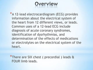
*A 12-lead electrocardiogram (ECG) provides
 information about the electrical system of
 the heart from 12 different views, or leads.
 Common uses of a 12-lead ECG include
 diagnosis of acute coronary syndromes,
 identification of dysrhythmias, and
 determination of the effects of medications
 or electrolytes on the electrical system of the
 heart.


*There are SIX chest ( precordial ) leads &
 FOUR limb leads.
*Check cables and lead wires for fraying, broken
 wires or discoloration before placement.

*It may be necessary to clip chest hair to ensure
 good skin contact with the electrode.

*Respiratory artifact can be common in doing the
 chest leads and may require position changes to
 ensure a good baseline. If sequential (serial) ECGs
 are to be obtained, chest lead sites should be
 marked to ensure that the same lead sites are used
 in subsequent ECGs. Some pregelled electrodes
 can be left in place for repeat ECGs.
*The supine position is best, but Fowler or other
 positions may be used for comfort. ECGs should be
 recorded in the same position each time to ensure
 that tracing changes are not caused by changes in
 body position. If another position is clinically
 required, note the position on the tracing or in the
 comments of the LCD input. Do not compromise
 lead placement because of a medication patch.
 Relocate the medication patch before applying the
 leads.

*Expose only the necessary parts of the patients
 legs, arms, and chest. This Provides privacy and
 warmth, which reduces anxiety, shivering and
 resulting artifacts.
*Clean the intended sites with alcohol pads,
 cleansing pads or soap and water, and dry
 thoroughly since moist skin is not conducive to
 electrode adherence.

*When applying the electrodes they must be secure
 to prevent external influences from affecting the
 ECG. The pregelled electrodes must be moist and
 adhesive to allow for appropriate conduction of
 impulses.

*Ensure that the patient is in the supine position, not
 touching the bedrails or footboard which may
 increase the chance of distortion of the trace.
*The following are the critical values that
 require reporting ( depends on your
 institution)
     1- VT/VF
     2- ST elevation, injury pattern
     3- High degree AVB ( AV dissociation, 2:1
        AV block, 3rd degree block )
     4- Severe bradycardia ( HR < 40 bpm )
*You can perform an ECG stat in
Extremely Urgent situations.
However, All ECGS need to have:
date of birth, tech initial ASAP
afterwards. You also need to hold
off transmitting the tracing until all
information is complete. So please
make sure you enter this info as
soon as you get it.
**NOTE: The leads are listed out of numerical
order,     intentionally because it facilitates easier
lead placement.


*Identify the angle of Louis or the sternal notch.
 This assists with identifying the second rib for
 correct placement of precordial leads in the
 appropriate intercostal space ( ICS ).


*Slight alterations in the position of any of the
 precordial leads may alter the ECG significantly
 and can have impact on diagnosis and treatment.
 V1 :
*Palpate the upper sternum to identify where the
clavicle joins the sternum (suprasternal notch).
Slide fingers down the center of the sternum to
the obvious bony prominence. This is the sternal
notch, or angle of Louis which serves as your
landmark. Move directly to the Right(of the
patient ) and you will feel a gap. That gap is the
second intercostal space. Slide your fingers
down over the rib until you feel the next gap.
This is the third intercostal space. Move down
over one more rib and into the next gap, the
fourth intercostal space.
This is where V1 is to be placed.
 V2 :
*Lead V2 is to placed directly across from V1 in
 the fourth ICS, on the left side of the sternum.


 V4 :
*Lead V4 is placed in the 5th intercostal space,
 midclavicular.


 V3 :
*Lead V3 is placed directly horizontally &
 equidistant in between Leads V2 and V4.
V6 :
*Lead V6 is placed in the 5th intercostal
 space, midaxillary horizontally level with V4 .


V5 :
*Lead V5 is placed horizontally & equidistant in
 between leads V4 and V6 (anterior axillary line )


 please see the following illustration
Diagram showing the correct placement for a 12-LEAD ECG.
            Source: University of Nottingham
 Limb leads should be placed in fleshy areas, and
 bony prominences should be avoided. The limb
  leads should be positioned in approximately the
  same place on each limb.


*Note : I have noticed the technicians in the
 Cardiopulmonary Care Center place the lower limb
 leads on the lower abdomen.


*please see the following illustration
Limb lead placement in 12-lead ECG
Source : Mosby’s Nursing procedures & skills
*The ECG must be marked accurately and have a
 clear baseline without artifact for correct
 interpretation. Three to six seconds are all that are
 needed for a permanent record; a longer strip may
 be obtained if a rhythm strip is needed. A rhythm
 strip is a long recording of a lead; lead II is
 commonly used.

*When connecting the wires to the Leads please
 double check the right wire to the matching lead.
 You will notice that the sequence is :
   RA, V1, V2, V3, V4, V5, V6, LA on one side &
   RL, LL on the other side.
*If you forget any of the steps or the sequence, a
 quick reference with 2 diagrams is printed on
 the ECG machine surface (most machines). In
 summary, you will enter the patient information
 first, then place the leads, connect them, then
 record a trace.

*Remember to plug the ECG machine after use
 please.

*Anyone in the health field setting can learn how
 to do a 12-Lead ECG. It just takes practice.
*Here is a link for a short video ( 4
 minutes 25 sec. ) that demonstrates
 the whole procedure. Copy this link &
 paste it in your window please.

http://elscontent.webinservice.com/NursingSkills
/Animations/CC_057/AnimationPlayer.html
*ECG stands for Electrocardiogram, which originally
 came from the German word elektrokardiogramm.
*this is where the acronym EKG comes from. The
 German elektrisch means “electrical,” kardio means
 “heart,” and the Hungarian gramm means “measure
 of mass.” The two acronyms ECG and EKG are used
 interchangeably; however, ECG tends to sound like
 another common medical acronym, EEG, or
 Electroencephalogram. For this reason some medical
 professionals prefer to say EKG.

*-LEAD EKGs are similar to standard EKGs ( monitor )
 except that they are more accurate & look at the
 heart through more angles.
*According to recent estimates by the American Heart
 Coronary heart disease caused approximately 1 of every
 6 deaths in the United States in 2006. Coronary heart
 disease mortality in 2006 was 425 425. In 2010, an
 estimated 785 000 Americans will have a new coronary
 attack, and approximately 470 000 will have a recurrent
 attack. It is estimated that an additional 195 000 silent
 first myocardial infarctions occur each year.
 Approximately every 25 seconds, an American will have
 a coronary event, and approximately every minute,
 someone will die of one. Prehospital 12-LEAD EKGs are
 allowing patients to reach definitive care immediately
 upon arrival at their local emergency department.
* Jacobson, C. (1996). Bedside cardiac monitoring. In: AACN Research
 Based Practice Protocol, Technology Series. Aliso Viejo, CA:
 American Association of Critical Care Publications, 1-32.

* Drew, B.J. (2002). Celebrating the 100th birthday of the
 electrocardiogram: Lessons learned from research in cardiac
 monitoring. Am J Crit Care, 11, 378-86.

* Circulation 2010, 121:e46-e215: Heart Disease and Stroke Statistics-
 -2010 Update : A Report From the American Heart Association
 originally published online December 17, 2009.

* Excerpted and adapted from AACN Procedure Manual for Critical
 Care, Fifth Edition, American Association of Critical-Care Nurses,
 edited by Debra J. Lynn-McHale Wiegand, PhD, RN, CCRN, FAAN,
 and Karen K. Carlson, MN, RN, CCNS, St. Louis: Elsevier/Saunders,
 2005.
Ahmad Amirdash BBA, BSN, CCRN
                       Clinical Resource Nurse
Emergency center, M.D Anderson Cancer Center
      1515 Holcombe Blvd, Houston TX 77030
                  aamirdash@mdanderson.org
                              T
      F                    V

12 lead electrocardiogram

  • 1.
    By: Ahmad Amirdash,BSN, CCRN Clinical Resource Nurse, EC
  • 2.
    *A 12-lead electrocardiogram(ECG) provides information about the electrical system of the heart from 12 different views, or leads. Common uses of a 12-lead ECG include diagnosis of acute coronary syndromes, identification of dysrhythmias, and determination of the effects of medications or electrolytes on the electrical system of the heart. *There are SIX chest ( precordial ) leads & FOUR limb leads.
  • 3.
    *Check cables andlead wires for fraying, broken wires or discoloration before placement. *It may be necessary to clip chest hair to ensure good skin contact with the electrode. *Respiratory artifact can be common in doing the chest leads and may require position changes to ensure a good baseline. If sequential (serial) ECGs are to be obtained, chest lead sites should be marked to ensure that the same lead sites are used in subsequent ECGs. Some pregelled electrodes can be left in place for repeat ECGs.
  • 4.
    *The supine positionis best, but Fowler or other positions may be used for comfort. ECGs should be recorded in the same position each time to ensure that tracing changes are not caused by changes in body position. If another position is clinically required, note the position on the tracing or in the comments of the LCD input. Do not compromise lead placement because of a medication patch. Relocate the medication patch before applying the leads. *Expose only the necessary parts of the patients legs, arms, and chest. This Provides privacy and warmth, which reduces anxiety, shivering and resulting artifacts.
  • 5.
    *Clean the intendedsites with alcohol pads, cleansing pads or soap and water, and dry thoroughly since moist skin is not conducive to electrode adherence. *When applying the electrodes they must be secure to prevent external influences from affecting the ECG. The pregelled electrodes must be moist and adhesive to allow for appropriate conduction of impulses. *Ensure that the patient is in the supine position, not touching the bedrails or footboard which may increase the chance of distortion of the trace.
  • 6.
    *The following arethe critical values that require reporting ( depends on your institution) 1- VT/VF 2- ST elevation, injury pattern 3- High degree AVB ( AV dissociation, 2:1 AV block, 3rd degree block ) 4- Severe bradycardia ( HR < 40 bpm )
  • 7.
    *You can performan ECG stat in Extremely Urgent situations. However, All ECGS need to have: date of birth, tech initial ASAP afterwards. You also need to hold off transmitting the tracing until all information is complete. So please make sure you enter this info as soon as you get it.
  • 8.
    **NOTE: The leadsare listed out of numerical order, intentionally because it facilitates easier lead placement. *Identify the angle of Louis or the sternal notch. This assists with identifying the second rib for correct placement of precordial leads in the appropriate intercostal space ( ICS ). *Slight alterations in the position of any of the precordial leads may alter the ECG significantly and can have impact on diagnosis and treatment.
  • 9.
     V1 : *Palpatethe upper sternum to identify where the clavicle joins the sternum (suprasternal notch). Slide fingers down the center of the sternum to the obvious bony prominence. This is the sternal notch, or angle of Louis which serves as your landmark. Move directly to the Right(of the patient ) and you will feel a gap. That gap is the second intercostal space. Slide your fingers down over the rib until you feel the next gap. This is the third intercostal space. Move down over one more rib and into the next gap, the fourth intercostal space. This is where V1 is to be placed.
  • 10.
     V2 : *LeadV2 is to placed directly across from V1 in the fourth ICS, on the left side of the sternum.  V4 : *Lead V4 is placed in the 5th intercostal space, midclavicular.  V3 : *Lead V3 is placed directly horizontally & equidistant in between Leads V2 and V4.
  • 11.
    V6 : *Lead V6is placed in the 5th intercostal space, midaxillary horizontally level with V4 . V5 : *Lead V5 is placed horizontally & equidistant in between leads V4 and V6 (anterior axillary line )  please see the following illustration
  • 12.
    Diagram showing thecorrect placement for a 12-LEAD ECG. Source: University of Nottingham
  • 13.
     Limb leadsshould be placed in fleshy areas, and bony prominences should be avoided. The limb leads should be positioned in approximately the same place on each limb. *Note : I have noticed the technicians in the Cardiopulmonary Care Center place the lower limb leads on the lower abdomen. *please see the following illustration
  • 14.
    Limb lead placementin 12-lead ECG Source : Mosby’s Nursing procedures & skills
  • 15.
    *The ECG mustbe marked accurately and have a clear baseline without artifact for correct interpretation. Three to six seconds are all that are needed for a permanent record; a longer strip may be obtained if a rhythm strip is needed. A rhythm strip is a long recording of a lead; lead II is commonly used. *When connecting the wires to the Leads please double check the right wire to the matching lead. You will notice that the sequence is : RA, V1, V2, V3, V4, V5, V6, LA on one side & RL, LL on the other side.
  • 16.
    *If you forgetany of the steps or the sequence, a quick reference with 2 diagrams is printed on the ECG machine surface (most machines). In summary, you will enter the patient information first, then place the leads, connect them, then record a trace. *Remember to plug the ECG machine after use please. *Anyone in the health field setting can learn how to do a 12-Lead ECG. It just takes practice.
  • 17.
    *Here is alink for a short video ( 4 minutes 25 sec. ) that demonstrates the whole procedure. Copy this link & paste it in your window please. http://elscontent.webinservice.com/NursingSkills /Animations/CC_057/AnimationPlayer.html
  • 18.
    *ECG stands forElectrocardiogram, which originally came from the German word elektrokardiogramm. *this is where the acronym EKG comes from. The German elektrisch means “electrical,” kardio means “heart,” and the Hungarian gramm means “measure of mass.” The two acronyms ECG and EKG are used interchangeably; however, ECG tends to sound like another common medical acronym, EEG, or Electroencephalogram. For this reason some medical professionals prefer to say EKG. *-LEAD EKGs are similar to standard EKGs ( monitor ) except that they are more accurate & look at the heart through more angles.
  • 19.
    *According to recentestimates by the American Heart Coronary heart disease caused approximately 1 of every 6 deaths in the United States in 2006. Coronary heart disease mortality in 2006 was 425 425. In 2010, an estimated 785 000 Americans will have a new coronary attack, and approximately 470 000 will have a recurrent attack. It is estimated that an additional 195 000 silent first myocardial infarctions occur each year. Approximately every 25 seconds, an American will have a coronary event, and approximately every minute, someone will die of one. Prehospital 12-LEAD EKGs are allowing patients to reach definitive care immediately upon arrival at their local emergency department.
  • 20.
    * Jacobson, C.(1996). Bedside cardiac monitoring. In: AACN Research Based Practice Protocol, Technology Series. Aliso Viejo, CA: American Association of Critical Care Publications, 1-32. * Drew, B.J. (2002). Celebrating the 100th birthday of the electrocardiogram: Lessons learned from research in cardiac monitoring. Am J Crit Care, 11, 378-86. * Circulation 2010, 121:e46-e215: Heart Disease and Stroke Statistics- -2010 Update : A Report From the American Heart Association originally published online December 17, 2009. * Excerpted and adapted from AACN Procedure Manual for Critical Care, Fifth Edition, American Association of Critical-Care Nurses, edited by Debra J. Lynn-McHale Wiegand, PhD, RN, CCRN, FAAN, and Karen K. Carlson, MN, RN, CCNS, St. Louis: Elsevier/Saunders, 2005.
  • 22.
    Ahmad Amirdash BBA,BSN, CCRN Clinical Resource Nurse Emergency center, M.D Anderson Cancer Center 1515 Holcombe Blvd, Houston TX 77030 aamirdash@mdanderson.org T F V