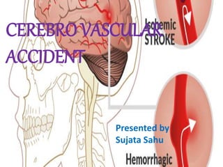
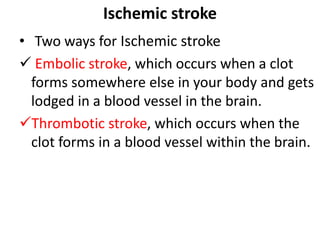
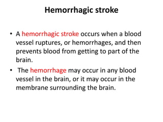
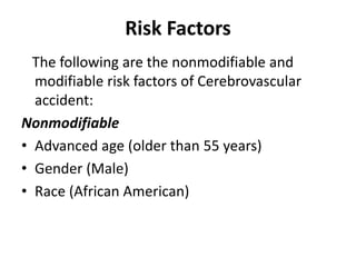
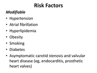
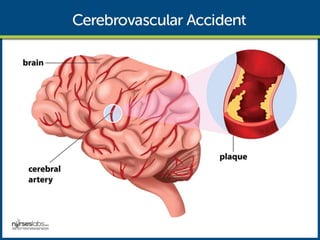
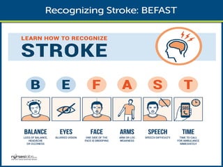
CEREBRO VASCULAR
ACCIDENT
Presented by
Sujata Sahu
Cerebrovascular Accident
Definition
• A cerebrovascular accident (CVA), an ischemic
stroke or “brain attack,” is a sudden loss of
brain function resulting from a disruption of
the blood supply to a part of the brain either
by a blockage or the rupture of a blood vessel.
• It is a functional abnormality
ofthe central nervous system.
Types of CVA
There are two main types of CVA or stroke:
• Ischemic stroke is caused by a blockage;
• Hemorrhagic stroke is caused by the rupture
of a blood vessel.
Both types of stroke deprive part
of the brain of blood and oxygen, causing
brain cells to die.
Ischemic stroke
• An ischemic stroke is the most common and
occurs when a blood clot blocks a blood
vessel and prevents blood and oxygen from
getting to a part of the brain.
Ischemic stroke
• Two ways for Ischemic stroke
 Embolic stroke, which occurs when a clot
forms somewhere else in your body and gets
lodged in a blood vessel in the brain.
Thrombotic stroke, which occurs when the
clot forms in a blood vessel within the brain.
Hemorrhagic stroke
• A hemorrhagic stroke occurs when a blood
vessel ruptures, or hemorrhages, and then
prevents blood from getting to part of the
brain.
• The hemorrhage may occur in any blood
vessel in the brain, or it may occur in the
membrane surrounding the brain.
Risk Factors
The following are the nonmodifiable and
modifiable risk factors of Cerebrovascular
accident:
Nonmodifiable
• Advanced age (older than 55 years)
• Gender (Male)
• Race (African American)
Risk Factors
Modifiable
• Hypertension
• Atrial fibrillation
• Hyperlipidemia
• Obesity
• Smoking
• Diabetes
• Asymptomatic carotid stenosis and valvular
heart disease (eg, endocarditis, prosthetic
heart valves)
Pathophysiology
• The disruption in the blood flow initiates a
complex series of cellular metabolic events.
• Decreased cerebral blood flow. The ischemic
cascade begins when cerebral blood flow
decreases to less than 25 mL per 100g of
blood per minute.
• Aerobic respiration. At this point, neurons are
unable to maintain aerobic respiration.
Pathophysiology
• Anaerobic respiration. The mitochondria
would need to switch to anaerobic respiration,
which generates large amounts of lactic acid,
causing a change in pH and rendering the
neurons incapable of producing sufficient
quantities of ATP.
• Loss of function. The membrane pumps that
maintain electrolyte balances fail and the cells
cease to function.
Pathophysiology
The disruption in the blood flow initiates a complex series of cellular metabolic events.
Causes
Strokes are caused by the following:
• Large artery thrombosis. Large artery
thromboses are caused by atherosclerotic
plaques in the large blood vessels of the brain.
• Small penetrating artery thrombosis. Small
penetrating artery thrombosis affects one or
more vessels and is the most common type of
ischemic stroke.
• Cardiogenic emboli. Cardiogenic emboli are
associated with cardiac dysrhythmias, usually
atrial fibrillation.
Clinical Manifestations
The quicker you can get a diagnosis and
treatment for a stroke, the better your
prognosis will be.
For this reason, it’s important to
understand and recognize the symptoms of a
stroke.
Symptoms of CVA
General sign & symptoms includes
• Difficulty walking
• Dizziness
• loss of balance and coordination
• Difficulty speaking or understanding others
who are speaking
• Numbness or paralysis in the face, leg, or arm,
most likely on just one side of the body
• Blurred or darkened vision
Symptoms of CVA
• A sudden headache, especially when
accompanied by nausea, vomiting, or
dizziness
• The symptoms of a stroke can vary depending
on the individual and where in the brain it has
happened. Symptoms usually appear
suddenly, even if they’re not very severe, and
they may become worse over time.
Symptoms of CVA
• Remembering the acronym “FAST” helps
people recognize the most common
symptoms of stroke:
• Face: Does one side of the face droop?
• Arm: If a person holds both arms out, does
one drift downward?
• Speech: Is their speech abnormal or slurred?
• Time: It’s time to call 911 and get to the
hospital if any of these symptoms are present.
Diagnosis of CVA
• Healthcare providers have a number of tools
to determine whether you’ve had a stroke.
• Healthcare provider will administer a full
physical examination, during which they’ll
check patient’s strength, reflexes, vision,
speech, and senses.
Diagnosis of CVA
• They’ll also check for a particular sound in the
blood vessels of your neck. This sound, which
is called a bruit, indicates abnormal blood
flow.
• Finally, they will check your blood pressure,
which may be high if you’ve had a stroke.
Diagnosis of CVA
• Blood tests: Blood test for clotting time, blood
sugar levels, or infection. These can all affect
the likelihood and progression of a stroke.
• Angiogram: An angiogram, which involves
adding a dye to your blood and taking an X-ray
of your head, can help your doctor find the
blocked or hemorrhaged blood vessel.
Diagnosis of CVA
• Carotid ultrasound: This test uses sound waves to
create images of the blood vessels in your neck.
This test can help your provider determine if
there’s abnormal blood flow toward your brain.
• CT scan: A CT scan is often performed soon after
symptoms of a stroke develop. The test can help
provider find the problem area or other problems
that might be associated with stroke.
Diagnosis of CVA
• MRI scan : An MRI can provide a more
detailed picture of the brain compared to CT
scan. It’s more sensitive than a CT scan in
being able to detect a stroke.
• Echocardiogram : This imaging technique uses
sound waves to create a picture of your heart.
It can help your provider find the source of
blood clots.
Diagnosis of CVA
• Electrocardiogram (EKG): This is an electrical
tracing of your heart. This will help your
healthcare provider determine if an abnormal
heart rhythm is the cause of a stroke.
Treatment for CVA
• The goal of treatment for ischemic stroke, for
instance, is to restore the blood flow.
• Treatment for stroke depends on the type of
stroke you’ve had.
• Treatments for hemorrhagic stroke are aimed
at controlling the bleeding.
Ischemic stroke treatment
• To treat an ischemic stroke, you may be given
a clot-dissolving drug or a blood thinner.
• You may also be given aspirin to prevent a
second stroke.
• Emergency treatment for this type of stroke
may include injecting medicine into the brain
or removing a blockage with a procedure.
Hemorrhagic stroke treatment
• For a hemorrhagic stroke, patient may be
given a drug that lowers the pressure in brain
caused by the bleeding.
• If the bleeding is severe, patient may need
surgery to remove excess blood.
• It’s also possible that patient will need
surgery to repair the ruptured blood vessel.
Medical Management
Medical management for secondary
prevention.
• Recombinant tissue plasminogen
activator would be prescribed unless
contraindicated, and there should be
monitoring for bleeding.
• Increased ICP. Management of increased ICP
includes osmotic diuretics, maintenance of
PaCO2 at 30-35 mmHg, and positioning to
avoid hypoxia through elevation of the head
of the bed.
Medical Management
• Endotracheal Tube. There is a possibility of
intubation to establish patent airway if
necessary.
• Hemodynamic monitoring. Continuous
hemodynamic monitoring should be
implemented to avoid an increase in blood
pressure.
• Neurologic assessment to determine if the
stroke is evolving and if other acute
complications are developing
Nursing Management
After the stroke is complete, management
focuses on the prompt initiation of
rehabilitation for any deficits.
Nursing Assessment
• During the acute phase, a neurologic flow
sheet is maintained to provide data about the
following important measures of the patient’s
clinical status:
• Change in level of consciousness or
responsiveness.
Nursing Assessment
• Presence or absence of voluntary or
involuntary movements of extremities.
• Stiffness or flaccidity of the neck.
• Eye opening, comparative size of pupils, and
pupillary reaction to light.
Nursing Assessment
• Color of the face and extremities; temperature
and moisture of the skin.
• Ability to speak.
• Presence of bleeding.
• Maintenance of blood pressure.
Nursing Assessment
• During the postacute phase, assess the
following functions:
• Mental status (memory, attention span,
perception, orientation, affect,
speech/language).
• Sensation and perception (usually the patient
has decreased awareness of pain and
temperature).
Nursing Assessment
• Motor control (upper and lower extremity
movement); swallowing ability, nutritional and
hydration status, skin integrity, activity
tolerance, and bowel and bladder function.
• Continue focusing nursing assessment on
impairment of function in patient’s daily
activities.
Nursing Diagnosis
• Based on the assessment data, the major
nursing diagnoses for a patient with stroke
may include the following:
• Impaired physical mobility elated to
hemiparesis, loss of balance and coordination,
spasticity, and brain injury.
• Acute pain related to hemiplegia and disuse.
• Disturbed sensory perception related to
altered sensory reception, transmission,
and/or integration.
Nursing Diagnosis
• Impaired urinary elimination related to
flaccid bladder detrusor instability, confusion,
or difficulty in communicating.
• Disturbed thought processes related to brain
damage.
• Impaired verbal communication related to
brain damage.
Nursing Diagnosis
• Risk for impaired skin integrity elated to
hemiparesis or hemiplegia and decreased
mobility.
• Interrupted family processes related to
catastrophic illness and caregiving burdens.
• Sexual dysfunction elated to neurologic
deficits or fear of failure.
Nursing plannings
The major nursing care planning goals for the
patient and family may include:
• Improve mobility.
• Avoidance of shoulder pain.
• Achievement of self-care.
• Relief of sensory and perceptual deprivation.
• Prevention of aspiration.
Nursing planning
• Continence of bowel and bladder.
• Improved thought processes.
• Achieving a form of communication.
• Maintaining skin integrity.
• Restore family functioning.
• Improve sexual function.
• Absence of complications.
Nursing Interventions
• Nursing care has a significant impact on the
patient’s recovery. In summary, here are some
nursing interventions for patients with stroke:
• Positioning. Position to prevent contractures,
relieve pressure, attain good body alignment,
and prevent compressive neuropathies.
• Prevent flexion. Apply splint at night to
prevent flexion of the affected extremity.
Nursing Interventions
• Prevent adduction. Prevent adduction of the
affected shoulder with a pillow placed in the
axilla.
• Prevent edema. Elevate affected arm to
prevent edema and fibrosis.
• Full range of motion. Provide full range of
motion four or five times a day to maintain
joint mobility.
Nursing Interventions
• Prevent venous stasis. Exercise is helpful in
preventing venous stasis, which may predispose
the patient to thrombosis and pulmonary
embolus.
• Regain balance. Teach patient to maintain
balance in a sitting position, then to balance
while standing and begin walking as soon as
standing balance is achieved.
• Personal hygiene. Encourage personal hygiene
activities as soon as the patient can sit up.
Nursing Interventions
• Manage sensory difficulties. Approach patient
with a decreased field of vision on the side where
visual perception is intact.
• Visit a speech therapist. Consult with a speech
therapist to evaluate gag reflexes and assist in
teaching alternate swallowing techniques.
• Voiding pattern. Analyze voiding pattern and
offer urinal or bedpan on patient’s voiding
schedule.
Nursing Interventions
• Be consistent in patient’s activities. Be
consistent in the schedule, routines, and
repetitions; a written schedule, checklists, and
audiotapes may help with memory and
concentration, and a communication board
may be used.
• Assess skin. Frequently assess skin for signs of
breakdown, with emphasis on bony areas
and dependent body parts.
Evaluation
Expected patient outcomes may include the
following:
• Improved mobility.
• Absence of shoulder pain.
• Self-care achieved.
• Relief of sensory and perceptual deprivation.
• Prevention of aspiration.
Evaluation
• Continence of bowel and bladder.
• Improved thought processes.
• Achieved a form of communication.
• Maintained skin integrity.
• Restored family functioning.
• Improved sexual function.
• Absence of complications.
Complications
• If cerebral oxygenation is still inadequate;
complications may occur.
• Tissue ischemia. If cerebral blood flow is
inadequate, the amount of oxygen supplied to
the brain is decreased, and tissue ischemia will
result.
• Cardiac dysrhythmias. The heart compensates
for the decreased cerebral blood flow, and with
too much pumping, dysrhythmias may occur.
Prevention of a CVA
• Maintain normal blood pressure.
• Refrain from smoking, and drink alcohol in
moderation
•
Prevention of a CVA
• DASH diet.
The DASH (Dietary Approaches to Stop
Hypertension) diet is high in fruits and
vegetables, moderate in low-fat dairy
products, and low in animal protein and can
lower the risk of stroke.
• Stroke risk screenings. Stroke risk screenings
are an ideal opportunity to lower stroke risk
by identifying people or groups of people who
are at high risk for stroke.
Prevention of a CVA
• Education. Patients and the community must
be educated about recognition and prevention
of stroke.
• Low-dose aspirin. Research findings suggest
that low-dose aspirin may lower the risk of
stroke in women who are at risk.
Prevention of a CVA
• Your healthcare provider may prescribe
medications for preventing stroke if they know
you’re at risk.
• Possible preventive medications for stroke
include drugs that thin the blood and prevent
clot formation.
Discharge and Home Care Guidelines
• Patient and family education is a fundamental
component of rehabilitation.
• Consult an occupational therapist. An
occupational therapist may be helpful in
assessing the home environment and
recommending modifications to help the patient
become more independent.
• Physical therapy. A program of physical therapy
may be beneficial, whether it takes place in the
home or in an outpatient program.
Discharge and Home Care Guidelines
• Antidepressant therapy. Depression is a
common and serious problem in the patient
who has had a stroke.
• Support groups. Community-based stroke
support groups may allow the patient and the
family to learn from others with similar
problems and to share their experiences.
Discharge and Home Care Guidelines
• Assess caregivers. Nurses should assess
caregivers for signs of depression, as
depression is also common among caregivers
of stroke survivors.
THANK YOU

Nursing management with cva patient

  • 2.
  • 3.
    Cerebrovascular Accident Definition • Acerebrovascular accident (CVA), an ischemic stroke or “brain attack,” is a sudden loss of brain function resulting from a disruption of the blood supply to a part of the brain either by a blockage or the rupture of a blood vessel. • It is a functional abnormality ofthe central nervous system.
  • 4.
    Types of CVA Thereare two main types of CVA or stroke: • Ischemic stroke is caused by a blockage; • Hemorrhagic stroke is caused by the rupture of a blood vessel. Both types of stroke deprive part of the brain of blood and oxygen, causing brain cells to die.
  • 5.
    Ischemic stroke • Anischemic stroke is the most common and occurs when a blood clot blocks a blood vessel and prevents blood and oxygen from getting to a part of the brain.
  • 6.
    Ischemic stroke • Twoways for Ischemic stroke  Embolic stroke, which occurs when a clot forms somewhere else in your body and gets lodged in a blood vessel in the brain. Thrombotic stroke, which occurs when the clot forms in a blood vessel within the brain.
  • 7.
    Hemorrhagic stroke • Ahemorrhagic stroke occurs when a blood vessel ruptures, or hemorrhages, and then prevents blood from getting to part of the brain. • The hemorrhage may occur in any blood vessel in the brain, or it may occur in the membrane surrounding the brain.
  • 8.
    Risk Factors The followingare the nonmodifiable and modifiable risk factors of Cerebrovascular accident: Nonmodifiable • Advanced age (older than 55 years) • Gender (Male) • Race (African American)
  • 9.
    Risk Factors Modifiable • Hypertension •Atrial fibrillation • Hyperlipidemia • Obesity • Smoking • Diabetes • Asymptomatic carotid stenosis and valvular heart disease (eg, endocarditis, prosthetic heart valves)
  • 10.
    Pathophysiology • The disruptionin the blood flow initiates a complex series of cellular metabolic events. • Decreased cerebral blood flow. The ischemic cascade begins when cerebral blood flow decreases to less than 25 mL per 100g of blood per minute. • Aerobic respiration. At this point, neurons are unable to maintain aerobic respiration.
  • 11.
    Pathophysiology • Anaerobic respiration.The mitochondria would need to switch to anaerobic respiration, which generates large amounts of lactic acid, causing a change in pH and rendering the neurons incapable of producing sufficient quantities of ATP. • Loss of function. The membrane pumps that maintain electrolyte balances fail and the cells cease to function.
  • 12.
    Pathophysiology The disruption inthe blood flow initiates a complex series of cellular metabolic events.
  • 13.
    Causes Strokes are causedby the following: • Large artery thrombosis. Large artery thromboses are caused by atherosclerotic plaques in the large blood vessels of the brain. • Small penetrating artery thrombosis. Small penetrating artery thrombosis affects one or more vessels and is the most common type of ischemic stroke. • Cardiogenic emboli. Cardiogenic emboli are associated with cardiac dysrhythmias, usually atrial fibrillation.
  • 14.
    Clinical Manifestations The quickeryou can get a diagnosis and treatment for a stroke, the better your prognosis will be. For this reason, it’s important to understand and recognize the symptoms of a stroke.
  • 15.
    Symptoms of CVA Generalsign & symptoms includes • Difficulty walking • Dizziness • loss of balance and coordination • Difficulty speaking or understanding others who are speaking • Numbness or paralysis in the face, leg, or arm, most likely on just one side of the body • Blurred or darkened vision
  • 16.
    Symptoms of CVA •A sudden headache, especially when accompanied by nausea, vomiting, or dizziness • The symptoms of a stroke can vary depending on the individual and where in the brain it has happened. Symptoms usually appear suddenly, even if they’re not very severe, and they may become worse over time.
  • 17.
    Symptoms of CVA •Remembering the acronym “FAST” helps people recognize the most common symptoms of stroke: • Face: Does one side of the face droop? • Arm: If a person holds both arms out, does one drift downward? • Speech: Is their speech abnormal or slurred? • Time: It’s time to call 911 and get to the hospital if any of these symptoms are present.
  • 19.
    Diagnosis of CVA •Healthcare providers have a number of tools to determine whether you’ve had a stroke. • Healthcare provider will administer a full physical examination, during which they’ll check patient’s strength, reflexes, vision, speech, and senses.
  • 20.
    Diagnosis of CVA •They’ll also check for a particular sound in the blood vessels of your neck. This sound, which is called a bruit, indicates abnormal blood flow. • Finally, they will check your blood pressure, which may be high if you’ve had a stroke.
  • 21.
    Diagnosis of CVA •Blood tests: Blood test for clotting time, blood sugar levels, or infection. These can all affect the likelihood and progression of a stroke. • Angiogram: An angiogram, which involves adding a dye to your blood and taking an X-ray of your head, can help your doctor find the blocked or hemorrhaged blood vessel.
  • 22.
    Diagnosis of CVA •Blood tests: Blood test for clotting time, blood sugar levels, or infection. These can all affect the likelihood and progression of a stroke. • Angiogram: An angiogram, which involves adding a dye to your blood and taking an X-ray of your head, can help your doctor find the blocked or hemorrhaged blood vessel.
  • 23.
    Diagnosis of CVA •Carotid ultrasound: This test uses sound waves to create images of the blood vessels in your neck. This test can help your provider determine if there’s abnormal blood flow toward your brain. • CT scan: A CT scan is often performed soon after symptoms of a stroke develop. The test can help provider find the problem area or other problems that might be associated with stroke.
  • 24.
    Diagnosis of CVA •MRI scan : An MRI can provide a more detailed picture of the brain compared to CT scan. It’s more sensitive than a CT scan in being able to detect a stroke. • Echocardiogram : This imaging technique uses sound waves to create a picture of your heart. It can help your provider find the source of blood clots.
  • 25.
    Diagnosis of CVA •Electrocardiogram (EKG): This is an electrical tracing of your heart. This will help your healthcare provider determine if an abnormal heart rhythm is the cause of a stroke.
  • 26.
    Treatment for CVA •The goal of treatment for ischemic stroke, for instance, is to restore the blood flow. • Treatment for stroke depends on the type of stroke you’ve had. • Treatments for hemorrhagic stroke are aimed at controlling the bleeding.
  • 27.
    Ischemic stroke treatment •To treat an ischemic stroke, you may be given a clot-dissolving drug or a blood thinner. • You may also be given aspirin to prevent a second stroke. • Emergency treatment for this type of stroke may include injecting medicine into the brain or removing a blockage with a procedure.
  • 28.
    Hemorrhagic stroke treatment •For a hemorrhagic stroke, patient may be given a drug that lowers the pressure in brain caused by the bleeding. • If the bleeding is severe, patient may need surgery to remove excess blood. • It’s also possible that patient will need surgery to repair the ruptured blood vessel.
  • 29.
    Medical Management Medical managementfor secondary prevention. • Recombinant tissue plasminogen activator would be prescribed unless contraindicated, and there should be monitoring for bleeding. • Increased ICP. Management of increased ICP includes osmotic diuretics, maintenance of PaCO2 at 30-35 mmHg, and positioning to avoid hypoxia through elevation of the head of the bed.
  • 30.
    Medical Management • EndotrachealTube. There is a possibility of intubation to establish patent airway if necessary. • Hemodynamic monitoring. Continuous hemodynamic monitoring should be implemented to avoid an increase in blood pressure. • Neurologic assessment to determine if the stroke is evolving and if other acute complications are developing
  • 31.
    Nursing Management After thestroke is complete, management focuses on the prompt initiation of rehabilitation for any deficits. Nursing Assessment • During the acute phase, a neurologic flow sheet is maintained to provide data about the following important measures of the patient’s clinical status: • Change in level of consciousness or responsiveness.
  • 32.
    Nursing Assessment • Presenceor absence of voluntary or involuntary movements of extremities. • Stiffness or flaccidity of the neck. • Eye opening, comparative size of pupils, and pupillary reaction to light.
  • 33.
    Nursing Assessment • Colorof the face and extremities; temperature and moisture of the skin. • Ability to speak. • Presence of bleeding. • Maintenance of blood pressure.
  • 34.
    Nursing Assessment • Duringthe postacute phase, assess the following functions: • Mental status (memory, attention span, perception, orientation, affect, speech/language). • Sensation and perception (usually the patient has decreased awareness of pain and temperature).
  • 35.
    Nursing Assessment • Motorcontrol (upper and lower extremity movement); swallowing ability, nutritional and hydration status, skin integrity, activity tolerance, and bowel and bladder function. • Continue focusing nursing assessment on impairment of function in patient’s daily activities.
  • 36.
    Nursing Diagnosis • Basedon the assessment data, the major nursing diagnoses for a patient with stroke may include the following: • Impaired physical mobility elated to hemiparesis, loss of balance and coordination, spasticity, and brain injury. • Acute pain related to hemiplegia and disuse. • Disturbed sensory perception related to altered sensory reception, transmission, and/or integration.
  • 37.
    Nursing Diagnosis • Impairedurinary elimination related to flaccid bladder detrusor instability, confusion, or difficulty in communicating. • Disturbed thought processes related to brain damage. • Impaired verbal communication related to brain damage.
  • 38.
    Nursing Diagnosis • Riskfor impaired skin integrity elated to hemiparesis or hemiplegia and decreased mobility. • Interrupted family processes related to catastrophic illness and caregiving burdens. • Sexual dysfunction elated to neurologic deficits or fear of failure.
  • 39.
    Nursing plannings The majornursing care planning goals for the patient and family may include: • Improve mobility. • Avoidance of shoulder pain. • Achievement of self-care. • Relief of sensory and perceptual deprivation. • Prevention of aspiration.
  • 40.
    Nursing planning • Continenceof bowel and bladder. • Improved thought processes. • Achieving a form of communication. • Maintaining skin integrity. • Restore family functioning. • Improve sexual function. • Absence of complications.
  • 41.
    Nursing Interventions • Nursingcare has a significant impact on the patient’s recovery. In summary, here are some nursing interventions for patients with stroke: • Positioning. Position to prevent contractures, relieve pressure, attain good body alignment, and prevent compressive neuropathies. • Prevent flexion. Apply splint at night to prevent flexion of the affected extremity.
  • 42.
    Nursing Interventions • Preventadduction. Prevent adduction of the affected shoulder with a pillow placed in the axilla. • Prevent edema. Elevate affected arm to prevent edema and fibrosis. • Full range of motion. Provide full range of motion four or five times a day to maintain joint mobility.
  • 43.
    Nursing Interventions • Preventvenous stasis. Exercise is helpful in preventing venous stasis, which may predispose the patient to thrombosis and pulmonary embolus. • Regain balance. Teach patient to maintain balance in a sitting position, then to balance while standing and begin walking as soon as standing balance is achieved. • Personal hygiene. Encourage personal hygiene activities as soon as the patient can sit up.
  • 44.
    Nursing Interventions • Managesensory difficulties. Approach patient with a decreased field of vision on the side where visual perception is intact. • Visit a speech therapist. Consult with a speech therapist to evaluate gag reflexes and assist in teaching alternate swallowing techniques. • Voiding pattern. Analyze voiding pattern and offer urinal or bedpan on patient’s voiding schedule.
  • 45.
    Nursing Interventions • Beconsistent in patient’s activities. Be consistent in the schedule, routines, and repetitions; a written schedule, checklists, and audiotapes may help with memory and concentration, and a communication board may be used. • Assess skin. Frequently assess skin for signs of breakdown, with emphasis on bony areas and dependent body parts.
  • 46.
    Evaluation Expected patient outcomesmay include the following: • Improved mobility. • Absence of shoulder pain. • Self-care achieved. • Relief of sensory and perceptual deprivation. • Prevention of aspiration.
  • 47.
    Evaluation • Continence ofbowel and bladder. • Improved thought processes. • Achieved a form of communication. • Maintained skin integrity. • Restored family functioning. • Improved sexual function. • Absence of complications.
  • 48.
    Complications • If cerebraloxygenation is still inadequate; complications may occur. • Tissue ischemia. If cerebral blood flow is inadequate, the amount of oxygen supplied to the brain is decreased, and tissue ischemia will result. • Cardiac dysrhythmias. The heart compensates for the decreased cerebral blood flow, and with too much pumping, dysrhythmias may occur.
  • 49.
    Prevention of aCVA • Maintain normal blood pressure. • Refrain from smoking, and drink alcohol in moderation •
  • 50.
    Prevention of aCVA • DASH diet. The DASH (Dietary Approaches to Stop Hypertension) diet is high in fruits and vegetables, moderate in low-fat dairy products, and low in animal protein and can lower the risk of stroke. • Stroke risk screenings. Stroke risk screenings are an ideal opportunity to lower stroke risk by identifying people or groups of people who are at high risk for stroke.
  • 51.
    Prevention of aCVA • Education. Patients and the community must be educated about recognition and prevention of stroke. • Low-dose aspirin. Research findings suggest that low-dose aspirin may lower the risk of stroke in women who are at risk.
  • 52.
    Prevention of aCVA • Your healthcare provider may prescribe medications for preventing stroke if they know you’re at risk. • Possible preventive medications for stroke include drugs that thin the blood and prevent clot formation.
  • 53.
    Discharge and HomeCare Guidelines • Patient and family education is a fundamental component of rehabilitation. • Consult an occupational therapist. An occupational therapist may be helpful in assessing the home environment and recommending modifications to help the patient become more independent. • Physical therapy. A program of physical therapy may be beneficial, whether it takes place in the home or in an outpatient program.
  • 54.
    Discharge and HomeCare Guidelines • Antidepressant therapy. Depression is a common and serious problem in the patient who has had a stroke. • Support groups. Community-based stroke support groups may allow the patient and the family to learn from others with similar problems and to share their experiences.
  • 55.
    Discharge and HomeCare Guidelines • Assess caregivers. Nurses should assess caregivers for signs of depression, as depression is also common among caregivers of stroke survivors.
  • 57.