Relationship between bme and cardiovascular system.Yasin Muh'd Yahya
relationship of BME and cardiovascular is a small is a presentation that will brief you in details about the relationship between cardiovascular and bme. hope you enjoy it.
An Exercise Tolerance Test or Stress Test records the heart's electrical activity (rate and rhythm) during exercise.
It is one of the commonest forms of stress tests used to induce provoke cardiac ischemia for diagnostic evaluation of coronary artery disease.
Cardiology
From Wikipedia, the free encyclopedia
Jump to navigationJump to search
This article is about the medical specialty. For the album, see Cardiology (album). For the medical journal, see Cardiology (journal).
Cardiology
Heart diagram blood flow en.svg
Blood flow diagram of the human heart. Blue components indicate de-oxygenated blood pathways and red components indicate oxygenated blood pathways.
System Cardiovascular
Subdivisions Interventional, Nuclear
Significant diseases Heart disease, Cardiovascular disease, Atherosclerosis,
earning websites that make the most money
Monitoring is essential in any kind of medical practice. It is the observation of disease, condition and several other parameters over time. Usually a medical monitor is used for continuously measuring vital signs.
Siva Hospitals
Ethamozhi Puthalam Road, Eathamozhi, Tamil Nadu 629501
Healthcare is no more about bringing just medical facilities to you. Its about integrating these facilities along with technology, know-how of advancing medical sciences, hospitality; functioning as a whole with humane at its core value in serving humanity.
Siva Hospitals understands this perfectly and constantly strives to bring you the best in healthcare advancements from around the world. To find out how we could serve you, read on
Cardiology
From Wikipedia, the free encyclopedia
Jump to navigationJump to search
This article is about the medical specialty. For the album, see Cardiology (album). For the medical journal, see Cardiology (journal).
Cardiology
Heart diagram blood flow en.svg
Blood flow diagram of the human heart. Blue components indicate de-oxygenated blood pathways and red components indicate oxygenated blood pathways.
System Cardiovascular
Subdivisions Interventional, Nuclear
Significant diseases Heart disease, Cardiovascular disease, Atherosclerosis,
earning websites that make the most money
Monitoring is essential in any kind of medical practice. It is the observation of disease, condition and several other parameters over time. Usually a medical monitor is used for continuously measuring vital signs.
Siva Hospitals
Ethamozhi Puthalam Road, Eathamozhi, Tamil Nadu 629501
Healthcare is no more about bringing just medical facilities to you. Its about integrating these facilities along with technology, know-how of advancing medical sciences, hospitality; functioning as a whole with humane at its core value in serving humanity.
Siva Hospitals understands this perfectly and constantly strives to bring you the best in healthcare advancements from around the world. To find out how we could serve you, read on
Ultrasonography of Heart or Cardiac ultrasonography or Echocardiogram or ultrasound of the heart is the production of two-dimensional cross-sectional images of intracardiac anatomy by stop-action compound scan pulse-echo ultrasound.
The images show the size and shape of the cardiac chambers in systole and diastole, the appearance of heart valves, and the orientation of the great vessels.
The stop-action display is created by repetitively activating the recording oscilloscope for a selected short segment of each cardiac cycle.
The activating signal is timed by the patient's electrocardiogram.
The asynchronous motion of the scanner accumulates additional echoes with each cycle.
As a non-invasive technique, it is without risk or morbidity.
invasive non invasive procedures.pdf for bsc nursing studentsshanmukhadevi
Chest X-ray:
The chest X-ray is a noninvasive tool used to visualize internal structures, such as the heart, lungs, soft tissues, and bones.
Most chest X-rays are taken while the patient is inhaling so that the lungs are fully expanded.
Several types of chest X-rays can be used to assess heart size, contour, and position; other types reveal cardiac and pericardial calcification as well as physiologic alterations in pulmonary circulation.
Exercise Stress Test Tredmill test by mohsin.pptxMohsinAli322520
1)An Exercise stress test helps determine how well your heart responds during times when it's working its hardest. It typically involves walking on a treadmill or pedaling on a stationary bike while hooked up to an EKG to monitor your heart’s activity.
2 )what is stress test ?
A stress test is a very commonly performed test to learn:
How well your heart pumps blood.
Whether your heart is receiving an adequate blood supply.
How you perform on physical activity (riding a treadmill or stationary bike) compared with other people your age and sex.
If your symptoms (chest discomfort, shortness of breath, feeling like your heart is racing or even dizziness) can be reproduced while performing physical activity.
3) how does Exercise stress test work ?
4 )why need of stress test indications ?
You may need this test to detect heart problems like:
Congenital heart disease.
Heart failure.
Coronary artery disease.
Heart valve disease.
Hypertrophic cardiomyopathy.
People with high-risk occupations (like pilots or professional athletes) may also need stress tests.
This test may be right for you if you have symptoms of heart disease, like:
Angina, which is chest pain or discomfort due to poor blood flow to the heart.
Arrhythmia, which is a rapid or irregular heartbeat.
Shortness of breath .
Feeling lightheaded or dizzy.
Stress tests are also for people with a heart disease diagnosis who:
Would like to start exercising.
Are undergoing treatment and healthcare providers need to determine how well it’s working.
Face a higher risk of complications due to a personal or family history of heart disease.
Have diabetes or other underlying conditions that increase your risk of heart disease.
different types of stress test ?
There are many methods for assessing heart function while it’s hard at work. All cardiac stress tests involve checking your heart rate, blood pressure, oxygen levels and electrical activity. But there are some differences.
Stress test types include:
Exercise stress test
Exercise stress echocardiogram
Nuclear stress test
Time taken by stress test ?
If you’re undergoing a basic stress test, the exercise portion lasts about 10 to 15 minutes. Additional time is necessary for getting ready to exercise and recovering afterward.
Stress tests that include echocardiography, nuclear imaging or MRI often are longer and may require you to be in the stress lab for several hours.
Preparation for exercise stress test ?
Not eat anything in the hours leading up to the test. If you’re having a nuclear stress test, you might not be able to eat until after your test.
Avoid caffeine for 24 hours before testing. This includes coffee, tea, energy drinks and certain over-the-counter medications.
Not smoke or use tobacco products.
Stop taking certain prescription medications the day of your test. These include beta-blockers and asthma inhalers. Talk to your healthcare provider before stopping any medications.
contraindication
Unstable angina
surgery
Uncontrolled Arrythmia
Similar to Heart diagnostic procedure in india at mumbai and delhi at affordable cost (20)
Lymphoma treatment in india at mumbai at affordable costpankaj nagpal
Lymphoma Treatment, Lymphoma Treatment India, Bone Marrow India, Lymph Nodes India, Bexxar India, Follicular Lymphoma India, Radiation Therapy India, India Hospital Tour India, Chemotherapy India, Immunotherapy India, Lymphoma Chemotherapy India, Lymphatic System India, Lymph Glands India, Lymphoma Treatment Hospitals India, Swollen Lymph Glands India, Lymphocytes India, B Lymphocytes India, NHL India, T Lymphocytes India, B Cells, Hospitals Of Lymphoma Treatment In India, T Cells, Lymph, Tumor, Uncommon Cancer, Blood Cancer, Hodgkin's Disease, General Treatment, Health Care, Fast-Growing
Gallbladder cancer treatment in india at low costpankaj nagpal
Gallbladder Cancer Treatment, Gallbladder Cancer India, Gall Bladder India, Gallbladder India, Bile Duct India, Biliary Tract India, Gallstones India, Cholesterol Gallstones India, India Hospital Tour India, Polyposis Coli India, Unresectable India, Cholangiocarcinomas India, Primary Sclerosing Cholangitis India, Cancer India, Diagnosis India, Abdominal Pain India, Jaundice, Vomiting Occur, Porcelain Gallbladder, Gall Bladder, Primary Cancers, Chronic, Gallbladder Neoplasms, Pear-Shaped Organ, small pear-shaped pouch, tissues
Prostatectomy Surgery abroad in India info on cost Prostatectomy Surgery India,Prostatectomy male Surgery hospitals India,Prostatectomy surgeons India.
NVBDCP.pptx Nation vector borne disease control programSapna Thakur
NVBDCP was launched in 2003-2004 . Vector-Borne Disease: Disease that results from an infection transmitted to humans and other animals by blood-feeding arthropods, such as mosquitoes, ticks, and fleas. Examples of vector-borne diseases include Dengue fever, West Nile Virus, Lyme disease, and malaria.
New Directions in Targeted Therapeutic Approaches for Older Adults With Mantl...i3 Health
i3 Health is pleased to make the speaker slides from this activity available for use as a non-accredited self-study or teaching resource.
This slide deck presented by Dr. Kami Maddocks, Professor-Clinical in the Division of Hematology and
Associate Division Director for Ambulatory Operations
The Ohio State University Comprehensive Cancer Center, will provide insight into new directions in targeted therapeutic approaches for older adults with mantle cell lymphoma.
STATEMENT OF NEED
Mantle cell lymphoma (MCL) is a rare, aggressive B-cell non-Hodgkin lymphoma (NHL) accounting for 5% to 7% of all lymphomas. Its prognosis ranges from indolent disease that does not require treatment for years to very aggressive disease, which is associated with poor survival (Silkenstedt et al, 2021). Typically, MCL is diagnosed at advanced stage and in older patients who cannot tolerate intensive therapy (NCCN, 2022). Although recent advances have slightly increased remission rates, recurrence and relapse remain very common, leading to a median overall survival between 3 and 6 years (LLS, 2021). Though there are several effective options, progress is still needed towards establishing an accepted frontline approach for MCL (Castellino et al, 2022). Treatment selection and management of MCL are complicated by the heterogeneity of prognosis, advanced age and comorbidities of patients, and lack of an established standard approach for treatment, making it vital that clinicians be familiar with the latest research and advances in this area. In this activity chaired by Michael Wang, MD, Professor in the Department of Lymphoma & Myeloma at MD Anderson Cancer Center, expert faculty will discuss prognostic factors informing treatment, the promising results of recent trials in new therapeutic approaches, and the implications of treatment resistance in therapeutic selection for MCL.
Target Audience
Hematology/oncology fellows, attending faculty, and other health care professionals involved in the treatment of patients with mantle cell lymphoma (MCL).
Learning Objectives
1.) Identify clinical and biological prognostic factors that can guide treatment decision making for older adults with MCL
2.) Evaluate emerging data on targeted therapeutic approaches for treatment-naive and relapsed/refractory MCL and their applicability to older adults
3.) Assess mechanisms of resistance to targeted therapies for MCL and their implications for treatment selection
Title: Sense of Taste
Presenter: Dr. Faiza, Assistant Professor of Physiology
Qualifications:
MBBS (Best Graduate, AIMC Lahore)
FCPS Physiology
ICMT, CHPE, DHPE (STMU)
MPH (GC University, Faisalabad)
MBA (Virtual University of Pakistan)
Learning Objectives:
Describe the structure and function of taste buds.
Describe the relationship between the taste threshold and taste index of common substances.
Explain the chemical basis and signal transduction of taste perception for each type of primary taste sensation.
Recognize different abnormalities of taste perception and their causes.
Key Topics:
Significance of Taste Sensation:
Differentiation between pleasant and harmful food
Influence on behavior
Selection of food based on metabolic needs
Receptors of Taste:
Taste buds on the tongue
Influence of sense of smell, texture of food, and pain stimulation (e.g., by pepper)
Primary and Secondary Taste Sensations:
Primary taste sensations: Sweet, Sour, Salty, Bitter, Umami
Chemical basis and signal transduction mechanisms for each taste
Taste Threshold and Index:
Taste threshold values for Sweet (sucrose), Salty (NaCl), Sour (HCl), and Bitter (Quinine)
Taste index relationship: Inversely proportional to taste threshold
Taste Blindness:
Inability to taste certain substances, particularly thiourea compounds
Example: Phenylthiocarbamide
Structure and Function of Taste Buds:
Composition: Epithelial cells, Sustentacular/Supporting cells, Taste cells, Basal cells
Features: Taste pores, Taste hairs/microvilli, and Taste nerve fibers
Location of Taste Buds:
Found in papillae of the tongue (Fungiform, Circumvallate, Foliate)
Also present on the palate, tonsillar pillars, epiglottis, and proximal esophagus
Mechanism of Taste Stimulation:
Interaction of taste substances with receptors on microvilli
Signal transduction pathways for Umami, Sweet, Bitter, Sour, and Salty tastes
Taste Sensitivity and Adaptation:
Decrease in sensitivity with age
Rapid adaptation of taste sensation
Role of Saliva in Taste:
Dissolution of tastants to reach receptors
Washing away the stimulus
Taste Preferences and Aversions:
Mechanisms behind taste preference and aversion
Influence of receptors and neural pathways
Impact of Sensory Nerve Damage:
Degeneration of taste buds if the sensory nerve fiber is cut
Abnormalities of Taste Detection:
Conditions: Ageusia, Hypogeusia, Dysgeusia (parageusia)
Causes: Nerve damage, neurological disorders, infections, poor oral hygiene, adverse drug effects, deficiencies, aging, tobacco use, altered neurotransmitter levels
Neurotransmitters and Taste Threshold:
Effects of serotonin (5-HT) and norepinephrine (NE) on taste sensitivity
Supertasters:
25% of the population with heightened sensitivity to taste, especially bitterness
Increased number of fungiform papillae
Couples presenting to the infertility clinic- Do they really have infertility...Sujoy Dasgupta
Dr Sujoy Dasgupta presented the study on "Couples presenting to the infertility clinic- Do they really have infertility? – The unexplored stories of non-consummation" in the 13th Congress of the Asia Pacific Initiative on Reproduction (ASPIRE 2024) at Manila on 24 May, 2024.
Acute scrotum is a general term referring to an emergency condition affecting the contents or the wall of the scrotum.
There are a number of conditions that present acutely, predominantly with pain and/or swelling
A careful and detailed history and examination, and in some cases, investigations allow differentiation between these diagnoses. A prompt diagnosis is essential as the patient may require urgent surgical intervention
Testicular torsion refers to twisting of the spermatic cord, causing ischaemia of the testicle.
Testicular torsion results from inadequate fixation of the testis to the tunica vaginalis producing ischemia from reduced arterial inflow and venous outflow obstruction.
The prevalence of testicular torsion in adult patients hospitalized with acute scrotal pain is approximately 25 to 50 percent
Ethanol (CH3CH2OH), or beverage alcohol, is a two-carbon alcohol
that is rapidly distributed in the body and brain. Ethanol alters many
neurochemical systems and has rewarding and addictive properties. It
is the oldest recreational drug and likely contributes to more morbidity,
mortality, and public health costs than all illicit drugs combined. The
5th edition of the Diagnostic and Statistical Manual of Mental Disorders
(DSM-5) integrates alcohol abuse and alcohol dependence into a single
disorder called alcohol use disorder (AUD), with mild, moderate,
and severe subclassifications (American Psychiatric Association, 2013).
In the DSM-5, all types of substance abuse and dependence have been
combined into a single substance use disorder (SUD) on a continuum
from mild to severe. A diagnosis of AUD requires that at least two of
the 11 DSM-5 behaviors be present within a 12-month period (mild
AUD: 2–3 criteria; moderate AUD: 4–5 criteria; severe AUD: 6–11 criteria).
The four main behavioral effects of AUD are impaired control over
drinking, negative social consequences, risky use, and altered physiological
effects (tolerance, withdrawal). This chapter presents an overview
of the prevalence and harmful consequences of AUD in the U.S.,
the systemic nature of the disease, neurocircuitry and stages of AUD,
comorbidities, fetal alcohol spectrum disorders, genetic risk factors, and
pharmacotherapies for AUD.
These simplified slides by Dr. Sidra Arshad present an overview of the non-respiratory functions of the respiratory tract.
Learning objectives:
1. Enlist the non-respiratory functions of the respiratory tract
2. Briefly explain how these functions are carried out
3. Discuss the significance of dead space
4. Differentiate between minute ventilation and alveolar ventilation
5. Describe the cough and sneeze reflexes
Study Resources:
1. Chapter 39, Guyton and Hall Textbook of Medical Physiology, 14th edition
2. Chapter 34, Ganong’s Review of Medical Physiology, 26th edition
3. Chapter 17, Human Physiology by Lauralee Sherwood, 9th edition
4. Non-respiratory functions of the lungs https://academic.oup.com/bjaed/article/13/3/98/278874
Flu Vaccine Alert in Bangalore Karnatakaaddon Scans
As flu season approaches, health officials in Bangalore, Karnataka, are urging residents to get their flu vaccinations. The seasonal flu, while common, can lead to severe health complications, particularly for vulnerable populations such as young children, the elderly, and those with underlying health conditions.
Dr. Vidisha Kumari, a leading epidemiologist in Bangalore, emphasizes the importance of getting vaccinated. "The flu vaccine is our best defense against the influenza virus. It not only protects individuals but also helps prevent the spread of the virus in our communities," he says.
This year, the flu season is expected to coincide with a potential increase in other respiratory illnesses. The Karnataka Health Department has launched an awareness campaign highlighting the significance of flu vaccinations. They have set up multiple vaccination centers across Bangalore, making it convenient for residents to receive their shots.
To encourage widespread vaccination, the government is also collaborating with local schools, workplaces, and community centers to facilitate vaccination drives. Special attention is being given to ensuring that the vaccine is accessible to all, including marginalized communities who may have limited access to healthcare.
Residents are reminded that the flu vaccine is safe and effective. Common side effects are mild and may include soreness at the injection site, mild fever, or muscle aches. These side effects are generally short-lived and far less severe than the flu itself.
Healthcare providers are also stressing the importance of continuing COVID-19 precautions. Wearing masks, practicing good hand hygiene, and maintaining social distancing are still crucial, especially in crowded places.
Protect yourself and your loved ones by getting vaccinated. Together, we can help keep Bangalore healthy and safe this flu season. For more information on vaccination centers and schedules, residents can visit the Karnataka Health Department’s official website or follow their social media pages.
Stay informed, stay safe, and get your flu shot today!
Lung Cancer: Artificial Intelligence, Synergetics, Complex System Analysis, S...Oleg Kshivets
RESULTS: Overall life span (LS) was 2252.1±1742.5 days and cumulative 5-year survival (5YS) reached 73.2%, 10 years – 64.8%, 20 years – 42.5%. 513 LCP lived more than 5 years (LS=3124.6±1525.6 days), 148 LCP – more than 10 years (LS=5054.4±1504.1 days).199 LCP died because of LC (LS=562.7±374.5 days). 5YS of LCP after bi/lobectomies was significantly superior in comparison with LCP after pneumonectomies (78.1% vs.63.7%, P=0.00001 by log-rank test). AT significantly improved 5YS (66.3% vs. 34.8%) (P=0.00000 by log-rank test) only for LCP with N1-2. Cox modeling displayed that 5YS of LCP significantly depended on: phase transition (PT) early-invasive LC in terms of synergetics, PT N0—N12, cell ratio factors (ratio between cancer cells- CC and blood cells subpopulations), G1-3, histology, glucose, AT, blood cell circuit, prothrombin index, heparin tolerance, recalcification time (P=0.000-0.038). Neural networks, genetic algorithm selection and bootstrap simulation revealed relationships between 5YS and PT early-invasive LC (rank=1), PT N0—N12 (rank=2), thrombocytes/CC (3), erythrocytes/CC (4), eosinophils/CC (5), healthy cells/CC (6), lymphocytes/CC (7), segmented neutrophils/CC (8), stick neutrophils/CC (9), monocytes/CC (10); leucocytes/CC (11). Correct prediction of 5YS was 100% by neural networks computing (area under ROC curve=1.0; error=0.0).
CONCLUSIONS: 5YS of LCP after radical procedures significantly depended on: 1) PT early-invasive cancer; 2) PT N0--N12; 3) cell ratio factors; 4) blood cell circuit; 5) biochemical factors; 6) hemostasis system; 7) AT; 8) LC characteristics; 9) LC cell dynamics; 10) surgery type: lobectomy/pneumonectomy; 11) anthropometric data. Optimal diagnosis and treatment strategies for LC are: 1) screening and early detection of LC; 2) availability of experienced thoracic surgeons because of complexity of radical procedures; 3) aggressive en block surgery and adequate lymph node dissection for completeness; 4) precise prediction; 5) adjuvant chemoimmunoradiotherapy for LCP with unfavorable prognosis.
Prix Galien International 2024 Forum ProgramLevi Shapiro
June 20, 2024, Prix Galien International and Jerusalem Ethics Forum in ROME. Detailed agenda including panels:
- ADVANCES IN CARDIOLOGY: A NEW PARADIGM IS COMING
- WOMEN’S HEALTH: FERTILITY PRESERVATION
- WHAT’S NEW IN THE TREATMENT OF INFECTIOUS,
ONCOLOGICAL AND INFLAMMATORY SKIN DISEASES?
- ARTIFICIAL INTELLIGENCE AND ETHICS
- GENE THERAPY
- BEYOND BORDERS: GLOBAL INITIATIVES FOR DEMOCRATIZING LIFE SCIENCE TECHNOLOGIES AND PROMOTING ACCESS TO HEALTHCARE
- ETHICAL CHALLENGES IN LIFE SCIENCES
- Prix Galien International Awards Ceremony
TEST BANK for Operations Management, 14th Edition by William J. Stevenson, Ve...kevinkariuki227
TEST BANK for Operations Management, 14th Edition by William J. Stevenson, Verified Chapters 1 - 19, Complete Newest Version.pdf
These lecture slides, by Dr Sidra Arshad, offer a quick overview of physiological basis of a normal electrocardiogram.
Learning objectives:
1. Define an electrocardiogram (ECG) and electrocardiography
2. Describe how dipoles generated by the heart produce the waveforms of the ECG
3. Describe the components of a normal electrocardiogram of a typical bipolar leads (limb II)
4. Differentiate between intervals and segments
5. Enlist some common indications for obtaining an ECG
Study Resources:
1. Chapter 11, Guyton and Hall Textbook of Medical Physiology, 14th edition
2. Chapter 9, Human Physiology - From Cells to Systems, Lauralee Sherwood, 9th edition
3. Chapter 29, Ganong’s Review of Medical Physiology, 26th edition
4. Electrocardiogram, StatPearls - https://www.ncbi.nlm.nih.gov/books/NBK549803/
5. ECG in Medical Practice by ABM Abdullah, 4th edition
6. ECG Basics, http://www.nataliescasebook.com/tag/e-c-g-basics
Ozempic: Preoperative Management of Patients on GLP-1 Receptor Agonists Saeid Safari
Preoperative Management of Patients on GLP-1 Receptor Agonists like Ozempic and Semiglutide
ASA GUIDELINE
NYSORA Guideline
2 Case Reports of Gastric Ultrasound
Ozempic: Preoperative Management of Patients on GLP-1 Receptor Agonists
Heart diagnostic procedure in india at mumbai and delhi at affordable cost
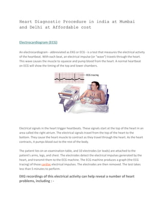
1. Heart Diagnostic Procedure in india at Mumbai
and Delhi at Affordable cost
Electrocardiogram (ECG)
An electrocardiogram - abbreviated as EKG or ECG - is a test that measures the electrical activity
of the heartbeat. With each beat, an electrical impulse (or "wave") travels through the heart.
This wave causes the muscle to squeeze and pump blood from the heart. A normal heartbeat
on ECG will show the timing of the top and lower chambers.
Electrical signals in the heart trigger heartbeats. These signals start at the top of the heart in an
area called the right atrium. The electrical signals travel from the top of the heart to the
bottom. They cause the heart muscle to contract as they travel through the heart. As the heart
contracts, it pumps blood out to the rest of the body.
The patient lies on an examination table, and 10 electrodes (or leads) are attached to the
patient's arms, legs, and chest. The electrodes detect the electrical impulses generated by the
heart, and transmit them to the ECG machine. The ECG machine produces a graph (the ECG
tracing) of those cardiac electrical impulses. The electrodes are then removed. The test takes
less than 5 minutes to perform.
EKG recordings of this electrical activity can help reveal a number of heart
problems, including : -
2. • Heart attack
• Lack of blood flow to the heart muscle
• A heart that is beating irregularly, or too fast or too slow
• A heart that does not pump forcefully enough.
Echocardiogram
An echocardiogram (echo) is a type of ultrasound examination that uses high-pitched sound
waves sent through a device called a transducer to produce an image of the heart and
sometimes the aorta.
An echocardiogram measures how well the heart is working by evaluating blood flow, heart
valves, and heart size, thickness, shape, and muscle movement.
Sticky patches or electrodes are attached to the chest and shoulders and connected to
electrodes or wires. These help to record the electrocardiogram (EKG or ECG) during the
echocardiography test. The EKG helps in the timing of various cardiac events (filling and
emptying of chambers).
3. A colorless gel is then applied to the chest and the echo transducer is placed on top of it. The
echo technologist then makes recordings from different parts of the chest to obtain several
views of the heart. You may be asked to move form your back and to the side. Instructions may
also be given for you to breathe slowly or to hold your breath. This helps in obtaining higher
quality pictures. The images are constantly viewed on the monitor. It is also recorded on
photographic paper and on videotape. The tape offers a permanent record of the examination
and is reviewed by the physician prior to completion of the final report.
The procedure is most often performed for the following reasons : -
1. Evaluate a heart murmur
2. Diagnose and determine the extent of valve conditions
3. Determine the presence of abnormalities in the structure of the heart
4. Measure the size and thickness of the heart and its chambers
5. Assess motion of the chamber walls and the extent of damage to the heart muscle after
a heart attack
6. Assess how different parts of the heart are functioning in patients with chronic heart
disease
7. Determine if fluid is collecting around the heart
8. Identify the presence of tumors in the heart
9. Assess for and monitor congenital defects
10. Evaluate a patient's response to treatment or a corrective procedure
11. Evaluate blood flow through the heart
12. Assess if the heart or major blood vessels coming and going from the heart have been
damaged by a traumatic injury, often done to determine a heart's condition before it is
donated for transplant
4. 13. Evaluate heart function and diagnose heart and lungs abnormalities in critically ill
patients in an intensive care unit
14. Evaluate chest pain
15. Evaluate for presence of blood clots within heart chambers
Holter Monitor
A Holter monitor, also called an ambulatory EKG, records the electrical signals of your heart for
a full 24- or 48-hour period. You wear small patches called electrodes on your chest that are
connected by wires to a small, portable recorder. The recorder can be clipped to a belt, kept in
a pocket, or hung around your neck. During the 24 or 48 hours, you do your usual daily
activities and keep a notebook, writing down any symptoms you have and the time they occur.
You then return both the recorder and the notebook to your doctor to read the results. Your
doctor can see how your heart was beating at the time you had symptoms.
The purpose of a Holter monitor is to record heart signals during typical daily activities and
while sleeping, and to find heart problems that may occur for only a few minutes out of the
day. Also, the Holter monitor can pick up irregular heartbeats that don't cause symptoms, but
are important to treat.
[ Holter monitor ]
5. A patient may wear a monitoring device, called a Holter monitor, for 24 or 48 hours while
performing normal daily activities at home. The monitor records the heart's rhythm during this
time. The device is connected with several long thin cables that adhere to the skin on the chest
with several sticky pads (similar to an ECG). The cables connect to a small, portable machine
that can be attached to a belt or a strap that is carried over the shoulder.
Stress Test
Stress testing provides your doctor with information about how your heart works during
physical stress. Some heart problems are easier to diagnose when your heart is working hard
and beating fast.
Norav Medical assures the ultimate exercise test ECG accuracy due to: In this test you will walk
or pedal on an exercise machine while the electrical activity of your heart is measured with an
electrocardiogram (ECG), and blood pressure readings are taken. This will measure your heart's
reaction to your body's increased need for oxygen.
The test continues until you reach a target heart rate, unless complications such as chest pain
or an exaggerated rise in blood pressure develop. You will continue to be monitored for 10 - 15
minutes after exercising, or until your heart rate returns to baseline.
This test will help the doctor evaluate the patient's cardiac condition related to :
-
1. Irregular heart rhythms
2. If there is a decreased supply of blood and oxygen to the heart with exercise.
6. 3. How hard the heart can work before symptoms develop
4. How quickly the heart recovers after exercise
5. The patient's overall level of cardiovascular conditioning
6. What his exercise target heart rate (THR) should be
Basic Facts about Stress Test
Stress testing is a painless, safe method to measure how well the heart
responds to an increase in the body's demand for oxygen.
Exercise is the most commonly used method of creating this increased
stress on the heart. For those who cannot exercise, a drug that
simulates the effect of exercise, such as dobutamine, may be used.
The electrocardiographic (ECG) stress test is the second most-
performed heart diagnostic test next to the resting, or standard, ECG.
The person taking the test may choose to stop the ECG stress test at any time.
Lipid Profile
The lipid profile is a group of tests that are often ordered together to determine risk of
coronary heart disease. The tests that make up a lipid profile are tests that have been shown to
be good indicators of whether someone is likely to have a heart attack or stroke caused by
blockage of blood vessels (hardening of the arteries).
A lipid profile measures total cholesterol, HDL cholesterol, LDL cholesterol, and triglycerides. A
physician may order a lipid profile as part of an annual exam or if there is specific concern
about CVD, especially coronary artery disease Total cholesterol comprises all the cholesterol
found in various lipoproteins such as high-density lipoproteins (HDL), low-density lipoproteins
(LDL), and very low-density lipoproteins (VLDL).
HDL helps to take cholesterol away from the cells and transport it back to the liver for removal.
It is thus called "good" cholesterol as persons with high levels of HDL may have a lower
7. incidence of heart disease.
LDL contains the greatest percentage of cholesterol and is responsible for cholesterol deposits
on the walls of the artery resulting in coronary artery disease. LDL is thus known as the "bad"
cholesterol. The cholesterol/HDL ratio is derived by dividing the total cholesterol by the HDL.
This ratio helps in assessing the risk of heart disease in individuals.
Triglycerides are neutral fats found in the tissue and blood. Triglycerides containing lipoproteins
may also contribute to the disorders related to coronary heart disease.
What are the desirable lipid profile values ?
Lipid profile values can be evaluated from the table below : -
Adult values Desirable Borderline High risk
Cholesterol < 200 mg/dl 200-240 mg/dl 240 mg/dl
Triglycerides <200 mg/dl 200-400 mg/dl 400-1000 mg/dl
HDL-cholesterol 60 mg/dl 35-45 mg/dl <35 mg/dl
LDL-cholesterol 130 mg/dl 130-160 mg/dl 160 mg/dl
Cholesterol/HDL 4.0 5.0 6.0
What preparations are required ?
The patient needs to be fasting for 12-14 hours before drawing the sample. He should also be
on his normal diet pattern. Intake of alcohol on the previous night should be avoided.
Please log on to : http://indiahospitaltour.com
Please log on to : http://indiahospitaltour.com/heart/heart-diagnostic-
procedures-india.html
contact Email :info@indiahospitaltour.com