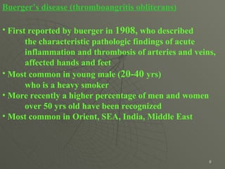
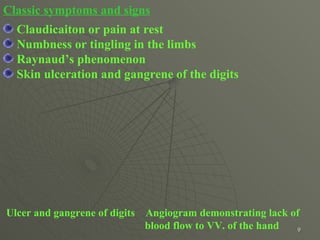
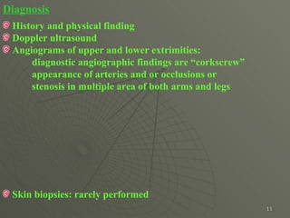
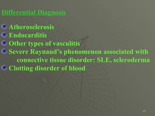
Downloaded 121 times
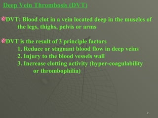
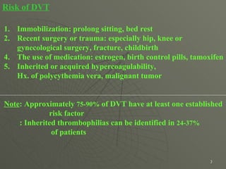
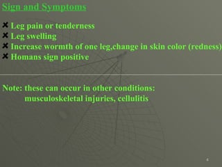
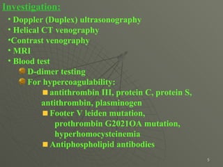
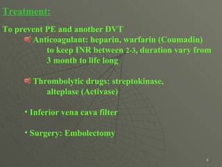


Deep Vein Thrombosis (DVT) is a blood clot that forms in the deep veins of the legs, thighs, pelvis or arms. Risk factors for DVT include immobilization, recent surgery or trauma, certain medications, and inherited or acquired hypercoagulability. Symptoms include leg pain, tenderness, swelling, warmth, and skin discoloration of one leg. Diagnosis is made through Doppler ultrasound, CT venography, or MRI imaging along with blood tests. Treatment involves anticoagulant drugs like heparin or warfarin to prevent pulmonary embolism and further clots.


![PERI-PROSTHETIC FRACTURE NAIL-PLATE CONSTRUCT [NPC].pptx](https://cdn.slidesharecdn.com/ss_thumbnails/drarunkumardrmohamedashrafperiprostheticfrasturenail-plateconstructnpc-260209164459-7e9d15a1-thumbnail.jpg?width=640&height=640&fit=bounds)


