Deep Vein Thrombosis-Dr. sharfuddin chowdhury
•
16 likes•5,341 views

This document discusses the modern management of deep vein thrombosis (DVT). It covers the epidemiology, classification, clinical presentation, diagnosis and imaging of DVT. The main diagnostic tools are the Wells criteria to determine pre-test probability, D-dimer testing, and compression ultrasound imaging of the legs. Compression ultrasound has high sensitivity and specificity for proximal DVT, while CT, MR and contrast venography can be used for more difficult cases or to diagnose pelvic DVT.

Recommended
More Related Content
What's hot
What's hot (20)
Viewers also liked
Viewers also liked (20)
Similar to Deep Vein Thrombosis-Dr. sharfuddin chowdhury
Similar to Deep Vein Thrombosis-Dr. sharfuddin chowdhury (20)
More from Shakila Rifat
Recently uploaded
Recently uploaded (20)
Deep Vein Thrombosis-Dr. sharfuddin chowdhury
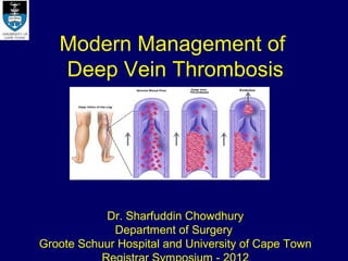
- 1. Modern Management of Deep Vein Thrombosis Dr. Sharfuddin Chowdhury Department of Surgery Groote Schuur Hospital and University of Cape Town
- 2. Introduction • Deep Vein Thrombosis (DVT) refers to the formation of clot in the deep veins of the extremities or pelvis due to a disordered balance between the thrombotic and fibrinolytic systems. • There are three possible sequele following a DVT: Acute Pulmonary Thrombo-Embolism (PTE) Complicated DVT (Phlegmasia; venous gangrene) The Post-Thrombotic Syndrome (PTS) .
- 3. Epidemiology • Worldwide 70-113 cases per 100,000 per year • No conclusive gender difference, however recurrence is M>F • Equally frequent among Whites and Blacks. • More common during winter months by 10% - 15% compared to summer months.
- 4. Epidemiology • Prevalence of DVT in hospitalized patient groups Spinal cord injury 60-80% Major trauma 40-80% Hip/Knee arthoplasty or hip fracture surgery 40-60% Critical care 10-80% Stroke 20-50% General surgery 15-40% Major gynaecological surgery 15-40% Neurosurgery 15-40% Major urologic surgery 15-40% Medical patients 10-20%
- 5. Classification • Acute (< 2 weeks) • Recurrent DVT • Anatomical Bilateral Ilio-femoral (L) ilio-femoral Multi segmental Profunda femoris vein Femoro-popliteal Proximal Knee Distal
- 6. Clinical Presentation Acute DVT may present as Asymptomatic Symptomatic Complicated Phlegmasia Caerulea dolens (PCD) Alba dolens (PAD) PTE Symptoms: Pain- exacerbated by ambulation, relieved by rest Swelling- below knee (distal DVT), Up to groin (proximal DVT) Erythema
- 8. Diagnosis of DVT • Pre-test probability: A meta analysis involving more than 5000 patients with suspected DVT from 12 clinical trials and systematic reviews showed that the combination of normal D- dimer and low clinical probability for DVT safely rules out the diagnosis of DVT. Level of Evidence: High (1A) Modified Wells Score Score Modified Wells Score Paresis, Paralysis or recent leg plaster immobilization 1 Active cancer (on going chemotherapy or within 6 months or palliative treatment) 1 Recent immobilization/ bed rest (> 3 days) or recent surgery (within 4 weeks) 1 calf circumference > 3 cm on affected side compared with normal side 1 Localized tenderness along the deep venous system distribution 1 Entire leg swollen 1 Pitting oedema of only the symptomatic leg 1 Collateral superficial veins (non-varicose) 1 Prior history of DVT 1 Alternative diagnosis at least as likely as DVT (ruptured Baker’s cyst,RA,Cellulitis -2 etc.) Score 0= Low risk, 1-2=Moderate risk, 3 or above = High risk
- 9. D-Dimer D-dimer is a specific degradation product of cross-linked fibrin. Sensitive but non specific marker False-positive D-dimers occur in patients with o Recent (within 10 days) surgery or trauma, o Recent myocardial infarction or stroke, o Acute infection, o Disseminated intravascular coagulation, o Pregnancy or recent delivery, o Active collagen vascular disease, or metastatic cancer
- 10. Imaging Imaging Advantages Disadvantages Modality Compression Sensitivity 97-100% and specificity of Difficult to perform in patients with Ultrasound 98-99% for proximal DVT morbid obesity, severe oedema, casts or other immobilization devices Non invasive, repeatable and widely available Decrease ability to visualize the Popliteal fossa in cases of distal DVT Higher sensitivity in detecting distal thrombosis Operator dependent CT Non invasive Limited data Venography Can diagnose Pelvic DVT Concurrently exclude PE MRV Highly accurate Expensive Safe during Pregnancy Not readily available Non-invasive Contrast “ Invasive Venography Requires specialized equipment Rare but serious side effects
- 11. Imaging Imaging Advantages Disadvantages Modality Compression Sensitivity 97-100% and specificity of Difficult to perform in patients with Ultrasound 98-99% for proximal DVT morbid obesity, severe oedema, casts or other immobilization devices Non invasive, repeatable and widely available Decrease ability to visualize the Popliteal fossa in cases of distal DVT Higher sensitivity in detecting distal thrombosis Operator dependent CT Non invasive Limited data Venography Can diagnose Pelvic DVT Concurrently exclude PE MRV Highly accurate Expensive Safe during Pregnancy Not readily available Non-invasive Contrast “Gold Standard” Invasive Venography Sensitivity approaches 100% Requires specialized equipment Easily interpretable Rare but serious side effects
- 12. Imaging Imaging Advantages Disadvantages Modality Compression Sensitivity 97-100% and specificity of Difficult to perform in patients with Ultrasound 98-99% for proximal DVT morbid obesity, severe oedema, casts or other immobilization devices Non invasive, repeatable and widely available Decrease ability to visualize the Popliteal fossa in cases of distal DVT Higher sensitivity in detecting distal thrombosis Operator dependent CT Non invasive Limited data Venography Can diagnose Pelvic DVT Concurrently exclude PE MRV Highly accurate Expensive Safe during Pregnancy Not readily available Non-invasive Contrast “Gold Standard” Invasive Venography Sensitivity approaches 100% Requires specialized equipment Easily interpretable Rare but serious side effects
- 13. Imaging Imaging Advantages Disadvantages Modality Compression Sensitivity 97-100% and specificity of Difficult to perform in patients with Ultrasound 98-99% for proximal DVT morbid obesity, severe oedema, casts or other immobilization devices Non invasive, repeatable and widely available Decrease ability to visualize the Popliteal fossa in cases of distal DVT Higher sensitivity in detecting distal thrombosis Operator dependent CT Non invasive Limited data Venography Can diagnose Pelvic DVT Concurrently exclude PE MRV Highly accurate Expensive Safe during Pregnancy Not readily available Non-invasive Contrast “Gold Standard” Invasive Venography Sensitivity approaches 100% Requires specialized equipment Easily interpretable Rare but serious side effects
- 14. Diagnostic Algorithms SUSPECTED DVT Pre-test clinical probability Low / moderate probability D-Dimer assay Negative Positive/Equivocal No Treatment Venous US Negative Positive Equivocal No Treatment Treatment MRI/CV Negative Positive No Treatment Treatment
- 15. Diagnostic Algorithms SUSPECTED DVT Pre-test clinical probability High probability Compression US Negative Positive Equivocal Serial US 5-7 days Negative Positive No Treatment Treatment Treatment MRI/CTV Negative Positive No Treatment Treatment
- 18. Factor Xa inhibitors • Fondaparinux • Rivaroxaban • Synthetic analogue of • Direct, selective and antithrombin-binding reversible inhibitors of Xa. pentasaccharide • Orally with acceptable sequence. safety profile. • Doesn’t cause • No monitoring needed. Thrombocytopenia, • Non-valvular Afib. Ortho Osteoporosis. VTE Proph. • Dosing • Dose: 10 mg OD 2.5mg od for prophylactic VTE 7.5mg od for established VTE 5mg <50kg,10mg >100kg
- 19. Direct thrombin inhibitors • Hirudin • Dabigatran • Heparin and LMWH are • Oral DTI indirect inhibitors of thrombin. • Indications: AF, • DTI do not require a Ortho VTE Prophylaxis plasma cofactor. Bind directly to thrombin and block its interaction with • Dose: 150-200mg od its substrates. • Monitoring not needed. • Use: HIT , PCI
- 20. Grades of Recommendation Grade Description of 2012 ACCP grade 1A Strong recommendation, high-quality evidence 1B Strong recommendation, moderate-quality evidence 1C Strong recommendation, low- or very-low-quality evidence 2A Weak recommendation, high-quality evidence 2B Weak recommendation, moderate-quality evidence 2C Weak recommendation, low- or very-low-quality evidence
- 21. Prophylaxis (ACCP Guidelines 2012) • Recommended Prophylaxis in surgical patients according to the estimated level of risks (ACCP Guidelines 2012) A. GENERAL AND ABDOMINO PELVIC SURGERY Risk level Prophylaxis Very low risk Early mobilization Low risk IPC (2C) Moderate risk LMWH, LDUH (2B) or IPC (2C) High risk LMWH / LDUH (1B) and IPC / ES (2C) Cancer surgery- LMWH 4 weeks (1B) IPC until risk of bleeding diminishes and LMWH / LDUH may be initiated
- 22. Prophylaxis (ACCP Guidelines 2012) • Major Trauma: Traumatic brain injury, acute spinal injury: UH, LMWH or IPC High risk: IPC and UH / LMWH (When not contraindicated by lower extremity injury) • Orthopaedic Surgery: Major Surgery (THA / TKA / HFS): > In hospital dual prophylaxis with antithrombotic agent (1B) and IPC (2C) > extended thromboprophylaxis in out patient period for up to 35 days (2B) > In case of increased risk of bleeding – IPC > Patients declining or uncooperative with injections: Apixaban, Dabigatran, Rivaroxiban, or dose adjusted VKA IVC Filter should not be used for primary VTE prevention (2C) Periodic surveillance with venous compression ultrasound should not be performed (2C)
- 23. Management of DVT Key recommendations according to ACCP Guidelines 2012 Distal DVT : A. No, mild or moderate symptoms and no risk for clot extension: No anticoagulation Serial doppler US over next 2 weeks to exclude clot extension (2C) B. Severe Symptoms: Anticoagulation 3 months (2C) Proximal DVT: > Anticoagulation 3 months (1B) Enoxaparin(bd) (2B) or Dalteparin/Tinzaparin/Fondaparinux (od) (2C) Followed by Warfarin rather than Dabigatran or Rivaroxaban > No routine use of thrombolytics or thrombectomy (2C) > Treat as an outpatient, if feasible
- 24. Management of DVT • Incidentally discovered (Asymptomatic DVT) > Leg, pelvic or vena cava thrombosis – Anticoagulation 3 months(2B) > Abdominal (portal, splenic, mesenteric or hepatic vein) thrombosis-no anticoagulation (2C) • Cancer associated DVT: > Anticoagulation at least 3 months, preferably long term unless bleeding risk is very high (1B) > LMWH is the preferred treatment, rather than Warfarin (2B)
- 25. Management of DVT • Arm DVT: > Axillary or more proximal veins- anticoagulation alone 3 months (1B) rather than thrombolysis (2C) > DVT associated with central venous catheter: Do not remove functional catheter and anticoagulate (2C) if catheter is removed continue anticoagulation 3 months thereafter. • Superficial thrombophlebitis: Superficial thrombophlebitis of leg of at least 5 cm length- Prophylactic dose of Fondaparinux or LMWH for 45 days (2B).
- 26. Management of DVT •Vena cava filter (=IVC filter): > should only be placed in the patient with acute DVT where anticoagulation is contraindicated (1B) > a permanent IVC filter, of itself, is not an indication for extended anticoagulation. •Compression stockings: at least 2 years to prevent or minimize PTS (2C) If at 2 years patient has bothersome symptoms of PTS (Pain, swelling), continue to wear for symptoms relief
- 27. IVC Filters • Indications: – Absolute contraindication to therapeutic anticoagulation – Failure of anticoagulation • Relative Indications: – recurrent VTE on anticoagulation, – Recurrent PE with pulmonary hypertension, – Extensive free-floating ilio-femoral thrombus, and – Post-thrombolysis of ilio-caval thrombus.
- 28. IVC Filters- Classification – Temporary filters are attached to a catheter that exits the skin – Retrievable filters are similar in design to permanent filters but are designed to be removed.
- 29. IVC Filters – Different Types A. Stainless steel Greenfield filter B. Modified hook titanium Greenfield filter C. Bird’s nest filter D. Simon Nitinol filter E. Vena tech filter
- 30. Filter Placement & Location • Angiographic imaging of the IVC: characterise IVC anatomy, exclude the presence of IVC thrombus. • Inserted percutaneously via the femoral or jugular approach under fluoroscopy guidance. INFRARENAL 90% of clinically significant PE originates from the lower extremity or pelvic veins, optimally immediately below the renal veins to minimize dead space if filter becomes thrombosed or IVC thrombus to form.
- 31. Filter Placement-Location Suprarenal placement: Duplicated IVC Large volume of thrombus within the infrarenal IVC Extrinsic compression (! Pregnancy/Young females)
- 32. Filter Placement-Location Bilateral Iliac Vein: Megacava Duplicated IVC Retroaortic left renal vein component that drains into the IVC close to the iliac venous confluence
- 33. Complications of IVC Filters Recurrent PE 2-5% Fatal PE 0.7% Venous Access site thrombosis 2-28% Filter migration 3-69% IVC wall penetration 9-24% IVC obstruction 6-30% Filter fracture 1% – Christopher J. Kwolek MD, Division of vascular and endo vascular surgery, Massachuatts General Hospital,
- 34. Anticoagulation after Filter Placement Whether concomitant anticoagulation therapy should be utilized following filter placement is unknown. Patients with IVC filters are at risk for – IVC thrombosis, – insertion site thrombosis and – recurrence of the initial thromboembolic event ACCP Guidelines suggest the use of conventional anticoagulation therapy if the risk of bleeding resolves. (2B)
- 35. IVC Filter-Retrieval Success depends on: • filter dwell time • amount of hook/strut/leg penetration and endothelialization • accessibility of the retrieval hook Turba et al report a 95% success rate for the GT Filter even with filters in place for more than 6 months. (Feb 2010 Endovascular today)
- 36. Methods of Thrombus Removal
- 37. Early Thrombus Removal -Ilio-femoral DVT • 2008 Guidelines- In favour of CDT (2B) • 2012 Guidelines- Against CDT (2C) BUT- DVT causes severe leg pain and swelling With anticoagulation, the period before improvement varies Difficulty ambulating and returning to full activity impair QOL
- 38. • Patient with ileo-femoral DVT (CFV and/or Iliac vein) develop PTS 60% of the time Author / year Journal N 2year PTS Prandoni 1996 Ann Intern Med 355 23% Brandjes 1997 Lancet 96 23% Prandoni 2004 Ann Intern Med 90 25% Partsch 2004 Int J Angiol 37 46% Van Dongen 2005 J Thromb Haemost 244 30% Kahn 2008 Ann Intern Med 387 60% Enden 2012 Lancet 99 56%
- 39. • Does immediate clot removal speed symptom relief, save valves, preserve patency and prevent PTS? Single- Centre RCTs Multicentre RCT - CaVenT Study A 35-patient RCT found CDT with 189 patients with femoral, common streptokinase to provide better 6- femoral, or iliac DVT: CDT + AC/comp month venous patency (72% vs 12%, p vs AC/comp alone < 0.01) and less valvular reflux (11% vs 41%, p = 0.042) – Enden T et al. Lancet 2012; 379:31-38 – Elsharawy M et al. Eur J Vasc Endovasc Surg 2-year PTS was significantly reduced 2002. with use of CDT (41.1% versus 55.6% A 183-patient RCT found CDT-PCDT to Control, p = 0.047) reduce 6-month PTS (3.4% vs 27.2%, p Limitations: sample size, used CDT (not PCDT) < 0.001) and recurrent VTE (2.3% versus 14.8%, p = 0.003) – Sharifi M et al. Cathet Cardiovasc Interv 2010.
- 40. A multicentre randomized trial on Acute venous Thrombosis : Thrombus Removal with Adjunctive Catheter directed Thrombolysis (ATTRACT) trial sponsored by The National Heart Lung and Blood Institute (NHLBI),U.S. As of:: July 3, 2012, 312 Patients have been enrolled
- 41. Catheter Directed Thrombolysis (CDT) • Indications: Acute ilio-femoral DVT Multi level DVT Massive DVT (Phlegmasia) < 10 days old • Contraindications: Absolute Strong Relative Other Relative Active bleeding / DIC Recent Major Surgery Renal failure Recent CVA / TIA Major Trauma(<10 days) Severe hepatic dysfunction Neurosurgery or Eye surgery (<3 months) Bacterial endocarditis Intracranial Trauma Major GIT bleed (<3 mo) Diabetic haemorrhagic (<3 months) Uncontrolled HPT(>180) Retinopathy Recent delivery (<10 d) Pregnancy or lactation ICSOL or seizure disorder Recent CPR (< 10 d)
- 42. CDT- Procedure • In cath lab • Under Local Anaesthesia • Straight flush angio-cath with multiple sideholes • The catheter tip is placed into the clot • Initial lysis with rt PA 10mg diluted in 200ml NS • Followed by rt PA infusion 1mg/hr(40 mg in 1L NS @ 25ml/hr)for 24hrs. • In addition Heparin infusion 500 U/hr to prevent catheter thrombosis • Monitor APTT (Therapeutic 2.5 X control) & Fibrinogen level 6hrly • Stop infusion if Fibrinogen levels < 2mg/dl • Ascending phlebogram before removal of catheter • Commence full anticoagulation & Warfarin after thrombus clearance
- 43. Angio Jet / Pulse Power Spray • The Angio Jet catheter system is comprised of a single – use catheter, single use pump set and a drive unit • Small pulses of high dose lytic agent delivered through a multi-side hole catheter with a guide wire occluding the end hole. • Jets create a localized low pressure zone at the catheter tip macerating thrombus and redirecting flow and debris into outflow channels directed behind the catheter tip for aspiration and removal. • Success in thrombus removal, restoration of venous patency, and preservation of valvular function and low haemorrhagic complications has been demonstrated. Example and principle of Angio Jet Power-Pulse spray for DVT (Courtesy of Possis Medical, Inc., • Expensive Minneapolis, MN.)
- 44. Trellis-8 Infusion System • The double balloon catheter is inserted into the thrombosed venous segment with the proximal balloon positioned at the upper edge of the thrombus. • Balloons are inflated and rtPA is infused into the thrombosed segment isolated by the balloons. • The intervening catheter spins at 1500 rpm for 15-20 mins. • The liquefied and fragmented thrombus is aspirated. Example of Trellis-8 infusion catheter for DVT. (Courtesy of Bacchus • Success evaluated by repeats segmental Vascular, Inc., Santa Clara, CA.) phlebography
- 45. Ultrasound Accelerated Thrombolysis • In combination with CDT • Does not directly macerate the clot • Create micro streams, increase thrombus permeability results in augmented lytic dispersion within the thrombus. • Parikh et al reported their initial experience with EKOS Endo wave system accelerated thrombolysis in 53 patients. Complete lysis (>90%)was observed in 70%, overall in 91%, median infusion time was 22 Example and principle of the EKOS hours, treatment time and the dose of ultrasound facilitated thrombolysis lytic agents were reduced. (Courtesy of EKOS Corp., Bothell, WA.) J Vasc Interv Radiol 2008 19:521-528
- 46. Adjunctive Venoplasty and Stenting • Adequate treatment of anatomic compression, stenosis, or persistent small thrombus after CDT or PMT requires angioplasty and stenting. • May Thurner syndrome (compression of left CIV by right CIA against L5) is the most common anatomic variant. • With anti coagulation alone, untreated iliac vein obstruction prevents vessel recanalization in 70-80% of patients and Venogram 6 weeks following EKOS clot propagation may continue up to 40%. and AngioJet with stent in left common iliac vein. Vein is widely patent without thrombus in filter which was removed
- 47. Venous Thrombectomy • Venous Thrombectomy is falling out of favour. • For pts. with CI to pharmacological thrombolysis or • Those in whom other modalities have failed in the setting of PCD • Under GA, Groin exposure, Venotomy, venous balloon catheter to extract thrombus. .
- 48. Conclusion • Acute DVT should be viewed as a chronic disease due to substantial long-term patient disability. • Anticoagulation remains the cornerstone of prevention and treatment of DVT. • Newer anti coagulants may simplify therapeutic paradigms and are expected to improve overall clinical outcome. • IVC filters should not be used routinely to prevent PE. • Most ACCP recommendations are weak 2B/C. • Catheter-based endovascular therapies offer the potential to preserve life and limb. • ATTRACT trial results awaited.
- 49. THANKYOU.
Editor's Notes
- WORLDWIDE estimated around 100/100,000 pa
- In hospitalized patients prevalence is highest in Spinal cord injury, Major trauma ,Hip/Knee arthoplasty or hip fracture surgery, critical care amongst others.
- Symptoms include…..
- Diagnosis of DVT starts with pre test clinical probability followed by D-dimer assay, followed by imaging that includes…..
- A combination of normal D- dimer and low clinical probability for DVT safely rules out the diagnosis of DVT. Level of Evidence: High (1A) According to the modified wells score, 1 point is given for the following…. A score of 0=low risk, 1-2=moderate risk, .3=high risk
- False positives occur in the following……
- Strategies to prevent DVT include:….. Mechanical methods comprise…
- Ill mention a bit about the newer anticoagulants….
- Grades of recomentations used in ACCP guidelines are divided into 1 and 2 1 stands for strong recommendation 2 for weak recommendation. 1 and 2 are further divided into a,b,c A for… B for…. C for…
- Recommendation for GENERAL AND ABDOMINO PELVIC SURGERY is as follows…. For….
- Recommendations for Management of distal DVT….. If no…. For proximal DVT….
- For….
- For…
- • There is a scarcity of robust evidence in favor of using IVC filters to manage DVT. • ACCP guidelines recommend IVC filter use in patients who cannot be anticoagulated due to bleeding risks. • There is insufficient data to support the use of IVC filters for such situations as recurrent VTE on anticoagulation, recurrent PE with pulmonary hypertension, extensive free-floating ilio-femoral thrombus, and post-thrombolysis of ilio-caval thrombus.
- IVC Filters can be classified as Permanent and Removable filters. Removable filters are again two types- Temporary and Retrievable filters. Temporary filters are attached to a catheter that exits the skin and therefore must be removed due to the risk of infection and embolization. Retrievable filters are similar in design to permanent filters but are designed to be removed. However, this must be done with caution, as neointimal hyperplasia can prevent removal or cause vessel wall damage upon removal
- The different types of IVC filters include….stainless steel greenfield filter……
- Right internal jugular or right common femoral The acute angle of the left iliac vein with the IVC directs the filter delivery sheath against the right lateral aspect of the IVC wall causing the filter to deployed in a tilted position
- For…….IVC filter can be placed suprarenally
- And bilteral iliac vein filters for…..
- Complications of IVC filters include…
- Whether concomitant anticoagulant therapy should be utilized following filter placement is unknown. Small thrombi are capable of passing through patent filters or through collaterals around obstructed filters; furthermore, direct thrombus extension can occur through the filter itself. Because patients with IVC filters are at risk for IVC thrombosis, insertion site thrombosis, and recurrence of the initial thromboembolic event, continued use of anticoagulants when there are no contraindications would seem to be a prudent recommendation
- For retrieval of IVC filter success depends on….. Here you can see….filter being hooked….and recaptured into sheath via Transjugular approach. Günther Tulip filter
- Thrombus removal can be done by open venous thrombectomy or endovascularly. Endovasscular techniques include…..
- The 2008 guidelines was in favour of CDT for early thrombus removal for ileofemoral DVT. The 2012 guidelines are against CDT.
- Different studies published in different journals over the past 15 years shows Patient with ileo-femoral DVT develop Post Thrombotic Syndrome 60% of the time in 2 years
- In 2002 , Elsharawy et al described a single centre Egyptian randomized trial comparing adjunctive CDT (with streptokinase) with anticoagulation alone in 35 patients with acute iliofemoral DVT. At 6 months, patient treated with CDT had a higher rate of normal venous function (72% vs 12%, p< 0.001) and less valvular reflux (11% vs 41% , p = 0.04)
- Indications for CDT includes….. Absolute contraindications are….
- CDT is performed in the endovascular suite under LA. Initial lysis by 10mg of actelase in 200ml saline given as a bolus. This is followed by….. In addition….
- Iliac vein obstruction are of 2 types: Thrombotic iliac vein lesion, Non thrombotic iliac vein lesion like may thurner syndrome which is compression of left CIV by right CIA against L5 Adequate treatment of anatomic compression, stenosis, or persistent small thrombus after CDT or PMT requires angioplasty and stenting. With anti coagulation alone, untreated iliac vein obstruction prevents vessel recanalization in 70-80% of patients and clot propagation may continue up to 40%.
- Is done by performing a venotomy and a venous balloon catheter to extract thrombus.