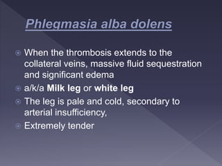
Deep Vein Thrombosis
Dr Mahmood
Emergency Medicine Resident
MEM
 A 67 old male patient presented with c/o painful swelling of left lower
limb since 5-6 days
 h/o trauma to left lower limb 3 days back
 Past history: nil
 No H/o: chest pain, SOB
 General examination:
-PR: 82/mins -RR:20/mins -Bp: 130/90mmhg
-Sp02: 98% on RA
 Systemic examination:
-RS: bilateral air entry
-CVS: s1+, s2+
-P/A: soft. BS+
L/E: swelling of left lower limb foot to thigh, tenderness +,
movements+, peripheral pulses+
 CBP: normal,
 TLC: 17,000
 Bleeding time and clotting time : normal
 2D-echo:
-no RWMA,
-good LV function ,
-IVC collapsing
 Color Doppler of venous system of left lower limb
performed
-Impression: intramural echogenic contents with absent
color flow uptake in distal SFV and popliteal vein s/o DVT
 A 45 years old male patient presented with complaints of right
lower limb painful swelling since 4 days.
- Sudden onset,
- Increasing gradually
- Sever cramping pain in calf and thigh
- increased on walking and movement of the limb
 h/o left sided nephrectomy 5 days back for left side renal cell carcinoma
(RCC)
 h/o chronic tobacco chewing
 No h/o of chest pan , SOB , abdomen pain
 No family history of VTE
 General examination:
-PR: 92/mins
-Bp: 140/90mmhg
-RR:20/mins
-Sp02: 98% on RA
 Systemic examination:
-RS: bilateral air entry
-CVS: s1+, s2+
-P/A: soft. BS+, scar of nephrectomy is seen over left lumbar
L/E: Swelling and redness over right lower limb from foot to thigh,
no dilated , engorged veins , skin is stretched and shiny
 Duplex ultrasound:
Impression: Common femoral ,deep femoral,
popliteal and posterior tibial vein show echogenic
thrombus and absent flow . s/o DVT
 2D-echo:
-no RWMA,
-good LV function ,
-IVC collapsing
 Surgical Management:
- Debridment of wound + IVC filter placement done
 Medical management :
-Tab ciplox (ciprofloxacin) 500 mg bd
-Tab ultracet (paracetamol+ tramadol) 1 tab bd
-Tab bevon tab od
-Tab pradaxa (dabigatran)150mg bd x 1month
-Tab dolo (paracetamol) 650 tid
 Advise:
-Left lower limb elevation
-High protein diet
 Introduction
 Etiology
 Risk factors
 Clinical features
 Complications
 Diagnostic procedures
 Management
 Prevention
 Differentials
 References
 Deep vein thrombosis (DVT) is the formation
of a blood clot in a deep vein most
commonly seen n the legs or in the arms
 Provoked DVT:
DVT where there is an identifiable risk factor
that likely caused the DVT.
 Unprovoked DVT:
DVT where there is no identifiable risk factor
that likely caused the DVT
 DVT usually originates in the lower extremity
venous system, starting at the calf and
progressing proximally to involve popliteal,
femoral or iliac system
 80-90% pulmonary emboli originates here
 An intimal defect often works as nidus for
clot formation
 Initially a platelet aggregate develops,
subsequently clotting factors through intrinsic
and extrinsic pathway fibrin and red cells
form a mesh until the lumen clot occludes
the vein wall.
 Age: incidence increase with advancing age
 Obesity: > BMI > is DVT
 Prolonged bed rest ( 4days or more)
 Lower limb fracture: a cast on the leg
 limb paralysis
 Extended travel (>4 hours)
 Acquired:
- Surgery and trauma are responsible for upto
40% of all thromboembolic disease
- Malignancy
- Hormonal therapy, OCP
- Pregnancy
- Nephrotic syndrome
- Antiphospholipid antibody syndrome
- SLE, IBD
 Inherited :
- Factor V Leiden mutation
- Antithrombin deficiency
- Protein C, S deficiency
- Prothrombin 20210 gene variant
- Homocysteinemia
 Trauma
 Surgery
 Peripheral and central lines
 Calf pain or tenderness or both
 Swelling wit pitting edema
 Increase in local temperature
 Redness or discoloration
 Dilatation of superficial veins
 Less frequent manifestations of DVT
includes
 Phlegmasia cerulea dolens
 Phlegmasia alba dolens
 Venous gangrene
 Extensive DVT of the major axial deep
venous channels of the lower extremity with
relative sparing if collateral veins
 The leg becomes blue in color, swollen and
painful
 Which may result in venous gangrene.
 When the thrombosis extends to the
collateral veins, massive fluid sequestration
and significant edema
 a/k/a Milk leg or white leg
 The leg is pale and cold, secondary to
arterial insufficiency,
 Extremely tender
 Homan’s sign:
- Pain in the calf region or knee with forced
dorsiflexion
 Moses / Bancroft’s sign:
- Gentle squeezing of the lower part of the calf from
- side to side causes sever pain
 Lowenberg sign:
- Pain is elicited rapidly when a blood pressure cuff is placed
and around the calf and inflated to 80mmhg
 Clinical examination alone is able to confirm
only 20-30%cases of DVT
 Blood test :
- D-Dimer test
- Coagulation profile
- Routine hematological investigation
 Imaging studies
 ECG and 2D echo
 D-dimer:
- D- dimer is a degradation product of fibrin cross-linking
- And elevated D- dimer level can occur when blood clots
are being formed. It can also be elevated in other
conditions unrelated to DVT (cancer, pregnancy, recent
surgery)
- The cutoff value for normal D-dimer is <500mcg/L
 Non invasive:
- Duplex ultrasound
- Plethysmography
- MRI techniques
Invasive:
- Venography
- Radio labeled fibrinogen
 Indications:
 When anticoagulant therapy is ineffective
 Unsafe
 Contraindicated
 The major surgical procedures for DVT are clot
removal and partial interruption of the inferior
vena cava to prevent pulmonary embolism.
 Inferior vena cave filter: it’s a type of vascular
filter into inferior vena cava to prevent life-thraetening
pulmonary embolism.
 Indication:
 Pulmonary embolism with contraindication to
anticoagulation.
 Recurrent pulmonary embolism despite adequate
anticoagulation.
 Pulmonary embolism (PE):
PE is a potentially life-threatening complication associated with DVT.
Sudden shortness of breath, chest pain while inhaling and coughing up
blood may occur with PE.
 Post-phlebitic syndrome:
 which manifest as leg pain and swelling, skin discoloration and skin
sores.
 Treatment complication: Bleeding ( hemorrhages).
 Recurrent DVT
 Chronic venous insufficiency
 Paradoxic emboli
 Do regular walking
 Limiting the amount of time you spend sitting
 Exercising daily, mainly walking, swimming or other
activities that promote good blood circulation.
 Avoid wearing tight- fitting clothes for extended periods.
 Drinking lots of fluids
 Consuming foods that acts as natural blood thinners to
reduce the risk of developing blood clots, such as
vitamin–E, ginger, cayenne pepper, garlic, turmeric and
cinnamon
 Wearing Elastic compression stockings
 Lower your blood pressure with dietary changes, like
reducing your salt and sugar intake.
 Lose weight if overweight
 Quit smoking
 Muscular tear
 Rupture of a Baker’s cyst
 Cellulites or other infection
 Thrombophlebitis
 Tumors
 Connective tissue disorders
 Tintinallis emergency medicine
 Rosens emergency medicine
 Dutton. Orthopedic examination, evaluation
and intervention
 Belly and love surery
 Medscape
Thank you

DVT ppt.pptx

  • 1.
    Deep Vein Thrombosis DrMahmood Emergency Medicine Resident MEM
  • 2.
     A 67old male patient presented with c/o painful swelling of left lower limb since 5-6 days  h/o trauma to left lower limb 3 days back  Past history: nil  No H/o: chest pain, SOB  General examination: -PR: 82/mins -RR:20/mins -Bp: 130/90mmhg -Sp02: 98% on RA  Systemic examination: -RS: bilateral air entry -CVS: s1+, s2+ -P/A: soft. BS+ L/E: swelling of left lower limb foot to thigh, tenderness +, movements+, peripheral pulses+
  • 3.
     CBP: normal, TLC: 17,000  Bleeding time and clotting time : normal  2D-echo: -no RWMA, -good LV function , -IVC collapsing  Color Doppler of venous system of left lower limb performed -Impression: intramural echogenic contents with absent color flow uptake in distal SFV and popliteal vein s/o DVT
  • 4.
     A 45years old male patient presented with complaints of right lower limb painful swelling since 4 days. - Sudden onset, - Increasing gradually - Sever cramping pain in calf and thigh - increased on walking and movement of the limb  h/o left sided nephrectomy 5 days back for left side renal cell carcinoma (RCC)  h/o chronic tobacco chewing  No h/o of chest pan , SOB , abdomen pain  No family history of VTE
  • 5.
     General examination: -PR:92/mins -Bp: 140/90mmhg -RR:20/mins -Sp02: 98% on RA  Systemic examination: -RS: bilateral air entry -CVS: s1+, s2+ -P/A: soft. BS+, scar of nephrectomy is seen over left lumbar L/E: Swelling and redness over right lower limb from foot to thigh, no dilated , engorged veins , skin is stretched and shiny
  • 6.
     Duplex ultrasound: Impression:Common femoral ,deep femoral, popliteal and posterior tibial vein show echogenic thrombus and absent flow . s/o DVT  2D-echo: -no RWMA, -good LV function , -IVC collapsing
  • 7.
     Surgical Management: -Debridment of wound + IVC filter placement done  Medical management : -Tab ciplox (ciprofloxacin) 500 mg bd -Tab ultracet (paracetamol+ tramadol) 1 tab bd -Tab bevon tab od -Tab pradaxa (dabigatran)150mg bd x 1month -Tab dolo (paracetamol) 650 tid  Advise: -Left lower limb elevation -High protein diet
  • 8.
     Introduction  Etiology Risk factors  Clinical features  Complications  Diagnostic procedures  Management  Prevention  Differentials  References
  • 9.
     Deep veinthrombosis (DVT) is the formation of a blood clot in a deep vein most commonly seen n the legs or in the arms
  • 11.
     Provoked DVT: DVTwhere there is an identifiable risk factor that likely caused the DVT.  Unprovoked DVT: DVT where there is no identifiable risk factor that likely caused the DVT
  • 12.
     DVT usuallyoriginates in the lower extremity venous system, starting at the calf and progressing proximally to involve popliteal, femoral or iliac system  80-90% pulmonary emboli originates here
  • 13.
     An intimaldefect often works as nidus for clot formation  Initially a platelet aggregate develops, subsequently clotting factors through intrinsic and extrinsic pathway fibrin and red cells form a mesh until the lumen clot occludes the vein wall.
  • 17.
     Age: incidenceincrease with advancing age  Obesity: > BMI > is DVT  Prolonged bed rest ( 4days or more)  Lower limb fracture: a cast on the leg  limb paralysis  Extended travel (>4 hours)
  • 18.
     Acquired: - Surgeryand trauma are responsible for upto 40% of all thromboembolic disease - Malignancy - Hormonal therapy, OCP - Pregnancy - Nephrotic syndrome - Antiphospholipid antibody syndrome - SLE, IBD
  • 19.
     Inherited : -Factor V Leiden mutation - Antithrombin deficiency - Protein C, S deficiency - Prothrombin 20210 gene variant - Homocysteinemia
  • 20.
     Trauma  Surgery Peripheral and central lines
  • 22.
     Calf painor tenderness or both  Swelling wit pitting edema  Increase in local temperature  Redness or discoloration  Dilatation of superficial veins
  • 24.
     Less frequentmanifestations of DVT includes  Phlegmasia cerulea dolens  Phlegmasia alba dolens  Venous gangrene
  • 25.
     Extensive DVTof the major axial deep venous channels of the lower extremity with relative sparing if collateral veins  The leg becomes blue in color, swollen and painful  Which may result in venous gangrene.
  • 27.
     When thethrombosis extends to the collateral veins, massive fluid sequestration and significant edema  a/k/a Milk leg or white leg  The leg is pale and cold, secondary to arterial insufficiency,  Extremely tender
  • 29.
     Homan’s sign: -Pain in the calf region or knee with forced dorsiflexion
  • 30.
     Moses /Bancroft’s sign: - Gentle squeezing of the lower part of the calf from - side to side causes sever pain  Lowenberg sign: - Pain is elicited rapidly when a blood pressure cuff is placed and around the calf and inflated to 80mmhg
  • 33.
     Clinical examinationalone is able to confirm only 20-30%cases of DVT  Blood test : - D-Dimer test - Coagulation profile - Routine hematological investigation  Imaging studies  ECG and 2D echo
  • 34.
     D-dimer: - D-dimer is a degradation product of fibrin cross-linking - And elevated D- dimer level can occur when blood clots are being formed. It can also be elevated in other conditions unrelated to DVT (cancer, pregnancy, recent surgery) - The cutoff value for normal D-dimer is <500mcg/L
  • 36.
     Non invasive: -Duplex ultrasound - Plethysmography - MRI techniques
  • 37.
  • 39.
     Indications:  Whenanticoagulant therapy is ineffective  Unsafe  Contraindicated  The major surgical procedures for DVT are clot removal and partial interruption of the inferior vena cava to prevent pulmonary embolism.
  • 40.
     Inferior venacave filter: it’s a type of vascular filter into inferior vena cava to prevent life-thraetening pulmonary embolism.  Indication:  Pulmonary embolism with contraindication to anticoagulation.  Recurrent pulmonary embolism despite adequate anticoagulation.
  • 42.
     Pulmonary embolism(PE): PE is a potentially life-threatening complication associated with DVT. Sudden shortness of breath, chest pain while inhaling and coughing up blood may occur with PE.  Post-phlebitic syndrome:  which manifest as leg pain and swelling, skin discoloration and skin sores.  Treatment complication: Bleeding ( hemorrhages).  Recurrent DVT  Chronic venous insufficiency  Paradoxic emboli
  • 43.
     Do regularwalking  Limiting the amount of time you spend sitting  Exercising daily, mainly walking, swimming or other activities that promote good blood circulation.  Avoid wearing tight- fitting clothes for extended periods.  Drinking lots of fluids
  • 44.
     Consuming foodsthat acts as natural blood thinners to reduce the risk of developing blood clots, such as vitamin–E, ginger, cayenne pepper, garlic, turmeric and cinnamon  Wearing Elastic compression stockings  Lower your blood pressure with dietary changes, like reducing your salt and sugar intake.  Lose weight if overweight  Quit smoking
  • 46.
     Muscular tear Rupture of a Baker’s cyst  Cellulites or other infection  Thrombophlebitis  Tumors  Connective tissue disorders
  • 48.
     Tintinallis emergencymedicine  Rosens emergency medicine  Dutton. Orthopedic examination, evaluation and intervention  Belly and love surery  Medscape
  • 49.