The document discusses the benefits of exercise for mental health. Regular physical activity can help reduce anxiety and depression and improve mood and cognitive functioning. Exercise causes chemical changes in the brain that may help boost feelings of calmness, happiness and focus.
Lymphoedema is an abnormal swelling of limb due to the collection of excessive amount of high protein fluid secondary to defective lymphatic drainage in the presence of normal capillary filteration.It is very disabiling condition to the patient. In this ppp I have discussed its clinical picture and management in a simple way
Venous ulcer is one of the commonest complication of varicose veins. It may also occur in a condition called post phlebitic limb which is a sequelae to acute deep vein thronbosis. Hurry in surgical treatment of this condition before the ulcer heals could lead to a failure. Good conservative treatment for healing of the ulcer followed by surgical intervention gives the best results.
Made by Ranjith R Thampi. A surgery powerpoint I made during internship for Management of Varicose Veins. Tried to cover as much as possible on the topic. Kindly comment before you download. Thanks!
anatomy of the lower extremity veins, CVI , ambulatory venous hypertension, varicose veins , clinical examination and performance of various tests of the varicose veins
This document include anatomy of venous system of lower limb , venous hypertension and venous pathology like varicose vein. ,DVT and venous ulcer useful for surgery postgraduate and graduate (MBBS ) students . Including pathophysiology ,management includes surgical and medical aspect
HOT NEW PRODUCT! BIG SALES FAST SHIPPING NOW FROM CHINA!! EU KU DB BK substit...GL Anaacs
Contact us if you are interested:
Email / Skype : kefaya1771@gmail.com
Threema: PXHY5PDH
New BATCH Ku !!! MUCH IN DEMAND FAST SALE EVERY BATCH HAPPY GOOD EFFECT BIG BATCH !
Contact me on Threema or skype to start big business!!
Hot-sale products:
NEW HOT EUTYLONE WHITE CRYSTAL!!
5cl-adba precursor (semi finished )
5cl-adba raw materials
ADBB precursor (semi finished )
ADBB raw materials
APVP powder
5fadb/4f-adb
Jwh018 / Jwh210
Eutylone crystal
Protonitazene (hydrochloride) CAS: 119276-01-6
Flubrotizolam CAS: 57801-95-3
Metonitazene CAS: 14680-51-4
Payment terms: Western Union,MoneyGram,Bitcoin or USDT.
Deliver Time: Usually 7-15days
Shipping method: FedEx, TNT, DHL,UPS etc.Our deliveries are 100% safe, fast, reliable and discreet.
Samples will be sent for your evaluation!If you are interested in, please contact me, let's talk details.
We specializes in exporting high quality Research chemical, medical intermediate, Pharmaceutical chemicals and so on. Products are exported to USA, Canada, France, Korea, Japan,Russia, Southeast Asia and other countries.
Explore natural remedies for syphilis treatment in Singapore. Discover alternative therapies, herbal remedies, and lifestyle changes that may complement conventional treatments. Learn about holistic approaches to managing syphilis symptoms and supporting overall health.
Recomendações da OMS sobre cuidados maternos e neonatais para uma experiência pós-natal positiva.
Em consonância com os ODS – Objetivos do Desenvolvimento Sustentável e a Estratégia Global para a Saúde das Mulheres, Crianças e Adolescentes, e aplicando uma abordagem baseada nos direitos humanos, os esforços de cuidados pós-natais devem expandir-se para além da cobertura e da simples sobrevivência, de modo a incluir cuidados de qualidade.
Estas diretrizes visam melhorar a qualidade dos cuidados pós-natais essenciais e de rotina prestados às mulheres e aos recém-nascidos, com o objetivo final de melhorar a saúde e o bem-estar materno e neonatal.
Uma “experiência pós-natal positiva” é um resultado importante para todas as mulheres que dão à luz e para os seus recém-nascidos, estabelecendo as bases para a melhoria da saúde e do bem-estar a curto e longo prazo. Uma experiência pós-natal positiva é definida como aquela em que as mulheres, pessoas que gestam, os recém-nascidos, os casais, os pais, os cuidadores e as famílias recebem informação consistente, garantia e apoio de profissionais de saúde motivados; e onde um sistema de saúde flexível e com recursos reconheça as necessidades das mulheres e dos bebês e respeite o seu contexto cultural.
Estas diretrizes consolidadas apresentam algumas recomendações novas e já bem fundamentadas sobre cuidados pós-natais de rotina para mulheres e neonatos que recebem cuidados no pós-parto em unidades de saúde ou na comunidade, independentemente dos recursos disponíveis.
É fornecido um conjunto abrangente de recomendações para cuidados durante o período puerperal, com ênfase nos cuidados essenciais que todas as mulheres e recém-nascidos devem receber, e com a devida atenção à qualidade dos cuidados; isto é, a entrega e a experiência do cuidado recebido. Estas diretrizes atualizam e ampliam as recomendações da OMS de 2014 sobre cuidados pós-natais da mãe e do recém-nascido e complementam as atuais diretrizes da OMS sobre a gestão de complicações pós-natais.
O estabelecimento da amamentação e o manejo das principais intercorrências é contemplada.
Recomendamos muito.
Vamos discutir essas recomendações no nosso curso de pós-graduação em Aleitamento no Instituto Ciclos.
Esta publicação só está disponível em inglês até o momento.
Prof. Marcus Renato de Carvalho
www.agostodourado.com
Title: Sense of Taste
Presenter: Dr. Faiza, Assistant Professor of Physiology
Qualifications:
MBBS (Best Graduate, AIMC Lahore)
FCPS Physiology
ICMT, CHPE, DHPE (STMU)
MPH (GC University, Faisalabad)
MBA (Virtual University of Pakistan)
Learning Objectives:
Describe the structure and function of taste buds.
Describe the relationship between the taste threshold and taste index of common substances.
Explain the chemical basis and signal transduction of taste perception for each type of primary taste sensation.
Recognize different abnormalities of taste perception and their causes.
Key Topics:
Significance of Taste Sensation:
Differentiation between pleasant and harmful food
Influence on behavior
Selection of food based on metabolic needs
Receptors of Taste:
Taste buds on the tongue
Influence of sense of smell, texture of food, and pain stimulation (e.g., by pepper)
Primary and Secondary Taste Sensations:
Primary taste sensations: Sweet, Sour, Salty, Bitter, Umami
Chemical basis and signal transduction mechanisms for each taste
Taste Threshold and Index:
Taste threshold values for Sweet (sucrose), Salty (NaCl), Sour (HCl), and Bitter (Quinine)
Taste index relationship: Inversely proportional to taste threshold
Taste Blindness:
Inability to taste certain substances, particularly thiourea compounds
Example: Phenylthiocarbamide
Structure and Function of Taste Buds:
Composition: Epithelial cells, Sustentacular/Supporting cells, Taste cells, Basal cells
Features: Taste pores, Taste hairs/microvilli, and Taste nerve fibers
Location of Taste Buds:
Found in papillae of the tongue (Fungiform, Circumvallate, Foliate)
Also present on the palate, tonsillar pillars, epiglottis, and proximal esophagus
Mechanism of Taste Stimulation:
Interaction of taste substances with receptors on microvilli
Signal transduction pathways for Umami, Sweet, Bitter, Sour, and Salty tastes
Taste Sensitivity and Adaptation:
Decrease in sensitivity with age
Rapid adaptation of taste sensation
Role of Saliva in Taste:
Dissolution of tastants to reach receptors
Washing away the stimulus
Taste Preferences and Aversions:
Mechanisms behind taste preference and aversion
Influence of receptors and neural pathways
Impact of Sensory Nerve Damage:
Degeneration of taste buds if the sensory nerve fiber is cut
Abnormalities of Taste Detection:
Conditions: Ageusia, Hypogeusia, Dysgeusia (parageusia)
Causes: Nerve damage, neurological disorders, infections, poor oral hygiene, adverse drug effects, deficiencies, aging, tobacco use, altered neurotransmitter levels
Neurotransmitters and Taste Threshold:
Effects of serotonin (5-HT) and norepinephrine (NE) on taste sensitivity
Supertasters:
25% of the population with heightened sensitivity to taste, especially bitterness
Increased number of fungiform papillae
Ozempic: Preoperative Management of Patients on GLP-1 Receptor Agonists Saeid Safari
Preoperative Management of Patients on GLP-1 Receptor Agonists like Ozempic and Semiglutide
ASA GUIDELINE
NYSORA Guideline
2 Case Reports of Gastric Ultrasound
Title: Sense of Smell
Presenter: Dr. Faiza, Assistant Professor of Physiology
Qualifications:
MBBS (Best Graduate, AIMC Lahore)
FCPS Physiology
ICMT, CHPE, DHPE (STMU)
MPH (GC University, Faisalabad)
MBA (Virtual University of Pakistan)
Learning Objectives:
Describe the primary categories of smells and the concept of odor blindness.
Explain the structure and location of the olfactory membrane and mucosa, including the types and roles of cells involved in olfaction.
Describe the pathway and mechanisms of olfactory signal transmission from the olfactory receptors to the brain.
Illustrate the biochemical cascade triggered by odorant binding to olfactory receptors, including the role of G-proteins and second messengers in generating an action potential.
Identify different types of olfactory disorders such as anosmia, hyposmia, hyperosmia, and dysosmia, including their potential causes.
Key Topics:
Olfactory Genes:
3% of the human genome accounts for olfactory genes.
400 genes for odorant receptors.
Olfactory Membrane:
Located in the superior part of the nasal cavity.
Medially: Folds downward along the superior septum.
Laterally: Folds over the superior turbinate and upper surface of the middle turbinate.
Total surface area: 5-10 square centimeters.
Olfactory Mucosa:
Olfactory Cells: Bipolar nerve cells derived from the CNS (100 million), with 4-25 olfactory cilia per cell.
Sustentacular Cells: Produce mucus and maintain ionic and molecular environment.
Basal Cells: Replace worn-out olfactory cells with an average lifespan of 1-2 months.
Bowman’s Gland: Secretes mucus.
Stimulation of Olfactory Cells:
Odorant dissolves in mucus and attaches to receptors on olfactory cilia.
Involves a cascade effect through G-proteins and second messengers, leading to depolarization and action potential generation in the olfactory nerve.
Quality of a Good Odorant:
Small (3-20 Carbon atoms), volatile, water-soluble, and lipid-soluble.
Facilitated by odorant-binding proteins in mucus.
Membrane Potential and Action Potential:
Resting membrane potential: -55mV.
Action potential frequency in the olfactory nerve increases with odorant strength.
Adaptation Towards the Sense of Smell:
Rapid adaptation within the first second, with further slow adaptation.
Psychological adaptation greater than receptor adaptation, involving feedback inhibition from the central nervous system.
Primary Sensations of Smell:
Camphoraceous, Musky, Floral, Pepperminty, Ethereal, Pungent, Putrid.
Odor Detection Threshold:
Examples: Hydrogen sulfide (0.0005 ppm), Methyl-mercaptan (0.002 ppm).
Some toxic substances are odorless at lethal concentrations.
Characteristics of Smell:
Odor blindness for single substances due to lack of appropriate receptor protein.
Behavioral and emotional influences of smell.
Transmission of Olfactory Signals:
From olfactory cells to glomeruli in the olfactory bulb, involving lateral inhibition.
Primitive, less old, and new olfactory systems with different path
The prostate is an exocrine gland of the male mammalian reproductive system
It is a walnut-sized gland that forms part of the male reproductive system and is located in front of the rectum and just below the urinary bladder
Function is to store and secrete a clear, slightly alkaline fluid that constitutes 10-30% of the volume of the seminal fluid that along with the spermatozoa, constitutes semen
A healthy human prostate measures (4cm-vertical, by 3cm-horizontal, 2cm ant-post ).
It surrounds the urethra just below the urinary bladder. It has anterior, median, posterior and two lateral lobes
It’s work is regulated by androgens which are responsible for male sex characteristics
Generalised disease of the prostate due to hormonal derangement which leads to non malignant enlargement of the gland (increase in the number of epithelial cells and stromal tissue)to cause compression of the urethra leading to symptoms (LUTS
These simplified slides by Dr. Sidra Arshad present an overview of the non-respiratory functions of the respiratory tract.
Learning objectives:
1. Enlist the non-respiratory functions of the respiratory tract
2. Briefly explain how these functions are carried out
3. Discuss the significance of dead space
4. Differentiate between minute ventilation and alveolar ventilation
5. Describe the cough and sneeze reflexes
Study Resources:
1. Chapter 39, Guyton and Hall Textbook of Medical Physiology, 14th edition
2. Chapter 34, Ganong’s Review of Medical Physiology, 26th edition
3. Chapter 17, Human Physiology by Lauralee Sherwood, 9th edition
4. Non-respiratory functions of the lungs https://academic.oup.com/bjaed/article/13/3/98/278874
Flu Vaccine Alert in Bangalore Karnatakaaddon Scans
As flu season approaches, health officials in Bangalore, Karnataka, are urging residents to get their flu vaccinations. The seasonal flu, while common, can lead to severe health complications, particularly for vulnerable populations such as young children, the elderly, and those with underlying health conditions.
Dr. Vidisha Kumari, a leading epidemiologist in Bangalore, emphasizes the importance of getting vaccinated. "The flu vaccine is our best defense against the influenza virus. It not only protects individuals but also helps prevent the spread of the virus in our communities," he says.
This year, the flu season is expected to coincide with a potential increase in other respiratory illnesses. The Karnataka Health Department has launched an awareness campaign highlighting the significance of flu vaccinations. They have set up multiple vaccination centers across Bangalore, making it convenient for residents to receive their shots.
To encourage widespread vaccination, the government is also collaborating with local schools, workplaces, and community centers to facilitate vaccination drives. Special attention is being given to ensuring that the vaccine is accessible to all, including marginalized communities who may have limited access to healthcare.
Residents are reminded that the flu vaccine is safe and effective. Common side effects are mild and may include soreness at the injection site, mild fever, or muscle aches. These side effects are generally short-lived and far less severe than the flu itself.
Healthcare providers are also stressing the importance of continuing COVID-19 precautions. Wearing masks, practicing good hand hygiene, and maintaining social distancing are still crucial, especially in crowded places.
Protect yourself and your loved ones by getting vaccinated. Together, we can help keep Bangalore healthy and safe this flu season. For more information on vaccination centers and schedules, residents can visit the Karnataka Health Department’s official website or follow their social media pages.
Stay informed, stay safe, and get your flu shot today!
Tom Selleck Health: A Comprehensive Look at the Iconic Actor’s Wellness Journeygreendigital
Tom Selleck, an enduring figure in Hollywood. has captivated audiences for decades with his rugged charm, iconic moustache. and memorable roles in television and film. From his breakout role as Thomas Magnum in Magnum P.I. to his current portrayal of Frank Reagan in Blue Bloods. Selleck's career has spanned over 50 years. But beyond his professional achievements. fans have often been curious about Tom Selleck Health. especially as he has aged in the public eye.
Follow us on: Pinterest
Introduction
Many have been interested in Tom Selleck health. not only because of his enduring presence on screen but also because of the challenges. and lifestyle choices he has faced and made over the years. This article delves into the various aspects of Tom Selleck health. exploring his fitness regimen, diet, mental health. and the challenges he has encountered as he ages. We'll look at how he maintains his well-being. the health issues he has faced, and his approach to ageing .
Early Life and Career
Childhood and Athletic Beginnings
Tom Selleck was born on January 29, 1945, in Detroit, Michigan, and grew up in Sherman Oaks, California. From an early age, he was involved in sports, particularly basketball. which played a significant role in his physical development. His athletic pursuits continued into college. where he attended the University of Southern California (USC) on a basketball scholarship. This early involvement in sports laid a strong foundation for his physical health and disciplined lifestyle.
Transition to Acting
Selleck's transition from an athlete to an actor came with its physical demands. His first significant role in "Magnum P.I." required him to perform various stunts and maintain a fit appearance. This role, which he played from 1980 to 1988. necessitated a rigorous fitness routine to meet the show's demands. setting the stage for his long-term commitment to health and wellness.
Fitness Regimen
Workout Routine
Tom Selleck health and fitness regimen has evolved. adapting to his changing roles and age. During his "Magnum, P.I." days. Selleck's workouts were intense and focused on building and maintaining muscle mass. His routine included weightlifting, cardiovascular exercises. and specific training for the stunts he performed on the show.
Selleck adjusted his fitness routine as he aged to suit his body's needs. Today, his workouts focus on maintaining flexibility, strength, and cardiovascular health. He incorporates low-impact exercises such as swimming, walking, and light weightlifting. This balanced approach helps him stay fit without putting undue strain on his joints and muscles.
Importance of Flexibility and Mobility
In recent years, Selleck has emphasized the importance of flexibility and mobility in his fitness regimen. Understanding the natural decline in muscle mass and joint flexibility with age. he includes stretching and yoga in his routine. These practices help prevent injuries, improve posture, and maintain mobilit
2. First in History
‘Siragranthi’-Varicose Veins
Sushrutha-Indian surgeon of antiquity is the first in history to
document ‘Siragranthi=Varicose veins’ as aneurysmal dilation of
Veins in ‘Samhit
Description of varicose vein as clinical entity can be traced back as
early as 5th century BC.
Forefathers of medicine including Hippocrates and Galen
described the disease and treatment modalities, which are still used.
Throughout centuries, surgical treatments have evolved from large, open
surgeries to minimally invasive approaches
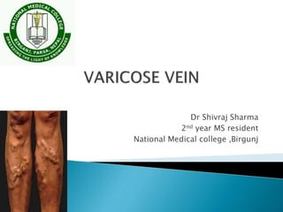
3. • Varicose veins are defined as dilated, elongated, tortuous
and palpable superficial veins(>3mm in diameter
measured in upright position with demonstrable reflux) as
a result of venous hypertension.
• It usually occurs due to permanent loss of valvular
mechanism and resultant venous hypertension.
4. Consists of:
• Deep system of veins which
lies below the deep fascia.
• Superficial system of veins which lies
outside the deep fascia (carry 10%
blood)
• Perforating veins which pass through
the deep fascia joining the superficial
to the deep system of veins.
5. 1: Superficial veins:
Long saphenous vein
Short saphenous vein
2: Deep veins :
Anterior & Posterior Tibial veins
Peroneal vein
Popliteal vein
Femoral vein
3: Perforator veins
6. • Valves present in superficial veins.
• Prevent flow of blood from proximal to distal and from deep to
superficial
• Absent from above groin level
• Valves can resist pressure up to 300 mm of Hg.
7. Largest and longest superficial
vein of the limb.
Begins on the dorsum of foot
from medial end of dorsal venous
arch.
Run 1 to 1.5 inch anterior to the
medial malleolus ,along the
medial side of the leg , and
behind knee .
8. At the ankle the position of the LSV
is constant , lying in the groove b/w
the anterior border of the medial
malleolus and tendon of tibialis
anterior.
In the thigh it inclines forwards to
reach the saphenous opening where
it pierces the cribriform fascia and
opens into the femoral vein 3-4 cm
below and lateral to the pubic
tubercle
9. In the lower 2/3 of leg and in
upper 2/3 of the thigh vein lie on
deep fascia .
Where the vein crosses the knee
joint it become more superficial
and often subcuticular .
10. Just below knee LSV receive posterior arch vein (Leonardo's
vein) which collect the blood from post-medial aspect of calf .
Anterior veins of leg(stocking vein) ascend across the shin
and join either LSV or posterior arch vein .
There is a free anastomosis b/w tributaries of short
saphenous vein and venous arch connecting medial ankle
perforating vein and this medial ankle perforating veins are
connected with LSV in lower third of leg .
11. In the thigh before entering in the saphenous opening it recieves
1. Anterolateral vein
2. Posteromedial vein of thigh
3. Superficial external pudendal vein
4. Superficial epigastric vein
5. Superficial circumflex iliac vein
6. Deep External Pudendal Vein
In the lower third of thigh long saphenous vein connect with
femoral vein in hunter’s canal by long perforating vein
( hunterian perforator)
12.
13. It begins by the fusion of
number of small veins below
and behind the lateral malleolus
. Here vein runs with the large
sural nerve up to lower third of
leg.
SSV runs upward up to the
middle of the popliteal space,
where it passes deep to fascia
to enter into popliteal vein .
14. These are communicating veins b/w superficial and deep
veins .
Two type:
1 Indirect veins
2 Direct veins
15. 1. Indirect perforating veins:
These consist of small
superficial veins which
penetrate the deep fascia to
connect with vessel in muscle
and in turn end in Deep vein.
16. Direct perforating veins :
These directly connect
superficial veins with deep
veins
17. Six Perforators joining the superficial
to deep venous system are located at
constant positions which are:
• 2, 4 and 6 inches above the
medial malleolus (Cockett’s
perforator)
• Just below the Tibial
tubercle(Boyd’s)
• In the adductor(Hunter’s) canal
of the thigh(Dodd’s perforator)
• Level of Mid-thigh
• Around 200 perforators are
described most of them
unnamed
18. • Negative pressure in thorax during inspiration to -6
mm.
• Calf muscle pump: Normal venous pressure in relaxed state 20mm of
Hg.Rises to 80-100 mm of Hg during muscle contraction.
• Vis a tergo : arterial pressure transmitted to venous side through
capillary bed
• Competent valves
• Venae commitants: lie by the side of artery, helped
by arterial pulsation to propel blood.
19. Primary
• Long hours of standing,
which increase the
hydrostatic pressure of
gravity
• Family history
• Pregnancy
• Ageing
Secondary
• Deep vein thrombosis
• Arterio venous
malformation- Parkes
Weber syndrome
• Hemangiomatous
malformation- Klippel
Trenaunay syndrome
• Pelvic mass
• Retro peritoneal fibrosis
23. • Bleeding
• Thrombophlebitis
• Venous Hypertension leading to
venous ulcer
• Calcification
• Talipes Equinovarus deformity of
foot
• Eczematoid dermatitis and
pigmentation
• Periostitis of subcutaneous
surface of tibia
• Carcinoma in long standing
venous ulcer-Marjolins ulcer
24.
25. Age : Any
Sex : F:M 10:1
Occupation : Jobs demanding prolong standing
person doing muscular work
Leg heaviness, exercise intolerance, pain in lower limb.
However, bursting pain means DVT
Ankle swelling usually at the end of day
Tortous dilated visible vein
Pruritus, restless legs, and paresthesias
Skin changes : pigmentation, ulcer
Dermal flare/thread veins
Reticular vein
26. Aims:
• Finding the system involved
• Extent of involvement
• Skin changes/ulcer around malleolus
• Trendelenberg test for patency of Sapheno-
femoral junction
• Perthe’s test for patency of deep veins
27. C
C
Clinical signs (grade0-6), supplemented by
(A) for asymptomatic and (S) for
symptomatic presentation
E
Etiologic Classification (Congenital,
Primary, Secondary)
A
Anatomic Distribution (Superficial, Deep,
or Perforator, alone or in combination)
P
Pathophysiologic Dysfunction (Reflux or
Obstruction, alone or in combination)
28. Staging
CEAP classification from American Venous Forum, last revised
2004
Used to standardize recording of venous disease
Clinical
C0 - No visible or palpable signs of venous disease
C1 - Telangiectases or reticular veins
C2 - Varicose veins
C3 - Edema
C4a - Pigmentation or eczema
C4b - Lipodermatosclerosis or atrophie blanche
C5 - Healed venous ulcer
C6 - Active venous ulcer
S – Symptomatic, includes: ache, pain, tightness, skin irritation,
heaviness, and muscle cramps
A – Asymptomatic
29. Etiologic classification
Ec - Congenital
Ep - Primary
Es - Secondary (post-thrombotic)
En - No venous cause identified
Anatomic classification
As - Superficial veins
Ap - Perforator veins
Ad - Deep veins
An - No venous location identified
Pathophysiologic classification
Pr - Reflux
Po - Obstruction
Pr,o – Reflux and obstruction
Pn - No venous pathophysiology identifiable
30. • Ambulatory venous pressure studies
• Venous Doppler study
• Air plethysmography
31. Ambulatory venous pressure more than 90 mm of Hg is associated with
venous ulceration.
Also regarded as GOLD STANDARD for diagnosis of chronic venous
insufficiency
Ulcer never occurs at AVP lesser than 30 mmof Hg.
Invasive procedure hence ideally not suitable for screening
32. • Indicated for diagnosis of calf muscle dysfunction
• Measures changes in leg volume in response to exercise and posture.
• Leg placed in 40 cm tubular Vinyl air chamber Leg volume measured in
supine, elevated , standing on opposite leg and after 10 tip toe jumps.
• Venous volume(VV), venous filling time90(VFT 90) and venous filling
index(VFI) and ejection fraction (EF)calculted
33.
34.
35. • If venous volume > 350 ml (normal 100-150 ml) Indicates
chronic venous insufficiency(CVI)
• If VFI is 7 ml per second(normal < 2ml per second)
indicates CVI
• If ejection fraction venous blood of calf muscle is less than
60 percent after one tip toe indicates Calf Muscle
dysfunction
• If remaining venous fraction(RVF) after 10 tip toes is more
than 40 percent indicates calf muscle dysfunction
• If RVF more than 40 percent and Venous filling index(VFI) > 2
ml per second then it indicates reflux
36. • To find patency of deep veins.
• To define the site of incompetent perforators & to
mark them preoperatively.
• To find out the competence of Saphenofemoral
junction & Sapheno popliteal junction.
• If Sapheno-popliteal junction is incompetent it should
be marked preoperatively because of its highly variable
& inconstant position.
• Ankle brachial index should be measured to rule out any
concomitant arterial disease.
37. • Avoiding prolonged standing, weight loss ,excercise
• Crepe bandaging and elastic stockings from toe to thigh, which
causes decreased edema, venous volume and reflux and
increases venous return.
• Compression stocking of the pressure of 18-24 mm is preferred
for varicose veins.
• Limb elevation above the level of heart while lying down
38. • Refusal for surgery
• Capillary veins, Venous
Stars (C1)
• Pregnant patients
• Waiting for surgery
• Early cases
Indications Contraindications
• Arterial Insufficiency
39. MICRONIZED PURIFIED FLAVONOID FRACTION: (MPFF)
DAFLON 500MG oral phlebotropic drug consisting of 90 %
micronized diosmin and 10% flavonoids expressed as
hesperidin.
Shown to improve venous tone and lymphatic drainage and
reduce capillary hyperpermeability by protecting the
microcirculation from inflammatory process.
CALCIUM DOBESILATE
PENTOXIFYLLINE : inhibits platelet aggregation hence reduce
blood viscosity and improves microcirculation
ASPIRIN
40. • Under Ultrasound guidance.
• Polidocanol is used
• Polidocanol converted in foam by mixing air using
three way tap.
• Spread of foam monitored under USG guidance as it
spreads.
• Apex of saphenous opening compressed by probe to prevent
foam entering deep veins.
• Leg also elevated
41.
42. Indications Contraindications
• Varicosity confined below
knee and caused by
incompetent perforators
• Recurrent/ residual
varicosities post-surgery
• Large Venous telangiectasia
• Dilated branch veins around
the knee following early long
saphenous incompetence
• Refusal for surgery
• Deep Venous thrombosis
• Sapheno Femoral
Incompetence
• Veins in lower 1/3rd ofleg
• Veins on the foot
• Veins in elderly
• Veins in fat legs
• Immobile patient
• Post thrombotic syndrome
• Dirty ulcer or extensive
eczema
43. • Complications:
• Extravenous Injection
• Deep vein thrombosis
• Hypersensitivity
• Skin pigmentation
• Gangrene of distal limb
44. • 5% monoethanolamine
with 2% benzyl alcohol
• 3% sodium
tetradecylsulphate in 2%
benzyl alcohol
• 25% glycerine with 2%
phenol
45. Types of surgeries done:
• Flush ligation of Sapheno femoral junction
with ligation of all tributaries ending at SFJ.
• Stripping of long saphenous upto the knee
joint.
• Flush Ligation of Short Saphenous vein.
• Subfascial ligation of perforators
46. • Curved or Hockey stick incision.
• Alternatively a 7-8 cm long Oblique incision .
• Femoral Vein is exposed 1 cm above and below the
Sapheno femoral junction.
• The all tributaries joining the termination of saphenous
vein are defined and ligated
• The end of the long saphenous vein is flush ligatedat
Saphenofemoral junction with silk and a second
ligature is transfixed to avoid haemorrhage.
• Femoral vein is inspected above and below the
junction and long saphenous divided.
47.
48. • An Oliers stripper is passed from the
groin Incision into the long saphenous
vein.
• A vertical incision is made just below
knee and vein exposed
• The stripper is extruded from the vein
and the acorn firmly tied in the vein.
• The stripper is firmly withdrawn with
the vein telescoped over it.
• The track is compressed with a large
sterile pad for 3 to 5 minutes.
49.
50. • Haemorrhage from torn varix
• Division or injury to the common Femoral Vein
• Sural Nerve or Saphenous nerve injury
• Postoperative Complications:
• Haematoma and bruising
• Wound infection
• Neuritis
• Lymphoedema
• Induration of stripper track
• Lymphatoma
• Deep Venous Thrombosis
51. • Maintain firm pressure over the limb
• Regular movement of the operated limb
• Limb elevation above heart level to reduce
venous pressure
• Removal of primary dressing after 7 to 10 days
52. Indications Contraindications
Chronic Venous
Insufficiency (C4-6)
Secondary varicose
veins
Arterial Insufficiency
Deep Vein Thrombosis
Subfascial Endoscopic Perforator Surgery is a
minimally invasive procedure where in
Incompetent perforators are ligated below the
deep fascia by creating space with CO2.
53. Insertion Of Ports for SEPS
A single 10 mm port for camera is inserted below the deep fascia at the
medial end of upper part of tibia. Another 5mm port inserted at junction
of upper 1/3rd and lower 2/3rd of thecalf.
54. • The intima of smaller veins can be destroyed by heat
generation and denaturation of collagen using a probe
consisting of a bipolar heat generator.
• Performed under ultrasound guidance and positionof
the probe is confirmed near the Saphenofemoral
junction.
• Probe is heated to 85 degrees and gradually retracted
down at a constant rate of 2-3cm/minute.
• must be avoided in presence of dilated veins, veins
with aneurysms and thrombosed veins.
55. • Employs diode laser for the destruction of
endothelial lining of the target vein.
• The ultrasound guides the location of probe,
which is placed 2 cm distal to the
Saphenofemoral junction.
• The probe is gradually withdrawn and ablates the
lumen as it regresses down the vein by boiling
the blood present within the lumen.
• Veins of all sizes can be treated with this
procedure.