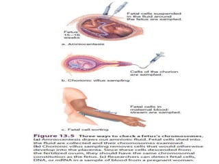
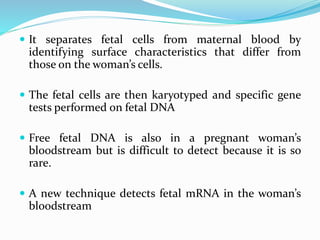
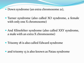
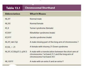
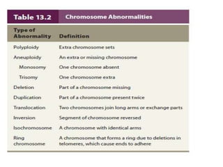
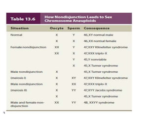
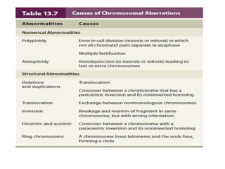
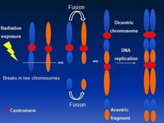
Portrait of a Chromosome
 Mutations range from single-base changes to entire
extra sets of chromosomes
 A mutation is considered a chromosomal aberration if
 it is large enough to see with a light microscope using
stains and/or fluorescent probes to highlight missing,
extra, or moved genetic material.
 Cytogenetics is the subdiscipline within genetics that
links chromosome variations to specific traits,
including illnesses
Required Parts: Telomeres and Centromeres
 A chromosome consists primarily of DNA and proteins
with a small amount of RNA
 Heterochromatin consists mostly of highly repetitive
DNA sequences, whereas euchromatin has more
protein-encoding sequences
 A chromosome must include structures that enable it
to replicate and remain intact
 Everything else is essentially informational cargo
(protein-encoding genes and their controls).
 The essential parts of a chromosome are:
 telomeres
 origin of replication sites, where replication forks begin to
form
 the centromere
 The centromere is the largest constriction of a
chromosome and it is where spindle fibers attach
when the cell divides
 Centromeres are replicated toward the end of S phase.
 A protein that may control their duplication is called
centromere protein A, or CENP-A
Karyotypes Chart Chromosomes
 A karyotype displays chromosomes in pairs by size and
by physical landmarks that appear during mitotic
metaphase, when DNA coils tightly
 The 24 human chromosome types are numbered from
largest to smallest—1 to 22. The other two
chromosomes are the X and the Y
 A chromosome is metacentric if the centromere
divides it into two arms of approximately equal length.
 It is submetacentric if the centromere establishes one
long arm and one short arm
 And acrocentric if it pinches off only a small amount
of material toward one end
 Telocentric chromosomes that have only one arm, but
humans do not.
 The long arm of a chromosome is designated q, and
the short arm p ( p stands for “petite”).
Visualizing Chromosomes
 Extra or missing chromosomes are detected by counting a
number other than 46.
 Identifying chromosome rearrangements, such as an
inverted sequence or an exchange of parts between two
chromosomes, requires a way to distinguish among the
chromosomes.
 A combination of stains and DNA probes applied to
chromosomes allows this.
 A DNA probe is a labeled piece of DNA that binds to its
complementary base sequence on a particular chromosome
Obtaining Cells for Chromosome Study
 Any cell other than a mature red blood cell (which lacks a
nucleus) can be used to examine chromosomes
 Amniocentesis and chorionic villus sampling have been
available for many years.
 They sample fetal cells from the amniotic fluid and chorionic
villi, respectively, and detect large-scale chromosomal
abnormalities.
 A newer technique called chromosome microarray analysis
can be paired with the older techniques to detect copy number
variants, which include extremely small sections of missing or
extra DNA.
 Chromosome microarray analysis probes and displays specific
sequences, detecting many disorders that other techniques miss
Amniocentesis
 A small sample of fetal cells and fluids is removed from the
uterus with a needle passed through the woman’s
abdominal wall
 The cellsare cultured for a week to 10 days, and typically 20
cells are karyotyped.
 The sampled amniotic fluid may also be examined for
deficient, excess, or abnormal biochemicals that could
indicate an inborn error of metabolism.
 Amniocentesis can detect approximately 1,000 of the more
than 5,000 known chromosomal and biochemical
problems
 The most common chromosomal abnormality
detected is one extra chromosome, called a trisomy.
 Amniocentesis is usually performed between 14 and 16
weeks gestation, when the fetus isn’t yet very large but
amniotic fluid is plentiful
 The procedure is also warranted if a couple has had
several spontaneous abortions or children with birth
defects or a known chromosome abnormality,
irrespective of maternal age
Chorionic Villus Sampling
 During the 10th through 12th week of pregnancy, chorionic
villus sampling (CVS) obtains cells from the chorionic villi
 Which are finger-like structures that develop into the
placenta
 A karyotype is prepared directly from the collected cells,
rather than first culturing them, as in amniocentesis
 Because chorionic villus cells descend from the fertilized
ovum, their chromosomes should be identical to those of
the embryo and fetus.
 Occasionally, a chromosomal aberration occurs only in a
cell of the embryo, or only in a chorionic villus cell
 This results in chromosomal mosaicism—the karyotype of
a villus cell differs from that of an embryo cell
 CVS is slightly less accurate than amniocentesis, and in
about 1 in 1,000 to 3,000 procedures
 It halts development of the feet and/or hands
 A condition termed transverse limb defects.
 Also, CVS does not sample amniotic fluid, so tests for
inborn errors of metabolism are not possible
Fetal Cells, DNA, and RNA
 Detecting fetal cells or nucleic acids in the pregnant woman’s
bloodstream is safer than amniocentesis and CVS
 The technique traces its roots to 1957, when a pregnant woman
died when cells from a very early embryo lodged in a major blood
vessel in her lung, blocking blood flow
 Researchers found that fetal cells enter the maternal circulation
in up to 70 percent of pregnancies.
 Cells from female embryos, however, cannot easily be
distinguished from the cells of the pregnant woman on the basis
of sex chromosome analysis.
 But fetal cells from either sex can be distinguished from
maternal cells using a device called a fluorescence-activated cell
sorter.
 It separates fetal cells from maternal blood by
identifying surface characteristics that differ from
those on the woman’s cells.
 The fetal cells are then karyotyped and specific gene
tests performed on fetal DNA
 Free fetal DNA is also in a pregnant woman’s
bloodstream but is difficult to detect because it is so
rare.
 A new technique detects fetal mRNA in the woman’s
bloodstream
 Down syndrome (an extra chromosome 21),
 Turner syndrome (also called XO syndrome, a female
with only one X chromosome)
 And Klinefelter syndrome (also called XXY syndrome,
a male with an extra X chromosome)
 Trisomy 18 is also called Edward syndrome
 and trisomy 13 is also known as Patau syndrome
Abnormal Chromosome Number
 A human karyotype is abnormal if the number of
chromosomes in a somatic cell is not 46
 Or if individual chromosomes have extra, missing, or
rearranged genetic material
 Abnormal chromosomes account for at least 50
percent of spontaneous abortions
Polyploidy
 The most drastic upset in chromosome number is an entire extra
set. A cell with extra sets of chromosomes is polyploid.
 An individual whose cells have three copies of each chromosome
is a triploid (designated 3N, for three sets of chromosomes).
 Two-thirds of all triploids result from fertilization of an oocyte
by two sperm
 The other cases arise from formation of a diploid gamete, such as
when a normal haploid sperm fertilizes a diploid oocyte
 However, certain human cells may be polyploid. The liver, for
example, has some tetraploid (4N) and even octaploid (8N) cells
Aneuploidy
 Cells missing a single chromosome or having an extra one are
aneuploid, which means “not good set.”
 A normal chromosome number is euploid, which means “good
set.”
 The meiotic error that causes aneuploidy is called
nondisjunction.
 Recall that in normal meiosis, homologs separate and each of
the resulting gametes receives only one member of each
chromosome pair.
 In nondisjunction, a chromosome pair fails to separate at
anaphase of either the first or second meiotic division.
 This produces a sperm or oocyte that has two copies of a
particular chromosome, or none, rather than the normal one
copy
 Different trisomies tend to be caused by nondisjunction in the
male or female, at meiosis I or II
 Aneuploidy and polyploidy also arise during mitosis, producing
groups of somatic cells with the extra or missing chromosome
 An individual with two chromosomally distinct cell populations
is a mosaic
 Another meiotic error that leads to unbalanced genetic material
is the formation of an isochromosome, which is a chromosome
that has identical arms
 Inheriting two chromosomes or chromosome segments from one
parent is called uniparental disomy (UPD) (“two bodies from
one parent”).
Cytogenetic analysis

Cytogenetic analysis

  • 2.
    Portrait of aChromosome  Mutations range from single-base changes to entire extra sets of chromosomes  A mutation is considered a chromosomal aberration if  it is large enough to see with a light microscope using stains and/or fluorescent probes to highlight missing, extra, or moved genetic material.  Cytogenetics is the subdiscipline within genetics that links chromosome variations to specific traits, including illnesses
  • 3.
    Required Parts: Telomeresand Centromeres  A chromosome consists primarily of DNA and proteins with a small amount of RNA  Heterochromatin consists mostly of highly repetitive DNA sequences, whereas euchromatin has more protein-encoding sequences  A chromosome must include structures that enable it to replicate and remain intact  Everything else is essentially informational cargo (protein-encoding genes and their controls).
  • 4.
     The essentialparts of a chromosome are:  telomeres  origin of replication sites, where replication forks begin to form  the centromere  The centromere is the largest constriction of a chromosome and it is where spindle fibers attach when the cell divides  Centromeres are replicated toward the end of S phase.  A protein that may control their duplication is called centromere protein A, or CENP-A
  • 6.
    Karyotypes Chart Chromosomes A karyotype displays chromosomes in pairs by size and by physical landmarks that appear during mitotic metaphase, when DNA coils tightly  The 24 human chromosome types are numbered from largest to smallest—1 to 22. The other two chromosomes are the X and the Y
  • 7.
     A chromosomeis metacentric if the centromere divides it into two arms of approximately equal length.  It is submetacentric if the centromere establishes one long arm and one short arm  And acrocentric if it pinches off only a small amount of material toward one end  Telocentric chromosomes that have only one arm, but humans do not.  The long arm of a chromosome is designated q, and the short arm p ( p stands for “petite”).
  • 9.
    Visualizing Chromosomes  Extraor missing chromosomes are detected by counting a number other than 46.  Identifying chromosome rearrangements, such as an inverted sequence or an exchange of parts between two chromosomes, requires a way to distinguish among the chromosomes.  A combination of stains and DNA probes applied to chromosomes allows this.  A DNA probe is a labeled piece of DNA that binds to its complementary base sequence on a particular chromosome
  • 10.
    Obtaining Cells forChromosome Study  Any cell other than a mature red blood cell (which lacks a nucleus) can be used to examine chromosomes  Amniocentesis and chorionic villus sampling have been available for many years.  They sample fetal cells from the amniotic fluid and chorionic villi, respectively, and detect large-scale chromosomal abnormalities.  A newer technique called chromosome microarray analysis can be paired with the older techniques to detect copy number variants, which include extremely small sections of missing or extra DNA.  Chromosome microarray analysis probes and displays specific sequences, detecting many disorders that other techniques miss
  • 11.
    Amniocentesis  A smallsample of fetal cells and fluids is removed from the uterus with a needle passed through the woman’s abdominal wall  The cellsare cultured for a week to 10 days, and typically 20 cells are karyotyped.  The sampled amniotic fluid may also be examined for deficient, excess, or abnormal biochemicals that could indicate an inborn error of metabolism.  Amniocentesis can detect approximately 1,000 of the more than 5,000 known chromosomal and biochemical problems
  • 12.
     The mostcommon chromosomal abnormality detected is one extra chromosome, called a trisomy.  Amniocentesis is usually performed between 14 and 16 weeks gestation, when the fetus isn’t yet very large but amniotic fluid is plentiful  The procedure is also warranted if a couple has had several spontaneous abortions or children with birth defects or a known chromosome abnormality, irrespective of maternal age
  • 14.
    Chorionic Villus Sampling During the 10th through 12th week of pregnancy, chorionic villus sampling (CVS) obtains cells from the chorionic villi  Which are finger-like structures that develop into the placenta  A karyotype is prepared directly from the collected cells, rather than first culturing them, as in amniocentesis  Because chorionic villus cells descend from the fertilized ovum, their chromosomes should be identical to those of the embryo and fetus.
  • 15.
     Occasionally, achromosomal aberration occurs only in a cell of the embryo, or only in a chorionic villus cell  This results in chromosomal mosaicism—the karyotype of a villus cell differs from that of an embryo cell  CVS is slightly less accurate than amniocentesis, and in about 1 in 1,000 to 3,000 procedures  It halts development of the feet and/or hands  A condition termed transverse limb defects.  Also, CVS does not sample amniotic fluid, so tests for inborn errors of metabolism are not possible
  • 16.
    Fetal Cells, DNA,and RNA  Detecting fetal cells or nucleic acids in the pregnant woman’s bloodstream is safer than amniocentesis and CVS  The technique traces its roots to 1957, when a pregnant woman died when cells from a very early embryo lodged in a major blood vessel in her lung, blocking blood flow  Researchers found that fetal cells enter the maternal circulation in up to 70 percent of pregnancies.  Cells from female embryos, however, cannot easily be distinguished from the cells of the pregnant woman on the basis of sex chromosome analysis.  But fetal cells from either sex can be distinguished from maternal cells using a device called a fluorescence-activated cell sorter.
  • 17.
     It separatesfetal cells from maternal blood by identifying surface characteristics that differ from those on the woman’s cells.  The fetal cells are then karyotyped and specific gene tests performed on fetal DNA  Free fetal DNA is also in a pregnant woman’s bloodstream but is difficult to detect because it is so rare.  A new technique detects fetal mRNA in the woman’s bloodstream
  • 18.
     Down syndrome(an extra chromosome 21),  Turner syndrome (also called XO syndrome, a female with only one X chromosome)  And Klinefelter syndrome (also called XXY syndrome, a male with an extra X chromosome)  Trisomy 18 is also called Edward syndrome  and trisomy 13 is also known as Patau syndrome
  • 22.
    Abnormal Chromosome Number A human karyotype is abnormal if the number of chromosomes in a somatic cell is not 46  Or if individual chromosomes have extra, missing, or rearranged genetic material  Abnormal chromosomes account for at least 50 percent of spontaneous abortions
  • 23.
    Polyploidy  The mostdrastic upset in chromosome number is an entire extra set. A cell with extra sets of chromosomes is polyploid.  An individual whose cells have three copies of each chromosome is a triploid (designated 3N, for three sets of chromosomes).  Two-thirds of all triploids result from fertilization of an oocyte by two sperm  The other cases arise from formation of a diploid gamete, such as when a normal haploid sperm fertilizes a diploid oocyte  However, certain human cells may be polyploid. The liver, for example, has some tetraploid (4N) and even octaploid (8N) cells
  • 25.
    Aneuploidy  Cells missinga single chromosome or having an extra one are aneuploid, which means “not good set.”  A normal chromosome number is euploid, which means “good set.”  The meiotic error that causes aneuploidy is called nondisjunction.  Recall that in normal meiosis, homologs separate and each of the resulting gametes receives only one member of each chromosome pair.  In nondisjunction, a chromosome pair fails to separate at anaphase of either the first or second meiotic division.  This produces a sperm or oocyte that has two copies of a particular chromosome, or none, rather than the normal one copy
  • 26.
     Different trisomiestend to be caused by nondisjunction in the male or female, at meiosis I or II  Aneuploidy and polyploidy also arise during mitosis, producing groups of somatic cells with the extra or missing chromosome  An individual with two chromosomally distinct cell populations is a mosaic  Another meiotic error that leads to unbalanced genetic material is the formation of an isochromosome, which is a chromosome that has identical arms  Inheriting two chromosomes or chromosome segments from one parent is called uniparental disomy (UPD) (“two bodies from one parent”).