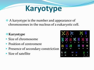
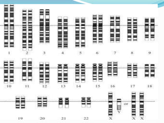
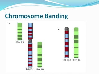
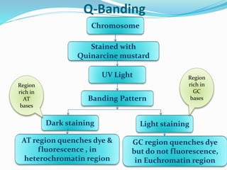
A karyotype is the number and appearance of chromosomes in a cell and can provide information about an organism's species. Key features used to characterize karyotypes include chromosome size, centromere position, and banding patterns. Karyotypes can be symmetric or asymmetric and are often represented visually using idiograms or karyograms. Analysis techniques like G-banding stain chromosomes to reveal identifying patterns. Comparing karyotypes across species provides insights into evolutionary mechanisms like centric fusion and fission that alter chromosome counts. In primates, chromosomal changes like the fusion that formed human chromosome 2 are important in lineage evolution.
Karyotype refers to chromosome appearance and number in eukaryotic cells, represented in idiograms with specific features.
Karyotypes can be symmetric or asymmetric, indicating differences in chromosome size and features in species.
Steps for analyzing karyotypes include cell collection, division induction, and staining. Techniques used are classical, spectral karyotype, and idiogram formation.
Classical analysis uses Giemsa dye for banding patterns to identify chromosomes. Spectral karyotype helps visualize human chromosomes; karyograms determine diploid numbers.
Human karyotype shows 22 autosome pairs categorized by morphology. Karyotyping helps in identifying chromosomal abnormalities, prenatal diagnosis, and evolutionary studies.
Nine banding techniques exist (G, R, C, N, T, and Q-Banding) for identifying chromosomal features, aiding in abnormality detection.
Karyotype evolution studies reveal chromosomal differences in species, particularly in primates, emphasizing fusion and fission mechanisms, illustrating genetic processes.
Karyotype
A karyotypeis the number and appearance of
chromosomes in the nucleus of a eukaryotic cell.
Karyotype
Size of chromosome
Position of centromere
Presence of secondary constriction
Size of satellite
3.
Each group ofplants or animals of species has a
set of chromosome with characteristic feature as
size , shape, position of centromere etc.
The karyotype is represented by a diagram called
Idiogram.
When the haploid set of chromosomes of an
organism are ordered in a series of decreasing size,
it is said to be an idiogram
Types of Karyotype
AsymmetricKaryotype Symmetric Karyotype
• Show larger difference
between smaller and larger
chromosome in a set.
Have more acrocentric
chromosomes.
Have relatively advanced
feature
Example- Ginkgo biloba,
flowering plants
Show lesser difference
between smaller and larger
chromosome in a set.
Have more metacentric
chromosomes.
Have no relatively advanced
feature
Example- Pinus
7.
Karyotype Analysis
To determinethe karyotype of an organism there are
following steps
Collection of cell from an organism
Induce the cell to divide
stop cell division at metaphase
Stain the chromosome
Study and analyze
8.
The studyof karyotype is called Karyology.
Three types techniques are used in karyotype
1. Classical karyotype analysis
2. Spectral karyotype
3. Formation of idiogram or karyogram
9.
Classical Karyotype analysis
Adye Giemsa is used to stain banding pattern on
chromosomes. Also called GC- bands
Each chromosome has a specific banding pattern so
bands help in identification of chromosomes.
The chromosomes of one pair must have same pattern.
10.
Spectral karyotype
This techniqueis used to visualize all the 23 pairs of
human chromosomes in different colours.
This technique is widely used to identify structural
chromosomal aberrations in cancer and other diseases.
11.
Formation of Karyogram
Thistechnique is used to determine diploid number of
chromosomes of an organism.
In this cells are locked in metaphase with colchicine
then stained and photographed.
They are arranged in a karyotype and karyogram.
In human female out of 2 X chromosome one remain
inactive and seem as bar body under microscope
12.
Analysis of HumanKaryotype
22 pairs of autosomes are arranged in 7 groups (A-G)
with different morphology.
14.
Advantages of Karyotyping
Revealsstructural features of each chromosomes.
Helps in studying chromosome banding pattern.
Helps in the identification of chromosomal
aberrations.
Diagnosis of prenatal genetic defects.
Aids in studying evolutionary changes
Classification of BandingTechniques
Based on
GC and AT rich regions
Constitutive heterochromatin region
The bands can be detected after treatment of
chromosomes with restriction enzyme(RI) so
also called- RE bands.
In chromosome banding we stain the
chromosome with different stains and study the
banding pattern.
The heterochromatin regionwhich are rich in A-T base pair
stain more darkly in G-Banding while Euchromatin which is
rich in G-C bands incorporate less stain.
The bands are same in homologous chromosome so
identification become easy .
Use to identify different chromosomal abnormalities as
chromosome number, deletion, inversion etc.
R-Banding is reverseof G-Banding that is light banded
region of G banded chromosomes become lightly stained
or vice versa.
R bands are produced in G-C chromosomes.
R- banding is not observed in plant chromosomes.
A-T regions are selectively denatured by heat leaving
G-C region intact
Helpful in analyzing the structure of chromosome ends
which are usually light stained with G band
C-Banding stains areasof heterochromatin which are
tightly packed and have repetitive DNA.
Useful in humans to stain centromere regions and other
regions containing constitutive heterochromatin as
secondary constriction and distal segment of Y
Chromosome long arm.
C-banding also studied in Vicia faba, Hordeum vulgare
etc.
N- Banding isused to find out location of NOR
(Nucleolar Organizer Region).
Banding involves staining with silver nitrate solution to
stain NOR rich in rRNA genes.
These bands are localized in the satellites of
chromosomes.
T-Banding involves stainingof Telomeric regions of
chromosomes.
T- Bands are smaller than R-Bands.
T-Bands are strictly observed at Telomeric site
28.
Q-Banding
Chromosome
Stained with
Quinarcine mustard
UVLight
Banding Pattern
Dark staining Light staining
AT region quenches dye &
fluorescence , in
heterochromatin region
GC region quenches dye
but do not fluorescence,
in Euchromatin region
Region
rich in
AT
bases
Region
rich in
GC
bases
29.
Q bands areespecially useful for distinguishing human Y
Chromosomes.
To identify the heterochromatin rich regions involving
satellites and centromere of specific chromosomes.
31.
Karyotype Evolution
Study ofKaryotypes of different species has revealed interesting
facts about both the plants and animal kingdom.
Reptiles and birds have large chromosomes
(Macrochromosomes) & small chromosomes
(Microchromosomes) that serve to differentiate them
cytogenetically.
Matthey distinguished between the basic chromosome number &
the number of chromosome arm, also called as Fundamental
number (FN). According to this concept, the metacentric
chromosome as two arms and the acrocentric chromosome and
telocentric chromosome has one arm. The number of arms in each
of different species can be compared .
32.
With regard toabsolute size of their chromosomes
mammals & birds constitute two independent groups. The
two orders have different DNA contents and different sex
determining mechanisms.
Two opposite changes in the number of chromosomes are
of particular importance in the evolution:-
In centric fusion, a process leads to decrease in
chromosome no. two acentric chromosomes join together
to produce a metacentric chromosome.
In dissociation/ fission, a process that leads to an increase
in chromosome no. a metacentric and a small
supernumerary metacentric chromosome become
translocated, so that acrocentric and submetacentric
chromosome are produced.
33.
Fusion and dissociation/fissionare the main mechanism by
which the chromosome no. can be decreased or increased
during evolution of the majority of animals and in some
group of plants.
Observations of chromosomal organization and of the
different Karyotypes in the individual the species, the
genes, and the major systematic groups indicates that a
chromosome mechanism are involved in evolution.
Evolution , however is a very complex and should be
considered different from different biochemical,
cytological, genetic, ecological and experimental aspects.
34.
Karyotype Evolution inPrimates
In recent years a great deal of work on evolutions has
focused on the possible cytogenetic relationship
between great apes and human species.
It was found that the primates have 48 chromosomes
and attempts were made to correlate the 24
chromosome pair with 23 pairs in humans. This
analysis was greatly facilitated by the use of banding
techniques that allow one to study the inner structure
of each chromosomes.
35.
These comparative studieshave been demonstrated:-
13 pairs of chromosomes in humans are identical with 13
pairs of chimpanzee.
Chromosome no.2 in humans has resulted from centric
fusion of two chromosomes present in hominoid apes.
The other chromosomes differ in occurrence of nine
pericentric inversion and two additions of chromatin
material.
The karyotype evolution in primates consists of the
modification of the morphology in the form of fusion,
fission, inversion,& reciprocal translocation.
36.
At present, pericentricinversion appears to be main
structural difference between individual chromosome
of great apes & humans.
It has been postulated that evolution of human
karyotype has occurred by series of pericentric
inversions which permitted the genetic isolation of
small breeding groups and selection of favorable gene
combination that give rise to homosapiens.