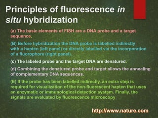
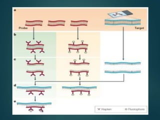
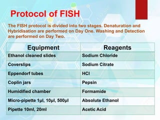
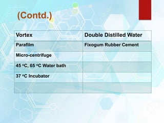
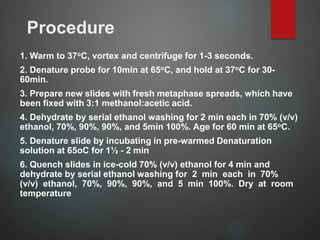
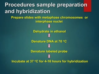
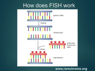
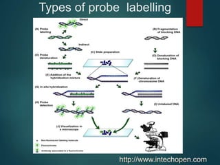
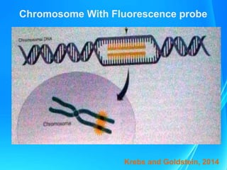
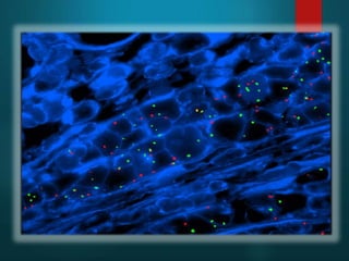
Department of Biotechnology,
Barkatullah University, Bhopal
Synopsi
s
Introduction
History and development
Procedure of FISH
Types of FISH
Requirement for FISH
Types of probes for FISH
Application of FISH
Advantages of FISH
Limitation of FISH
Recent research
Reference
Introduction
FISH is a method that can be used to detect small
deletions and duplications that are not visible using
microscope analysis. It can also be used to detect how
many chromosomes of a certain type are present in each
cell and to confirm rearrangements that are suspected
after microscope analysis
FISH gained widespread recognition as a physical
mapping technique to support large-scale mapping and
sequencing efforts related to the human genome project
www.abnova.com
Principles of fluorescence in
situ hybridization
(a) The basic elements of FISH are a DNA probe and a target
sequence.
(B) Before hybridization the DNA probe is labelled indirectly
with a hapten (left panel) or directly labelled via the incorporation
of a fluorophore (right panel).
(c) The labeled probe and the target DNA are denatured.
(d) Combining the denatured probe and target allows the annealing
of complementary DNA sequences.
(E) If the probe has been labelled indirectly, an extra step is
required for visualization of the non-fluorescent hapten that uses
an enzymatic or immunological detection system. Finally, the
signals are evaluated by fluorescence microscopy
History and development
(1969)- In situ hybridization technique was
developed by joseph G Gall and Mary lou Pardue
and John et al.(1969)
(1985)- The non isotopic in situ hybridization
using biotin labeled DNA probes was
first introduced in plant species
by Rayburn and Gill
(1991)-The first application of FISH to plant
cytogenetics was the work of Leitch et al.(1991)
Protocol of FISH
The FISH protocol is divided into two stages. Denaturation and
Hybridisation are performed on Day One. Washing and Detection
are performed on Day Two.
Equipment Reagents
Ethanol cleaned slides Sodium Chloride
Coverslips Sodium Citrate
Eppendorf tubes HCl
Coplin jars Pepsin
Humidified chamber Formamide
Micro-pipette 1μl, 10μl, 500μl Absolute Ethanol
Pipette 10ml, 20ml Acetic Acid
(Contd.)
Vortex Double Distilled Water
Parafilm Fixogum Rubber Cement
Micro-centrifuge
45 oC, 65 oC Water bath
37 oC Incubator
Procedure
1. Warm to 37oC, vortex and centrifuge for 1-3 seconds.
2. Denature probe for 10min at 65oC, and hold at 37oC for 30-
60min.
3. Prepare new slides with fresh metaphase spreads, which have
been fixed with 3:1 methanol:acetic acid.
4. Dehydrate by serial ethanol washing for 2 min each in 70% (v/v)
ethanol, 70%, 90%, 90%, and 5min 100%. Age for 60 min at 65oC.
5. Denature slide by incubating in pre-warmed Denaturation
solution at 65oC for 1½ - 2 min
6. Quench slides in ice-cold 70% (v/v) ethanol for 4 min and
dehydrate by serial ethanol washing for 2 min each in 70%
(v/v) ethanol, 70%, 90%, 90%, and 5 min 100%. Dry at room
temperature
Procedure(Contd,)
7. Apply probe (15μl*) onto the slide. Apply coverslip and remove
air bubbles by gently pushing on coverslip with a pencil. Seal
with rubber cement.
8. Place slide in an air tight, prewarmed humidified chamber and
incubate overnight in the dark at 37 oC.
Procedures sample preparation
and hybridization
Prepare slides with metaphase chromosomes or
interphase nuclei
Dehydrate in ethanol
Denature DNA at 70 oC
Denature labeled probe
Incubate at 37 oC for 4-16 hours for hybridization
FISH prep device
Volpi and Bridger , 2008)
www.abnova.com
Types of FISH
ACM-FISH- Alpha (centromere),classical and midi
satalite of fish
Cat-FISH- Cellular compartment analtsis of temporal
(cat) activity by FISH
COD-FISH- Chromosomal orientation and direction-
FISH
D-FISH- Double Fusioin-FISH
Cryo-FISH- Cryosections -FISH
LNA-FISH- Locked nucleic acids (LNAs)
Volpi and Bridger, 2008
Types of FISH (Contd.)
Q-FISH- Quantitative-FISH has been used mainly for measuring
the number of telomere repeats on a particular chromosome.
Fusion-Signal FISH- FS-FISH techniques was initially
devised for the identification of the Phildelphia translocation
Immuno-FISH- immune-FISH is a combination of two
Techniques.
M-FISH- Multiplex-FISH a protocol for 24-color Karyotyping
Multilocus or ML-FISH- This FISH assay was initially
desigened to screen for multiple microdeletion syndromes in
patients.
Volpi and Bridger, 2008
How does FISH work
Requirements for FISH
There are 3 main component for FISH
Sample
Fluorescent Probe
Fluorescence microscope
Types of probes for
FISH
Scientists use three different types of FISH probes, each of which has a
different application:
Locus specific probes bind to a particular region of a chromosome.
This type of probe is useful when scientists have isolated a small portion of a
gene and want to determine on which chromosome the gene is located, or how
many copies of a gene exist within a particular genome.
Alphoid or centromeric repeat probes are generated from
repetitive sequences found in the middle of each chromosome These probes
can also be used in combination with "locus specific probes" to determine
whether an individual is missing genetic material from a particular
chromosome.
Whole chromosome probes are actually collections of smaller
probes, each of which binds to a different sequence along the length of a
given chromosome
https://www.genome.gov/
TYPES OF FISH PROBES
…………………………..
http://www.carolguze.com
Types of probe labelling
http://www.intechopen.com
Chromosome With Fluorescence probe
Krebs and Goldstein, 2014
Selected FISH probes or cellular stains
FISH probes or
cellular stains
Contaminants Target
microorganism(s)
DAPI NA DNA of all
microorganisms (live
and dead)
Acridine orange NA DNA of all
microorganisms (live
and dead)
Ac627BR Naphthalene Naphthalene dioxygenase
(nahAc)
mRNA
RhLu s-Triazine
herbicides
Rhodococcus
wratislaviensis 16S
rRNA
KT1phe Trichloroethene Ralstonia eutropha KT1
16S rRNA
Fluorescence microscope
Light source:
High-pressure mercury vapor lamps, tungsten-halogen
lamps or xenon lamps.
Filters:
1. Exciting filter: To let a certain wave
length of light passes so that can
excite the given fluorochrome
carried on sample.
2. Barrier filter: To allow the visible
light passes so that the fluorescence can
be seen by eyes or the image can be
captured.
How are the data reported
Depending on the method, FISH results can be
presented in two different ways:
• If FISH is evaluated using a microscope and manual
counting of labeled cells, the results are presented as cells per
unit (liter of liquid or gram of solid) analyzed.
• If FISH is evaluated with advanced microscopy techniques
and digital image processing, the results are usually presented
on a relative volume or area basis, which can be converted by the
laboratory to cells per unit of liquid or solid
Action of FISH
Low-resolution FISH With Metaphase
chromosomes- Fish was originally used with metaphase
chromosomes. These chromosomes, prepared from nuclei that are
undergoing division, are highly condensed with each chromosome in a
set taking up a recognizable appearance, characterized by position of
its centromere and the banding pattern that emerges after the
chromosome preparation is stained.
High-resolution FISH with extended
chromosomes and DNA fibers - The resolution of
FISH is determined not by the hybridization technique itself but by the
nature of the chromosomal preparation being studied .if metaphase
chromosomes are too condensed for fine scale mapping then we must
use chromosomes
Applications
Detection of numerical and structural Chromosomal
abnormality.
Identification of marker chromosomes.
Monitoring the effect of therapy.
Detection of early relapse or minimal residual
diseases.
Detection gene deletion and gene amplification.
Field of investigation
FISH-based assays have been developed within different field of
investigation, including.
Clinical genetics
Toxicology
Microbial ecology
Evolutionary biology
Comparative genomics
Cellular genomics
Chromosomal biology
Volpi and Bridger, 2008
Advantages of fish
Rapid technique and large number of cells can be
scored in a short period.
Efficiency of Hybridization and deletion is high.
Sensitivity and specificity is high.
Cytogenetic data can be obtained from non-dividing or
terminally differentiated cells.
Cytogenetic data can be obtained from poor samples
that contain too few cells for routine cytogenetic
analysis.
Methode has been adapted for automated systems.
Limitation of fish
1. Restricted to those abnormalities that can be detected
with currently available probe.
2. Only one or a few abnormalities can be assessed
simultaneously.
3. Due to Failuer to detect signal FISH is higher sensitive
for trisomy but less sensitive foe detecting chromosome
loss or deletion.
4. Cytogenetic data can be obtained only for the target
chromosomes thus FISH is not a good screening tool for
cytogenetically heterogeneous disease.
5. Requires fluorescence Microscopy and an image
analysis system.
Recent Research
Clonal evolution as detected by interphase FISH is
associated with worse overall survival in a population-
based analysis of patients with chronic lymphocytic
leukemia in British Columbia, Canada
Huang S.J.,Bergin k., Smith A.C.,Gerrie A.S., Bruyere H., Dalal C.B.,
Sugika D.K.
Cancer Genetics 210, 1–8 (2017)
Department of Haematology at The Alfred, Melbourne,
Australia
References
Brown T.A.(2007); Mapping Genomes by physical
Techniques; Genomes; BIOS scientific
publishers Ltd.; 9 Newtec Place, Oxford OX4
IRE,UK; Ed 3rd; pp 89-93
Krebs J.E., Goldstein E.S., Kilpatrick S.T.(2014);
Methods in molecular biology and genetic
engineering; Lrwin’s Genomes 11; Jones and
barrlett publicat; B-1 4262/3, Ansari Road,
Daryaganj, New Delhi, India; Ed 1st; pp 53-54
References
Abbasi F.M., Khan M.T., Perveen F., Masood R., Inamullah,et.al
(2010); Historical perspective of ISH for the analysis Of genetics;
African Journal of Biotechnology Vol. 9(54),pp 9142-9147
Muller R. (2011).; Fluorescence in situ hybridization; Interstate
Technology and Regulatory Council; Us department of energy US
(609) 948-960
Shah p., Vedarethinam I., Kwasny D., Andresen L., Skov S., et.al
(2011); FISH prep: A novel Intergrated device for metaphase FISH
sample prepration; Licensee MDPI Basel, Switzerland 2, 116-128
Volpi V.V., Bridger J.M. (2008).; An overview of the FISH techniques;
school of health sciences and social care, Brunel University UX
bridge UK 45: 385-409
Contd…
https://www.genome.gov/10000206/fish-fact-sheet/
www.abnova.com
http://www.carolguze.com/text/442-4-chromosome_analysis.shtml
http://www.nature.com/scitable content/principles-of-fluorescence-in-
situ-hybridization35120
http://www.leica-microsystems.com/applications/life-
science/fluorescence
http://www.intechopen.com/books/plant-breeding-from-
laboratories-to-fields/genomic-in-situ-hybridization-in-
triticeae-a-methodological-approach

fish- Fluorescence in situ hybridization

  • 1.
  • 2.
    Synopsi s Introduction History and development Procedureof FISH Types of FISH Requirement for FISH Types of probes for FISH Application of FISH Advantages of FISH Limitation of FISH Recent research Reference
  • 3.
    Introduction FISH is amethod that can be used to detect small deletions and duplications that are not visible using microscope analysis. It can also be used to detect how many chromosomes of a certain type are present in each cell and to confirm rearrangements that are suspected after microscope analysis FISH gained widespread recognition as a physical mapping technique to support large-scale mapping and sequencing efforts related to the human genome project www.abnova.com
  • 4.
    Principles of fluorescencein situ hybridization (a) The basic elements of FISH are a DNA probe and a target sequence. (B) Before hybridization the DNA probe is labelled indirectly with a hapten (left panel) or directly labelled via the incorporation of a fluorophore (right panel). (c) The labeled probe and the target DNA are denatured. (d) Combining the denatured probe and target allows the annealing of complementary DNA sequences. (E) If the probe has been labelled indirectly, an extra step is required for visualization of the non-fluorescent hapten that uses an enzymatic or immunological detection system. Finally, the signals are evaluated by fluorescence microscopy
  • 6.
    History and development (1969)-In situ hybridization technique was developed by joseph G Gall and Mary lou Pardue and John et al.(1969) (1985)- The non isotopic in situ hybridization using biotin labeled DNA probes was first introduced in plant species by Rayburn and Gill (1991)-The first application of FISH to plant cytogenetics was the work of Leitch et al.(1991)
  • 7.
    Protocol of FISH TheFISH protocol is divided into two stages. Denaturation and Hybridisation are performed on Day One. Washing and Detection are performed on Day Two. Equipment Reagents Ethanol cleaned slides Sodium Chloride Coverslips Sodium Citrate Eppendorf tubes HCl Coplin jars Pepsin Humidified chamber Formamide Micro-pipette 1μl, 10μl, 500μl Absolute Ethanol Pipette 10ml, 20ml Acetic Acid
  • 8.
    (Contd.) Vortex Double DistilledWater Parafilm Fixogum Rubber Cement Micro-centrifuge 45 oC, 65 oC Water bath 37 oC Incubator
  • 9.
    Procedure 1. Warm to37oC, vortex and centrifuge for 1-3 seconds. 2. Denature probe for 10min at 65oC, and hold at 37oC for 30- 60min. 3. Prepare new slides with fresh metaphase spreads, which have been fixed with 3:1 methanol:acetic acid. 4. Dehydrate by serial ethanol washing for 2 min each in 70% (v/v) ethanol, 70%, 90%, 90%, and 5min 100%. Age for 60 min at 65oC. 5. Denature slide by incubating in pre-warmed Denaturation solution at 65oC for 1½ - 2 min 6. Quench slides in ice-cold 70% (v/v) ethanol for 4 min and dehydrate by serial ethanol washing for 2 min each in 70% (v/v) ethanol, 70%, 90%, 90%, and 5 min 100%. Dry at room temperature
  • 10.
    Procedure(Contd,) 7. Apply probe(15μl*) onto the slide. Apply coverslip and remove air bubbles by gently pushing on coverslip with a pencil. Seal with rubber cement. 8. Place slide in an air tight, prewarmed humidified chamber and incubate overnight in the dark at 37 oC.
  • 11.
    Procedures sample preparation andhybridization Prepare slides with metaphase chromosomes or interphase nuclei Dehydrate in ethanol Denature DNA at 70 oC Denature labeled probe Incubate at 37 oC for 4-16 hours for hybridization
  • 12.
    FISH prep device Volpiand Bridger , 2008)
  • 14.
  • 15.
    Types of FISH ACM-FISH-Alpha (centromere),classical and midi satalite of fish Cat-FISH- Cellular compartment analtsis of temporal (cat) activity by FISH COD-FISH- Chromosomal orientation and direction- FISH D-FISH- Double Fusioin-FISH Cryo-FISH- Cryosections -FISH LNA-FISH- Locked nucleic acids (LNAs) Volpi and Bridger, 2008
  • 16.
    Types of FISH(Contd.) Q-FISH- Quantitative-FISH has been used mainly for measuring the number of telomere repeats on a particular chromosome. Fusion-Signal FISH- FS-FISH techniques was initially devised for the identification of the Phildelphia translocation Immuno-FISH- immune-FISH is a combination of two Techniques. M-FISH- Multiplex-FISH a protocol for 24-color Karyotyping Multilocus or ML-FISH- This FISH assay was initially desigened to screen for multiple microdeletion syndromes in patients. Volpi and Bridger, 2008
  • 17.
  • 18.
    Requirements for FISH Thereare 3 main component for FISH Sample Fluorescent Probe Fluorescence microscope
  • 19.
    Types of probesfor FISH Scientists use three different types of FISH probes, each of which has a different application: Locus specific probes bind to a particular region of a chromosome. This type of probe is useful when scientists have isolated a small portion of a gene and want to determine on which chromosome the gene is located, or how many copies of a gene exist within a particular genome. Alphoid or centromeric repeat probes are generated from repetitive sequences found in the middle of each chromosome These probes can also be used in combination with "locus specific probes" to determine whether an individual is missing genetic material from a particular chromosome. Whole chromosome probes are actually collections of smaller probes, each of which binds to a different sequence along the length of a given chromosome https://www.genome.gov/
  • 20.
    TYPES OF FISHPROBES ………………………….. http://www.carolguze.com
  • 21.
    Types of probelabelling http://www.intechopen.com
  • 22.
    Chromosome With Fluorescenceprobe Krebs and Goldstein, 2014
  • 24.
    Selected FISH probesor cellular stains FISH probes or cellular stains Contaminants Target microorganism(s) DAPI NA DNA of all microorganisms (live and dead) Acridine orange NA DNA of all microorganisms (live and dead) Ac627BR Naphthalene Naphthalene dioxygenase (nahAc) mRNA RhLu s-Triazine herbicides Rhodococcus wratislaviensis 16S rRNA KT1phe Trichloroethene Ralstonia eutropha KT1 16S rRNA
  • 25.
    Fluorescence microscope Light source: High-pressuremercury vapor lamps, tungsten-halogen lamps or xenon lamps. Filters: 1. Exciting filter: To let a certain wave length of light passes so that can excite the given fluorochrome carried on sample. 2. Barrier filter: To allow the visible light passes so that the fluorescence can be seen by eyes or the image can be captured.
  • 26.
    How are thedata reported Depending on the method, FISH results can be presented in two different ways: • If FISH is evaluated using a microscope and manual counting of labeled cells, the results are presented as cells per unit (liter of liquid or gram of solid) analyzed. • If FISH is evaluated with advanced microscopy techniques and digital image processing, the results are usually presented on a relative volume or area basis, which can be converted by the laboratory to cells per unit of liquid or solid
  • 27.
    Action of FISH Low-resolutionFISH With Metaphase chromosomes- Fish was originally used with metaphase chromosomes. These chromosomes, prepared from nuclei that are undergoing division, are highly condensed with each chromosome in a set taking up a recognizable appearance, characterized by position of its centromere and the banding pattern that emerges after the chromosome preparation is stained. High-resolution FISH with extended chromosomes and DNA fibers - The resolution of FISH is determined not by the hybridization technique itself but by the nature of the chromosomal preparation being studied .if metaphase chromosomes are too condensed for fine scale mapping then we must use chromosomes
  • 28.
    Applications Detection of numericaland structural Chromosomal abnormality. Identification of marker chromosomes. Monitoring the effect of therapy. Detection of early relapse or minimal residual diseases. Detection gene deletion and gene amplification.
  • 29.
    Field of investigation FISH-basedassays have been developed within different field of investigation, including. Clinical genetics Toxicology Microbial ecology Evolutionary biology Comparative genomics Cellular genomics Chromosomal biology Volpi and Bridger, 2008
  • 30.
    Advantages of fish Rapidtechnique and large number of cells can be scored in a short period. Efficiency of Hybridization and deletion is high. Sensitivity and specificity is high. Cytogenetic data can be obtained from non-dividing or terminally differentiated cells. Cytogenetic data can be obtained from poor samples that contain too few cells for routine cytogenetic analysis. Methode has been adapted for automated systems.
  • 31.
    Limitation of fish 1.Restricted to those abnormalities that can be detected with currently available probe. 2. Only one or a few abnormalities can be assessed simultaneously. 3. Due to Failuer to detect signal FISH is higher sensitive for trisomy but less sensitive foe detecting chromosome loss or deletion. 4. Cytogenetic data can be obtained only for the target chromosomes thus FISH is not a good screening tool for cytogenetically heterogeneous disease. 5. Requires fluorescence Microscopy and an image analysis system.
  • 32.
    Recent Research Clonal evolutionas detected by interphase FISH is associated with worse overall survival in a population- based analysis of patients with chronic lymphocytic leukemia in British Columbia, Canada Huang S.J.,Bergin k., Smith A.C.,Gerrie A.S., Bruyere H., Dalal C.B., Sugika D.K. Cancer Genetics 210, 1–8 (2017) Department of Haematology at The Alfred, Melbourne, Australia
  • 33.
    References Brown T.A.(2007); MappingGenomes by physical Techniques; Genomes; BIOS scientific publishers Ltd.; 9 Newtec Place, Oxford OX4 IRE,UK; Ed 3rd; pp 89-93 Krebs J.E., Goldstein E.S., Kilpatrick S.T.(2014); Methods in molecular biology and genetic engineering; Lrwin’s Genomes 11; Jones and barrlett publicat; B-1 4262/3, Ansari Road, Daryaganj, New Delhi, India; Ed 1st; pp 53-54
  • 34.
    References Abbasi F.M., KhanM.T., Perveen F., Masood R., Inamullah,et.al (2010); Historical perspective of ISH for the analysis Of genetics; African Journal of Biotechnology Vol. 9(54),pp 9142-9147 Muller R. (2011).; Fluorescence in situ hybridization; Interstate Technology and Regulatory Council; Us department of energy US (609) 948-960 Shah p., Vedarethinam I., Kwasny D., Andresen L., Skov S., et.al (2011); FISH prep: A novel Intergrated device for metaphase FISH sample prepration; Licensee MDPI Basel, Switzerland 2, 116-128 Volpi V.V., Bridger J.M. (2008).; An overview of the FISH techniques; school of health sciences and social care, Brunel University UX bridge UK 45: 385-409
  • 35.