Download to read offline

![Molecular Diagnostics 4th year
3 Mr. Omer Yahia Elhussein
A. Genetic illness that caused by chromosomal anomalies, abnormalities of
chromosomal structure or number.
1. Down syndrome (Trisomy 21).
2. Patau syndrome (Trisomy 13)
3. Edwards syndrome (Trisomy 18)
4. Klinefelter Syndrome
(XXY male, XXY syndrome, XXXY syndrome, XXYY syndrome,
XXXXY syndrome, XXXYY syndrome.
5. DiGeorge Syndrome (a deletion on the long arm of chromosome 22)
6. Cri-du-chat Syndrome (chromosome deletion 5p syndrome).
7. Turner syndrome (Absence of one copy of X chromosome).
B. Genetic ill health has many facets. Many inherited genetic diseases are
caused by abnormal forms, mutations, of single genes inherited through
the gametes (sperm and egg).
1. Sickle cell anemia.
2. Cystic fibrosis.
3. Phenylketonuria.
4. Muscular dystrophy.
5. Thalassaemia.
6. Huntington disease
7. Hemophilia
C. Some cancer genetic syndromes:
1. Familial adenomatous polyposis [FAP].
2. Retinoblastoma [RB].
3. Multiple endocrine neoplasia type 2A.
4. Li-Fraumeni Syndrome.](https://image.slidesharecdn.com/pre-nataltest-160323140430/85/Pre-natal-test-3-320.jpg)
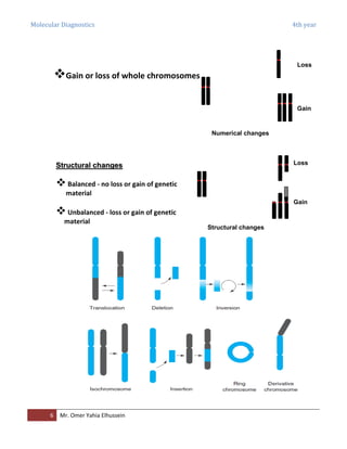

The document discusses molecular diagnostics and clinical genetics, focusing on techniques for prenatal testing to identify genetic disorders in fetuses. It outlines the purposes of genetic testing, such as prenatal diagnosis and carrier detection, as well as common chromosomal abnormalities associated with inherited conditions. Additionally, it describes the structure and classification of human chromosomes and the nature of chromosomal aberrations, including numerical and structural changes.


