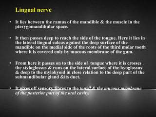
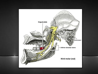
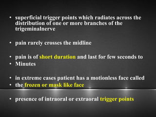
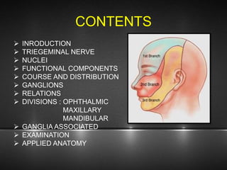
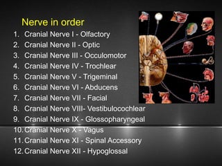
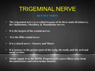
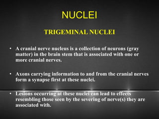
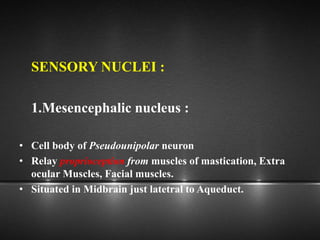
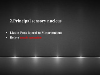
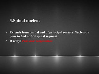
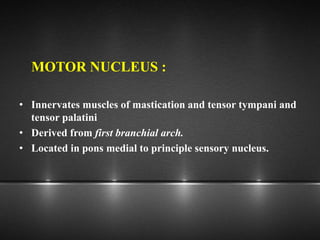
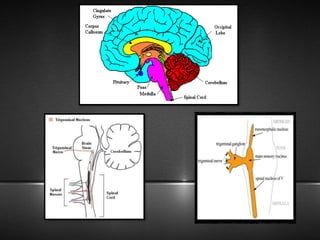
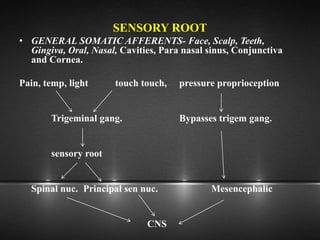
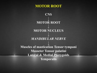
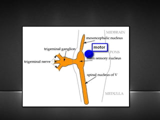
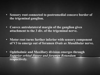
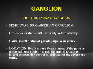
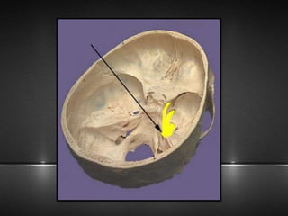
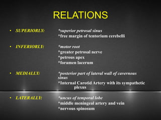
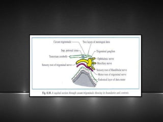
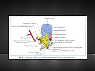
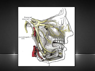
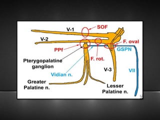
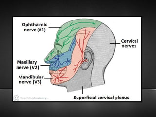
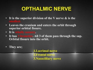
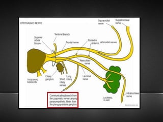
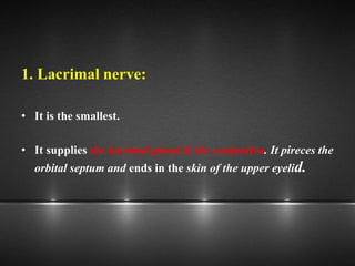
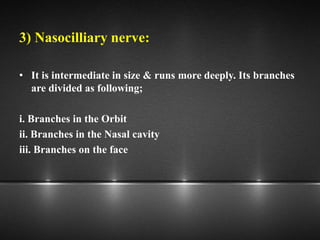
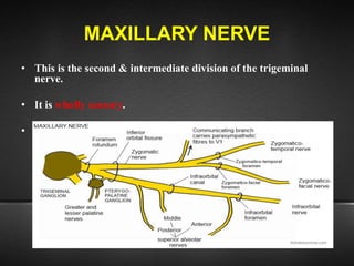
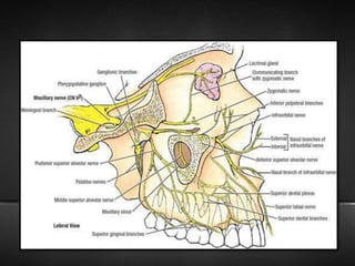
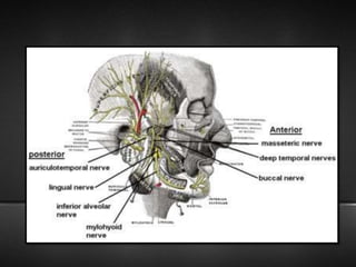
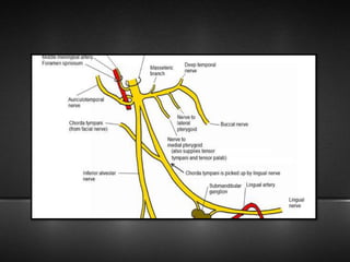
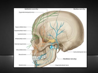
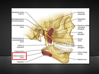
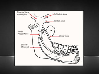
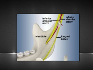
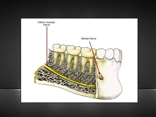
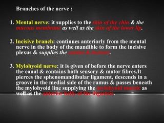
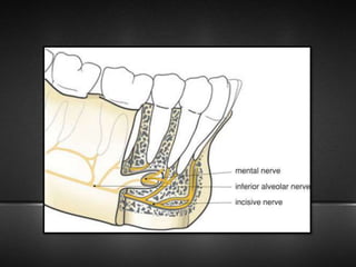
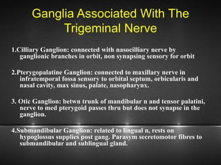
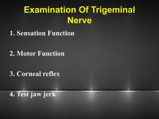
The document provides information about the trigeminal nerve (CN V), which is the largest of the cranial nerves. It has both sensory and motor components. The trigeminal nerve has three main divisions - the ophthalmic, maxillary, and mandibular nerves. It innervates most of the face and provides sensory innervation to the teeth and oral cavity. The trigeminal nerve nuclei are located in the pons and midbrain. The trigeminal ganglion contains the cell bodies of pseudounipolar neurons. The branches and distribution of the three divisions of the trigeminal nerve are described in detail.
Trigeminal nerve maxillary nerve and clinical implicationDr Ravneet Kour
The document discusses the maxillary branch of the trigeminal nerve and its clinical implications. It begins by describing the basic anatomy of neurons, nerves and cranial nerves. It then focuses on the trigeminal nerve as the fifth cranial nerve, describing its nuclei, ganglion and three main branches - the ophthalmic, maxillary and mandibular nerves. Most of the document details the anatomy and branches of the maxillary nerve, including those in the pterygopalatine fossa, orbit, infraorbital canal and face. It concludes by discussing three clinical implications - trigeminal neuralgia, herpes zoster ophthalmicus and Wallenberg syndrome.
This document provides an overview of the trigeminal nerve (CN V), including its nuclei, functional components, course and distribution, the trigeminal ganglion, and the three divisions of the trigeminal nerve - ophthalmic, maxillary, and mandibular nerves. It describes the sensory and motor nuclei of the trigeminal nerve in the brainstem and discusses the sensory and motor roots. It also outlines the anatomy and branches of the three divisions of the trigeminal nerve.
The maxillary nerve is the second division of the trigeminal nerve. It originates in the lateral wall of the cavernous sinus and passes through the foramen rotundum into the pterygopalatine fossa. It gives off several branches in the pterygopalatine fossa and infratemporal fossa, including the posterior superior alveolar nerve, zygomatic nerve, and anterior and middle superior alveolar nerves. The maxillary nerve terminates by innervating structures in the face, providing sensation to the maxillary region. A maxillary nerve block can provide anesthesia for dental procedures involving the maxillary area, such as when a large canine space abscess is
The document discusses the trigeminal nerve (CN V), which has three main divisions - the ophthalmic, maxillary, and mandibular nerves. It describes the anatomy and branches of each division. The ophthalmic nerve is the smallest and is wholly sensory. It has three branches - the lacrimal, frontal, and nasociliary nerves. The maxillary nerve is the second division and is also wholly sensory. It has several branches including the zygomatic and posterior superior alveolar nerves. The mandibular nerve is the third and largest division and has both sensory and motor components.
The mandibular nerve is the largest of the three divisions of the trigeminal nerve. It is made up of both sensory and motor roots. It supplies sensation to the lower face, teeth, gums, lower lip, chin, and anterior two-thirds of the tongue. It also innervates the muscles of mastication. The mandibular nerve divides into anterior and posterior branches which further divide to innervate the muscles and skin of the face and mouth.
The trigeminal nerve is the 5th cranial nerve and is a mixed nerve responsible for sensation in the face and motor function of muscles of mastication. It has 3 main divisions - the ophthalmic, maxillary, and mandibular nerves. The mandibular nerve is the largest division and supplies motor innervation to the muscles of mastication as well as sensory innervation to parts of the face and scalp.
The document discusses the anatomy and branches of the mandibular nerve, including its course, distribution, and supply. It describes various anesthetic techniques for blocking branches of the mandibular nerve, including the inferior alveolar nerve block, lingual nerve block, buccal nerve block, and mental nerve block. It also discusses potential local complications from anesthetic techniques such as needle breakage, prolonged anesthesia, and soft tissue injury.
This document provides an overview of the trigeminal nerve, which is the fifth cranial nerve. It begins with an introduction to cranial nerves and then discusses the specific nuclei, ganglia, and divisions of the trigeminal nerve. The three divisions - ophthalmic, maxillary, and mandibular nerves - are described in detail regarding their course, branches, and sensory and motor functions. Key structures discussed include the trigeminal ganglion, gasserian ganglion, pterygopalatine ganglion, and submandibular ganglion. The document concludes with a recap of the main branches of the trigeminal nerve.
Innervation of the face
The nervvous system
Nerve transmission
Definition of Pain
Pain Receptors
Pain nerve fibers
Reaction to pain
Pain Pathway
Control of Pain
Mode of action of local anesthesia
This document provides an overview of the trigeminal nerve (cranial nerve V) in 12 sections. It discusses the elementary structure of neurons, classification of cranial nerves, embryology and nuclei of the trigeminal nerve, the trigeminal ganglion, and the three divisions (ophthalmic, maxillary, mandibular) and their branches. Diagrams are included to illustrate the course and branches of the trigeminal nerve. The presentation provides detailed anatomical information about the trigeminal nerve and related structures.
This document describes the anatomy and branches of the mandibular nerve (CN V3). It originates from the trigeminal ganglion and pons and exits the skull through the foramen ovale. Its main branches innervate the muscles of mastication and provide sensory innervation to the lower face and oral cavity. The anterior and posterior divisions each give off motor and sensory branches with specific distributions.
The Indian Dental Academy is the Leader in continuing dental education , training dentists in all aspects of dentistry and
offering a wide range of dental certified courses in different formats.
The document discusses the trigeminal nerve, including its anatomy and branches. It describes the maxillary nerve in detail, including its origin, course, branches both within the cranium and on the face. It discusses several clinical implications and conditions related to trigeminal nerve injury or involvement, such as trigeminal neuralgia, maxillary sinus infections, herpes zoster ophthalmicus, and tumors affecting the trigeminal nerve ganglia or branches.
The maxillary nerve originates from the trigeminal ganglion and provides sensory innervation to much of the face and upper teeth. It gives off several branches that innervate the nasal cavity, palate, gums, upper teeth, lower eyelid, upper lip and cheek. Maxillary nerve blocks are commonly used in dental procedures to anesthetize these areas by injecting local anesthetic near branches of the nerve. The document discusses the anatomy and branches of the maxillary nerve in detail.
This document provides an overview of the trigeminal nerve (CN V) in 12 sections. It discusses the structure of neurons and nerves, lists and classifies the 12 cranial nerves, describes the embryological development and nuclei of the trigeminal nerve, details the trigeminal ganglion and course of the trigeminal nerve, and outlines its three main branches (ophthalmic, maxillary, mandibular) and their distributions. The document provides a comprehensive anatomical description of the trigeminal nerve in under 3 sentences.
The document discusses the facial artery, which arises from the external carotid artery. It has both cervical and facial parts. The cervical part runs upwards in the neck, allowing movement of neck structures. It gives off branches like the ascending palatine and tonsillar arteries. The facial part enters the face by piercing the mandible. In the face, it gives branches like the inferior and superior labial arteries and terminates by anastomosing with the ophthalmic artery. The document also discusses the common carotid artery and its branches.
The ophthalmic nerve is the smallest of the three divisions of the trigeminal nerve. It arises from the upper part of the semi lunar ganglion and passes forward along the lateral wall of the cavernous sinus before dividing into three branches - the lacrimal, frontal, and nasociliary nerves. These branches innervate sensory structures of the eye, upper face, and nasal cavity. The ophthalmic nerve also transmits parasympathetic fibers that control functions of the iris, ciliary muscle, and lacrimal gland.
The maxillary artery arises from the external carotid artery and divides into three parts - the mandibular, pterygoid, and pterygopalatine parts. It supplies structures in the face like the maxilla and mandible. The maxillary artery and its branches anastomose with other vessels and are clinically significant for conditions like nosebleeds, epidural hematomas, and complications during procedures like Le Fort I osteotomies when the branches can be injured. Precise surgical techniques are important to avoid damaging branches like the descending palatine artery.
Development,anatomy and applied anatomy of MaxillaSoni Bista
This document provides an overview of the maxilla bone including its development, features, blood supply, age changes, and maxillary sinus elevation procedures. Some key points:
- The maxilla develops from ossification centers and contributes to the formation of the face, nose, mouth, orbit, and palate.
- Features include the body, frontal process, zygomatic process, alveolar process, and palatine process. The maxillary sinus is located within the body.
- The maxillary sinus expands with age, sometimes exposing tooth roots. Sinus elevation procedures use grafts to augment the bone for dental implants.
- The second molar is often close to the floor of the
Growth & development of maxilla and mandiblePiyush Verma
The document summarizes the growth and development of the maxilla and mandible. It discusses the prenatal growth of the maxilla, including how the maxillary process develops from the first branchial arch and fuses with other structures to form the primitive palate. It also describes the development of the primary and secondary palate, with the palatal shelves growing horizontally to fuse and form the completed palate. The prenatal growth of the mandible is also discussed briefly.
The document summarizes the trigeminal nerve, including its nuclear columns, trigeminal ganglion, three divisions of the nerve, and clinical considerations. The trigeminal nerve has three divisions - the ophthalmic, maxillary, and mandibular nerves. It discusses the branches and distributions of each division. Clinically, examination of the trigeminal nerve involves sensory and motor testing as well as trigeminal reflexes. Common conditions involving the trigeminal nerve like trigeminal neuralgia and postherpetic neuralgia are also mentioned.
The document summarizes the development and growth of the mandible. It begins with the development of the body, rami, and alveolar process from mesenchyme and Meckel's cartilage. Growth occurs through secondary cartilage in the condyle and subperiosteal bone formation. The mandible changes with age from a shell-like bone at birth to a reduced size in old age due to absorption of the alveolar process after tooth loss.
The temporomandibular joint (TMJ) connects the jaw to the skull. TMJ disorders are commonly caused by muscular problems or issues with the TMJ elements. Diagnosis involves x-rays or CT/MRI scans of the joint. Conservative treatments include rest, warm compresses, splints, gentle exercises, and injections. More invasive procedures include washing out the joint or cortisone injections. Surgery is a last resort to replace the jaw joints.
This document describes the anatomy and branches of the mandibular nerve (CN V3). It begins in the middle cranial fossa and has both sensory and motor components. Its main branches include the buccal, masseteric, deep temporal, lateral pterygoid nerves anteriorly, and the lingual, inferior alveolar, and auriculotemporal nerves posteriorly. The otic ganglion is also discussed, which relays parasympathetic fibers to the parotid gland via connections with the mandibular and glossopharyngeal nerves.
The anatomy of the nerve supply of the head and neck has many significant applications in maxillofacial surgery. Understanding these important anatomic relations- variations enables surgeons to perform the surgical procedures safely. Knowledge of these concepts helps us to recognize the problems and complications as and when they occur and manage them accordingly.
The trigeminal nerve is the 5th cranial nerve and is a mixed nerve responsible for sensation in the face and motor function of the muscles of mastication. It has 3 major divisions - the ophthalmic, maxillary, and mandibular nerves. The ophthalmic nerve provides sensory innervation to the upper face and eye region. The maxillary nerve provides sensory innervation to the mid face region including the maxillary teeth. The mandibular nerve provides sensory innervation to the lower face and motor innervation to the muscles of mastication.
this presentation consist of introduction to types of nerves, structure of nerve and cranial nerves. there is a detail description about, origin , course of the trigeminal nerve and its branches and the structures supplying the nerve. it also contains applied anatomy of the nerve and its importance of the nerve in oral and maxillofacial surgeries. a detail description about the examination of the trigeminal nerve is also mentioned in the presentation. hoping that it would be useful to the students and people seeking for knowledge about the trigeminal nerve.
The trigeminal nerve is the largest of the cranial nerves. It has both sensory and motor functions. The trigeminal nerve divides into three main branches - the ophthalmic, maxillary, and mandibular nerves. The ophthalmic nerve is purely sensory and innervates parts of the face including the eye, forehead, and nose. It divides further into the lacrimal, frontal, and nasociliary nerves. The frontal nerve gives off the supraorbital and supratrochlear nerves which supply the forehead.
The trigeminal nerve is the largest of the cranial nerves and provides sensory and motor innervation to the face. It has three major branches - the ophthalmic, maxillary, and mandibular nerves. The ophthalmic nerve further divides into the frontal, lacrimal, and nasociliary nerves. The nasociliary nerve branches into the anterior and posterior ethmoidal nerves which supply sensory innervation to the paranasal sinuses and nasal cavity.
This document provides an overview of the trigeminal nerve (CN V), including its origins, branches, and functions. It notes that the trigeminal nerve has three main divisions - the ophthalmic, maxillary, and mandibular nerves. The cell bodies of the trigeminal nerve's sensory neurons are located in the trigeminal ganglion. The document then describes the individual branches and functions of the ophthalmic, maxillary, and mandibular nerves. It provides details on the sensory distributions and innervations of each branch.
The facial nerve emerges from the brainstem between the pons and medulla. It has motor, sensory, and parasympathetic secretomotor components. During embryonic development, it arises from the second branchial arch. The nerve passes through the internal acoustic meatus and facial canal within the temporal bone. It gives off several branches within the facial canal before exiting at the stylomastoid foramen. Its main branches in the face include the temporal, zygomatic, buccal, marginal mandibular, and cervical branches which innervate the muscles of facial expression.
The document provides information about the anatomy and function of the trigeminal nerve (CN V) and the condition of trigeminal neuralgia. It discusses the embryology, nuclei, course and branches of the trigeminal nerve. It also describes trigeminal neuralgia as a condition involving sudden, severe pain in the face triggered by light touch. The document summarizes treatment options for trigeminal neuralgia which include medications and surgical procedures. It also briefly discusses herpes zoster ophthalmicus and Wallenberg syndrome in relation to the trigeminal nerve.
The document provides information on the trigeminal nerve (CN V), including its anatomy, branches, and distribution. Some key points:
- CN V is the largest cranial nerve, supplying sensation to the face and motor function to the muscles of mastication.
- It has three main branches - the ophthalmic, maxillary, and mandibular nerves. The ophthalmic nerve innervates the forehead and orbit. The maxillary nerve innervates the midface, and the mandibular nerve innervates the lower face and jaw.
- Each branch has smaller divisions that provide both sensory and motor function to the face, mucosa, and muscles of the head and neck
The trigeminal nerve is the largest cranial nerve. It has both sensory and motor functions. Sensory fibers carry sensations from the face and head to nuclei in the pons and medulla. Motor fibers innervate muscles of mastication. The trigeminal nerve divides into three main branches - the ophthalmic, maxillary, and mandibular nerves - which further branch to innervate regions of the face, scalp and oral cavity.
The trigeminal nerve is the largest cranial nerve, providing sensory and motor functions. It has three major divisions - ophthalmic, maxillary, and mandibular. The trigeminal ganglion contains the cell bodies of pseudounipolar neurons which relay sensory information from the face to the brainstem nuclei. Trigeminal neuralgia is a painful condition characterized by sudden, severe facial pain that may be triggered by light touch. Herpes zoster ophthalmicus affects the ophthalmic division and can cause eye and skin lesions. Wallenberg syndrome results in loss of sensation in patterns due to a stroke affecting the trigeminal nerve tracts.
Indian Dental Academy: will be one of the most relevant and exciting training center with best faculty and flexible training programs for dental professionals who wish to advance in their dental practice,Offers certified courses in Dental implants,Orthodontics,Endodontics,Cosmetic Dentistry, Prosthetic Dentistry, Periodontics and General Dentistry.
The document discusses the trigeminal nerve (cranial nerve V) in three sentences: It describes the trigeminal nerve as the largest cranial nerve, a mixed nerve with both motor and sensory components. It originates from the trigeminal ganglion and divides into three main branches - the ophthalmic, maxillary, and mandibular nerves - which innervate the face and associated structures. The document provides detailed information on the embryology, nuclei, course and branches of the trigeminal nerve.
FACIAL NERVE SEMINAR BY DIPESH MDS1.pptxdipeshmadge6
The document discusses the facial nerve (CN VII), including its nuclei of origin, functional components, course through the skull, branches and distribution. It provides motor innervation to the muscles of facial expression and special sensory innervation for taste. Within the facial canal it gives off the chorda tympani, nerve to stapedius and greater petrosal nerve. Its five terminal branches innervate muscles of the face and scalp.
Indian Dental Academy: will be one of the most relevant and exciting training center with best faculty and flexible training programs for dental professionals who wish to advance in their dental practice,Offers certified courses in Dental implants,Orthodontics,Endodontics,Cosmetic Dentistry, Prosthetic Dentistry, Periodontics and General Dentistry.
The trigeminal nerve is the largest of the 12 cranial nerves. It has both sensory and motor functions, supplying sensation to the face and motor innervation to the muscles of mastication. The trigeminal nerve has three main divisions - the ophthalmic, maxillary, and mandibular nerves. These divisions and their branches innervate different regions of the face and head. Injuries or disorders of the trigeminal nerve can result in numbness, pain, or muscle dysfunction in the territories it supplies. Trigeminal neuralgia is a painful condition characterized by episodes of intense, stabbing pain in areas innervated by the trigeminal nerve.
The document discusses the trigeminal and facial nerves, which provide sensory and motor innervation to the face. It covers their anatomy, clinical assessment, and common injuries. The trigeminal nerve has three main branches - the ophthalmic, maxillary, and mandibular nerves. It provides sensory innervation and some motor functions. The facial nerve innervates the muscles of facial expression. Common causes of injury include local anesthetic injections, surgery like third molar removal or dental implants, and traumatic injuries. Assessing trigeminal nerve function involves testing sensation and motor functions like the jaw jerk reflex.
The trigeminal nerve is the 5th cranial nerve that has both sensory and motor functions. It has three major divisions - the ophthalmic, maxillary, and mandibular nerves. The ophthalmic and maxillary nerves are purely sensory and innervate parts of the face, while the mandibular nerve contains both sensory and motor fibers that innervate muscles of mastication. The trigeminal ganglion contains the cell bodies of the sensory fibers and relays information to various sensory nuclei in the brainstem.
This document provides an overview of the trigeminal nerve (CN V) including its:
- Intracranial course and branches originating from the trigeminal ganglion
- Three main divisions - ophthalmic, maxillary, and mandibular nerves
- Branches of each division and the areas they innervate, such as the face, nasal cavity, and oral cavity
- Applied anatomy and clinical significance of parts like the trigeminal ganglion
The trigeminal nerve is the largest of the cranial nerves. It has both sensory and motor functions. It has three main divisions: the ophthalmic, maxillary, and mandibular nerves. The ophthalmic nerve innervates the eye and forehead. The maxillary nerve supplies sensation to the cheek, upper teeth, and nose. The mandibular nerve provides motor input to the muscles of mastication and sensation to the lower face, teeth, and chin.
Similar to Trigeminal Nerve and Applied Anatomy (20)
TEST BANK For Community Health Nursing A Canadian Perspective, 5th Edition by...Donc Test
TEST BANK For Community Health Nursing A Canadian Perspective, 5th Edition by Stamler, Verified Chapters 1 - 33, Complete Newest Version Community Health Nursing A Canadian Perspective, 5th Edition by Stamler, Verified Chapters 1 - 33, Complete Newest Version Community Health Nursing A Canadian Perspective, 5th Edition by Stamler Community Health Nursing A Canadian Perspective, 5th Edition TEST BANK by Stamler Test Bank For Community Health Nursing A Canadian Perspective, 5th Edition Pdf Chapters Download Test Bank For Community Health Nursing A Canadian Perspective, 5th Edition Pdf Download Stuvia Test Bank For Community Health Nursing A Canadian Perspective, 5th Edition Study Guide Test Bank For Community Health Nursing A Canadian Perspective, 5th Edition Ebook Download Stuvia Test Bank For Community Health Nursing A Canadian Perspective, 5th Edition Questions and Answers Quizlet Test Bank For Community Health Nursing A Canadian Perspective, 5th Edition Studocu Test Bank For Community Health Nursing A Canadian Perspective, 5th Edition Quizlet Test Bank For Community Health Nursing A Canadian Perspective, 5th Edition Stuvia Community Health Nursing A Canadian Perspective, 5th Edition Pdf Chapters Download Community Health Nursing A Canadian Perspective, 5th Edition Pdf Download Course Hero Community Health Nursing A Canadian Perspective, 5th Edition Answers Quizlet Community Health Nursing A Canadian Perspective, 5th Edition Ebook Download Course hero Community Health Nursing A Canadian Perspective, 5th Edition Questions and Answers Community Health Nursing A Canadian Perspective, 5th Edition Studocu Community Health Nursing A Canadian Perspective, 5th Edition Quizlet Community Health Nursing A Canadian Perspective, 5th Edition Stuvia Community Health Nursing A Canadian Perspective, 5th Edition Test Bank Pdf Chapters Download Community Health Nursing A Canadian Perspective, 5th Edition Test Bank Pdf Download Stuvia Community Health Nursing A Canadian Perspective, 5th Edition Test Bank Study Guide Questions and Answers Community Health Nursing A Canadian Perspective, 5th Edition Test Bank Ebook Download Stuvia Community Health Nursing A Canadian Perspective, 5th Edition Test Bank Questions Quizlet Community Health Nursing A Canadian Perspective, 5th Edition Test Bank Studocu Community Health Nursing A Canadian Perspective, 5th Edition Test Bank Quizlet Community Health Nursing A Canadian Perspective, 5th Edition Test Bank Stuvia
Histololgy of Female Reproductive System.pptxAyeshaZaid1
Dive into an in-depth exploration of the histological structure of female reproductive system with this comprehensive lecture. Presented by Dr. Ayesha Irfan, Assistant Professor of Anatomy, this presentation covers the Gross anatomy and functional histology of the female reproductive organs. Ideal for students, educators, and anyone interested in medical science, this lecture provides clear explanations, detailed diagrams, and valuable insights into female reproductive system. Enhance your knowledge and understanding of this essential aspect of human biology.
8 Surprising Reasons To Meditate 40 Minutes A Day That Can Change Your Life.pptxHolistified Wellness
We’re talking about Vedic Meditation, a form of meditation that has been around for at least 5,000 years. Back then, the people who lived in the Indus Valley, now known as India and Pakistan, practised meditation as a fundamental part of daily life. This knowledge that has given us yoga and Ayurveda, was known as Veda, hence the name Vedic. And though there are some written records, the practice has been passed down verbally from generation to generation.
Does Over-Masturbation Contribute to Chronic Prostatitis.pptxwalterHu5
In some case, your chronic prostatitis may be related to over-masturbation. Generally, natural medicine Diuretic and Anti-inflammatory Pill can help mee get a cure.
These lecture slides, by Dr Sidra Arshad, offer a quick overview of the physiological basis of a normal electrocardiogram.
Learning objectives:
1. Define an electrocardiogram (ECG) and electrocardiography
2. Describe how dipoles generated by the heart produce the waveforms of the ECG
3. Describe the components of a normal electrocardiogram of a typical bipolar lead (limb II)
4. Differentiate between intervals and segments
5. Enlist some common indications for obtaining an ECG
6. Describe the flow of current around the heart during the cardiac cycle
7. Discuss the placement and polarity of the leads of electrocardiograph
8. Describe the normal electrocardiograms recorded from the limb leads and explain the physiological basis of the different records that are obtained
9. Define mean electrical vector (axis) of the heart and give the normal range
10. Define the mean QRS vector
11. Describe the axes of leads (hexagonal reference system)
12. Comprehend the vectorial analysis of the normal ECG
13. Determine the mean electrical axis of the ventricular QRS and appreciate the mean axis deviation
14. Explain the concepts of current of injury, J point, and their significance
Study Resources:
1. Chapter 11, Guyton and Hall Textbook of Medical Physiology, 14th edition
2. Chapter 9, Human Physiology - From Cells to Systems, Lauralee Sherwood, 9th edition
3. Chapter 29, Ganong’s Review of Medical Physiology, 26th edition
4. Electrocardiogram, StatPearls - https://www.ncbi.nlm.nih.gov/books/NBK549803/
5. ECG in Medical Practice by ABM Abdullah, 4th edition
6. Chapter 3, Cardiology Explained, https://www.ncbi.nlm.nih.gov/books/NBK2214/
7. ECG Basics, http://www.nataliescasebook.com/tag/e-c-g-basics
Here is the updated list of Top Best Ayurvedic medicine for Gas and Indigestion and those are Gas-O-Go Syp for Dyspepsia | Lavizyme Syrup for Acidity | Yumzyme Hepatoprotective Capsules etc
Promoting Wellbeing - Applied Social Psychology - Psychology SuperNotesPsychoTech Services
A proprietary approach developed by bringing together the best of learning theories from Psychology, design principles from the world of visualization, and pedagogical methods from over a decade of training experience, that enables you to: Learn better, faster!
Basavarajeeyam is an important text for ayurvedic physician belonging to andhra pradehs. It is a popular compendium in various parts of our country as well as in andhra pradesh. The content of the text was presented in sanskrit and telugu language (Bilingual). One of the most famous book in ayurvedic pharmaceutics and therapeutics. This book contains 25 chapters called as prakaranas. Many rasaoushadis were explained, pioneer of dhatu druti, nadi pareeksha, mutra pareeksha etc. Belongs to the period of 15-16 century. New diseases like upadamsha, phiranga rogas are explained.
3. INTRODUCTION
• The nervous system of man is made up of innumerable neurons
which further constitute the nerve fibres
• Nerve :
A bundle of fibers that uses chemical and electrical signals to
transmit sensory and motor information from one body part of
the body to another.
• Neurons :
These are specialized cells that constitute the functional units of
the nervous system and has a special property of being able to
conduct impulses rapidly from onepart of the body to another.
4.
5. • The cranial nerves are composed of twelve pairs of nerves
that originate from the nervous tissue of the brain.
• In order to reach their targets they must ultimately
exit/enter the cranium through openings in the skull.
• Hence, their name is derived from their association with
the cranium.
6. Nerve in order
1. Cranial Nerve I - Olfactory
2. Cranial Nerve II - Optic
3. Cranial Nerve III - Occulomotor
4. Cranial Nerve IV - Trochlear
5. Cranial Nerve V - Trigeminal
6. Cranial Nerve VI - Abducens
7. Cranial Nerve VII - Facial
8. Cranial Nerve VIII- Vestibulocochlear
9. Cranial Nerve IX - Glossopharyngeal
10.Cranial Nerve X - Vagus
11.Cranial Nerve XI - Spinal Accessory
12.Cranial Nerve XII - Hypoglossal
7. TRIGEMINAL NERVE
DENTIST NERVE
• The trigeminal nerve is so called because of its three main divisions i.e.
the Ophthalmic, Maxillary & Mandibular nerves.
• It is the largest of the cranial nerves.
• It is the fifth cranial nerve
• It is a mixed nerve : Sensory and Motor
• It is sensory to the greater part of the scalp, the teeth, and the oral and
nasal cavities.
• Motor supply is to the MOM. Proprioceptive nerve fibres arise from
the masticatory and extra-ocular muscles.
8. NUCLEI
TRIGEMINAL NUCLEI
• A cranial nerve nucleus is a collection of neurons (gray
matter) in the brain stem that is associated with one or
more cranial nerves.
• Axons carrying information to and from the cranial nerves
form a synapse first at these nuclei.
• Lesions occurring at these nuclei can lead to effects
resembling those seen by the severing of nerve(s) they are
associated with.
9. SENSORY NUCLEI :
1.Mesencephalic nucleus :
• Cell body of Pseudounipolar neuron
• Relay proprioception from muscles of mastication, Extra
ocular Muscles, Facial muscles.
• Situated in Midbrain just latetral to Aqueduct.
11. 3.Spinal nucleus
• Extends from caudal end of principal sensory Nucleus in
pons to 2nd or 3rd spinal segment
• It relays Pain and Temperature
12. MOTOR NUCLEUS :
• Innervates muscles of mastication and tensor tympani and
tensor palatini
• Derived from first branchial arch.
• Located in pons medial to principle sensory nucleus.
15. SENSORY ROOT
• GENERAL SOMATIC AFFERENTS- Face, Scalp, Teeth,
Gingiva, Oral, Nasal, Cavities, Para nasal sinus, Conjunctiva
and Cornea.
Pain, temp, light touch touch, pressure proprioception
Trigeminal gang. Bypasses trigem gang.
sensory root
Spinal nuc. Principal sen nuc. Mesencephalic
CNS
16. MOTOR ROOT
CNS
MOTOR ROOT
MOTOR NUCLEUS
MANDIBULAR NERVE
Muscles of mastication Tensor tympani
Masseter Tensor palatini
Lateral & Medial Pterygoids
Temporalis
17. COURSE & DISTRIBUTION
• Both motor and sensory root are attached ventrally to
junction of pons and middle cerebellar peduncle with
motor root lying ventromedially to the sensory root.
• Pass anteriorly in middle cranial fossa to lie below
tentorium cerebelli in cavum trigeminale, here motor root
lies inferior to sensory root.
18.
19. • Sensory root connected to postromedial concave border of
the trigeminal ganglion.
• Convex antrolatateral margin of the ganglion gives
attachment to the 3 div. of the trigeminal nerve.
• Motor root turns further inferior with sensory component
of V3 to emerge out of foramen Ovale as Mandibular nerve.
• Ophthalmic and Maxillary division emerges through
Superior orbital fissure and foramen Rotundum
respectively.
20. GANGLION
THE TRIGEMINAL GANGLION
• SEMILUNAR OR GASSERIAN GANGLION.
• Cresentric in shape with convexity anterolaterally.
• Contains cell bodies of pseudounipolar neurons.
• LOCATION: lies in a bony fossa at apex of the petrous
temporal bone on floor of middle cranial fossa, just
lateral to posterior part of lateral wall of the cavernous
sinus.
21. • COVERINGS: covered by dural pouch = MECKLES
CAVE or CAVUM TRIGEMINALE cave lined by pia and
arachnoid thus the ganglion is bathed in CSF.
• ARTERIAL SUPPLY: Ganglionic branches of Internal
Carotid Artery, middle meningeal artery and accessory
meningeal artery.
22.
23.
24. RELATIONS
• SUPERIORLY: *superior petrosal sinus
*free margin of tentorium cerebelli
• INFERIORLY: *motor root
*greater petrosal nerve
*petrous apex
*foramen lacerum
• MEDIALLY: *posterior part of lateral wall of cavernous
sinus
*Internal Carotid Artery with its sympathetic
plexus
• LATERALLY: *uncus of temporal lobe
*middle meningeal artery and vein
*nervous spinosum
31. OPTHALMIC NERVE
• It is the superior division of the V nerve & is the
smallest.
• Leaves the cranium and enters the orbit through
superior orbital fissure.
• It is wholly sensory.
• It has 3 branches. All 3 of them pass through the sup.
Orbital fissure into the orbit.
• They are;
1.Lacrimal nerve
2.Frontal nerve
3.Nasocilliary nerve
32.
33. 1. Lacrimal nerve:
• It is the smallest.
• It supplies the lacrimal gland & the conjuntiva. It pireces the
orbital septum and ends in the skin of the upper eyelid.
34. 2) Frontal nerve:
• It is the largest branch & appears to be the direct continuation
of the ophthalmic division.
• It enters the orbit through the SOF divides into 2 branches.
i. The supra orbital branch: It is larger & more laterally placed.
It supplies the skin of the forehead &
scalp as far back as the vertex.
It also supplies the mucous membrane
of the frontal sinus & pericranium
ii. The supra trochlear branch: It is smaller & more medially
placed. It curves upward on the
forehead , close to the bone.
It supplies the skin of the upper
eyelid & lower part of theforehead.
35. 3) Nasocilliary nerve:
• It is intermediate in size & runs more deeply. Its branches
are divided as following;
i. Branches in the Orbit
ii. Branches in the Nasal cavity
iii. Branches on the face
36. (I) Branches in the Orbit:
i. Long root of the cilliary ganglion: It is sensory &
passes through the ganglion without synapsing and
supplies the eyeball.
ii. Long ciliary nerve: Supplies the Iris & Cornea.
iii. Posterior ethmoidal nerve: It enters the post
ethmoidal canal & supplies to the mucous membrane
lining of the Post. Etmoidal & Sphenoidal paranasal air
cells.
37. iv. Anterior ethmoidal nerve: It supplies to the Ant.ethmoidal
& frontal paranasal air cells. In the upper part of the nasal
cavity, it further divides into:
1) Internal nasal branches: It has medialseptal branches to
the septal membrane. It also has lateral branches, which
supply the nasal conchae & the ant. nasal wall
2) External nasal branches: supplies the skin on the tip & ala
of the nose.
38. 2) Branches in the nasal cavity:
The branches arising here supply the mucous membrane of
the nasal cavity.
3) Terminal branches on the face:
They supply sensory nerves to the skin of the medial parts
of the both eyelids, the lacrimal sac. They also supply skin on
the bridge of the nose.
39. MAXILLARY NERVE
• This is the second & intermediate division of the trigeminal
nerve.
• It is wholly sensory.
• Course: It begins at the middle of the trigeminal ganglion as a
flattened, plexiform band, passes horizontally forwards along
the lateral wall of the cavernous sinus. It leaves the skull
through the foramen rotundum & becomes more cylindrical &
firmer in texture.It crosses the upper part of the pterygopalatine
fossa, inclines laterally on the posterior part of the orbital
process of the maxilla & enters the orbit through the inferior
orbital fissure.It is now termed as the infra orbital nerve. It passes
through the infra orbital groove & canal in the floor of the orbit
& appears on the face through the infra orbital foramen.
40.
41.
42. • The branches of the maxillary nerve can be divided into
the following 4 groups:
1) In the cranium: Meningeal
2) In the pterygopalatine fossa: Ganglionic, Zygomatic,
Post.superior alveolar
3) In the infra orbital canal: Middle sup. alveolar,
Anterior superior/ Greater alveolar
4) On the face: Palpebral, nasal, superior labial
43. I. Branch given off on the cranium
1. Meningeal branch: It is given off near the foramen
rotundum. It supplies the duramater of the anterior &
middle cranial fossae.
44. II. Branches in the pterygopalatine fossa.
1. The ganglionic branches: They connect the maxillary
nerve to the pterygopalatine ganglion.They contain secretomotor
fibres to the lacrimal gland. They provide sensory fibres to the
orbital periosteum & mucous membrane of the nose, palate &
pharynx.
2. The zygomatic nerve: It arises in the pterygopalatine
fosssa from the maxillary nerve and travels anteriorly ,
entering through the inferior orbital fissure where it divides into 2
branches. The Zygomaticofacial nerve perforates the facial
surfaces & supplies the skin over the zygomatic bone..
45. • The Zygomaticotemporal nerve perforates the temporal surface of
the zygomatic bone , pierces the temporalis fascia, & supplies the
skin over the anterior temporal fossa region.
3. Posterior superior alveolar nerve:
• It begins in the pterygopalatine fossa but divides into 3 branches
which emerge through the pterygomaxillary fissure.
• 2 branches enter the posterior wall of the maxilla above the
tuberosity & supply the 3 molar teeth(except the mesiobuccal root
of first molar).
• The third branch pierces the buccinator & supplies the adjoining
part of the gingiva & cheek along the buccal side of the upper
molar teeth.
46. The branches of the Pterygopalatine ganglion are:
I. Orbital branches: periosteum of the orbit.Posterior ethmoidal &
sphenoidal air cells.
II. Palatine branches:
1.Anterior/greater palatine : mucosa of the hard palate & palatal gingiva.
2.Middle palatine : mucous membrane of the soft palate
3.Posterior palatine : mucous membrane of the Tonsillar area.
III. Nasal branches:
1.Posterior superior lateral : posterior part of the nasal conchae
2.Nasopalatine/Sphenopalatine : mucous membrane of the premaxilla.
IV. Pharyngeal branch: mucous membrane of the nasopharynx
47.
48. III. Branches in the Infraorbital canal
( Infraorbital nerve)
1. Middle superior alveolar nerve:
• It arises from the Infra orbital nerve & runs downwards & forwards
along the infraorbital groove along the lateral wall of the maxillary
sinus.
• It divides into branches which supply the maxillary premolars &
mesiobuccal root of the first molar teeth.
2. Anterior superior alveolar nerve:
• It also arises in the infraorbital canal near the mid point. It runs in the
anterior wall of the maxillary antrum. It runs inferiorly & divides into
the branches, which supply the canine & incisors.
• A nasal branch from this nerve, given off from the superior dental
plexus supplies the mucous membrane of the anterior part of the lateral
wall & floor of the nasal cavity. It ends in the nasal septum.
49. • IV. Branches given on the face:
1. The palpebral branches:
• They arise deep to the orbicularis oculi & pierce the muscle, supplying
the skin over the lower eyelid& lateral angle of the eye along with the
Zygomaticofacial & Facial nerves.
2. The nasal branches:
• They supply the skin of the nose & tip of the nasal septum & join the
External nasal branch of the anterior ethmoidal nerve.
3. The superior labial branches:
•
• These are large & numerous. They supply the skin over the anterior
part of the cheek & upper lip including the mucous membrane & labial
glands. They are joined by the facial nerve & form the infraorbital
plexus.
50.
51. MANDIBULAR NERVE
• It is the third & largest division of the trigeminal nerve.
• It is made up of 2 roots: a large sensory root which proceeds
from the lateral part of the trigeminal ganglion & almost
immediately emerges out through the foramen ovale & a small
motor root which passes below the ganglion, & unites with the
sensory root just outside the foramen.
• Immediately beyond the junction of the 2 roots, the nerve sends
off the meningeal branch & the nerve to the medial pterygoid.
Now the main trunk divides into a small anterior & a large
posterior trunk.
• As it descends from the foramen, the mandibular nerve lies at a
distance of 4 cm from the surface & a little in front of the neck
of the mandible.
52.
53. The braches of the Mandibular nerve:
I. Branches of the undivided nerve.
i. Meningeal branch/nervus spinosus.
ii. Nerve to the medial pterygoid
II. Branches of the divided nerve:
(A) Anterior division: (B) Posterior division:
1.Buccal nerve 1. Auriculotemporal nerve
2.Massetric nerve 2.Lingual nerve
3.Deep temporal nerve 3.Inferior alveolar nerve
4.Nerve to the lateral pterygoid
54.
55.
56. BRANCHES OF THE UNDIVIDED NERVE
1.Meningeal nerve:
• It enters the skull through the foramen spinosum along
with MMA.
• It has anterior & posterior divisions that supply the dura
of the middle & anterior cranial fossae.
2. Nerve to the medial pterygoid:
• It is a slender branch that supplies to the deep surface of
the muscle.
• It also gives 1-2 filaments to the tensor tympani & the
tensor veli palati muscles.
57. • BRANCHES OF THE DIVIDED NERVE
I. Anterior division
1.The buccal nerve:
• It passes between the 2 heads of the lateral pterygoid &
descends beneath or through the temporalis. It emerges
from under cover of the ramus & ant. border of the
masseter & unites with the buccal branches of he facial
nerve.
• It supplies the skin over the ant. part of the buccinator &
mucous membrane lining the buccal surface of the gum.
58. 2.The massetric nerve:
• Passes laterally above the lateral pterygoid in front of the TMJ &
behind the tendon of temporalis.
• It passes through the mandibular notch to sink into the masseter
muscle.
• It also gives a branch to the TMJ.
3.The deep temporal nerves:
• They are 2 in number.
• They pass above the upper head of the lateral pterygoid, turn above
the infra temporal crest & sink into the deep part of the temporalis
muscle.
4.The nerve to the lateral pterygoid
• These are 2 in number; one supplying each muscle head.
59. II.Posterior Division
1.The Auriculotemporal nerve:
Course of the nerve
• The auriculotemporal nerve arises by a medial & lateral roots,
that enclircle the MMA & unite behind it just below the
foramen spinosum.
• The united nerve passes backwards, deep to the lateral
pterygoid muscle & passes between the sphenomandibular
ligament & the neck of the condyle.
• It then passes laterally behind the TMJ i.r.t. to the upper part of
the parotid. It emerges from behind the TMJ, ascends posterior
to the superficial temporal vessels & crosses the posterior root of
the zygomatic arch.
60.
61. Branches of the Auriculotemporal nerve:
1. Parotid branches: secretomotor, vasomotor.
2. Articular branches: to the TMJ.
3. Auricular branches: to the skin of the helix & tragus.
4. Meatal branches: Meatus of the tymphanic
membrane
5. Terminal branches: Scalp over the temporal region
62.
63. Lingual nerve
• It lies between the ramus of the mandible & the muscle in the
pterygomandibular space.
• It then passes deep to reach the side of the tongue. Here it lies in
the lateral lingual sulcus against the deep surface of the
mandible on the medial side of the roots of the third molar tooth
where it is covered only by mucous membrane of the gum.
• From here it passes on to the side of tongue where it is crosses
the styloglossus & runs on the lateral surface of the hyoglossus
& deep to the mylohyoid in close relation to the deep part of the
submandibular gland &its duct.
• It gives off sensory fibres to the tonsil & the mucous membrane
of the posterior part of the oral cavity.
64.
65. Branches of lingual nerve and its communications:
1.Chorda tympani
2.Communications with submandibular ganglion
3.Hypoglossal nerve
66. Inferior alveolar nerve
• It is the largest terminal branch of the posterior division of the
mandibular nerve.
• The nerve descends deep to the lateral pterygoid muscle at the
lower border of the muscle, it passes b/n the sphenomandibular
ligament & the ramus to enter the mandibular foramen.
• In the canal the nerve runs alongside the inferior alveolar artery
as far as the mental foramen where it emerges out& gives off the
mental & incisive branches.
• From here the nerve runs in the canal giving of branches to the
mandibular teeth as apical fibres & enters the apical foramina
of the teeth to supply mainly the pulp as well as the
periodontium.
67.
68.
69.
70.
71. Branches of the nerve :
1. Mental nerve: it supplies to the skin of the chin & the
mucous membrane as well as the skin of the lower lip.
2. Incisive branch: continues anteriorly from the mental
nerve in the body of the mandible to form the incisive
plexus & supplies the canine & incisors.
3. Mylohyoid nerve: it is given of before the nerve enters
the canal & contains both sensory & motor fibres.It
pierces the sphenomandibular ligament, descends in a
groove in the medial side of the ramus & passes beneath
the mylohyoid line supplying the mylohyoid muscle as
well as the anterior belly of the digastric.
72.
73. Ganglia Associated With The
Trigeminal Nerve
1.Cilliary Ganglion: connected with nasocilliary nerve by
ganglionic branches in orbit, non synapsing sensory for orbit
2.Pterygopalatine Ganglion: connected to maxillary nerve in
infratemporal fossa sensory to orbital septum, orbicularis and
nasal cavity, max sinus, palate, nasopharynx.
3. Otic Ganglion: betwn trunk of mandibular n and tensor palatini,
nerve to med pterygoid passes thru but does not synapse in the
ganglion.
4.Submandibular Ganglion: related to lingual n, rests on
hypoglossus supplies post gang. Parasym secretomotor fibres to
submandibular and sublingual gland.
75. • Sensation function
Use sterile sharp item on forehead, cheek,
and jaw
If any abnormality present we test the
thermal sensation and light touch
76. • Corneal reflex
A clean piece of cotton wool and ask the
patient to look away gently touch the cornea
with the cotton wool and the patient will
blink.
77. • Test jaw jerk
Doctor finger on tip of
jaw, grip patellar
hammer halfway up shaft
and tap finger lightly
usually nothing happens,
or just a slight closure.
79. Trigeminal Neuralgia
• Also known as Fothergill’s disease,Tic douloureux
(painful jerking)
• It is defined as , sudden ,usually ,unilateral ,severe
,brief ,stabbing , lancinating , recurring pain in the
distribution of one or more branches of trigeminal
nerve.
• Mean age: 50 y onwards
• Female predominance (male : female = 1:2 ~2:3)
80. Pathogenesis of trigeminal neuralgia:
• It is usualy idiopathic.
• The probable etiologic factors are:
Intra cranial tumors: Traumatic compression of the
trigeminal nerve by neoplastic (cerebellopontine
angle tumor) or vascular anomalies eg arteriovenous
malformations
: granulomatous and non granulomatous infections
involving 5th cranial nerve.
82. • Pulsation of vessels upon the
trigeminal nerve root do not
visibly damage the nerve.
• However, irritation from
repeated pulsations may lead
to changes of nerve function,
and delivery of abnormal
signals to the trigeminal
nerve nucleus.
• Over time, this is thought to
cause hyperactivity of the
trigeminal nerve nucleus,
resulting in the generation of
TN pain.
83. General characteristics
• Incidence: seen in about 4 in 100000 persons
• Age of occurrence: 5th to 6th decade
• Sex predilection: female predisposition
• Side involved more frequently: right side
• Division of trigeminal nerve involved; most
commonly : max> mand> oph
86. • superficial trigger points which radiates across the
distribution of one or more branches of the
trigeminalnerve
• pain rarely crosses the midline
• pain is of short duration and last for few seconds to
• Minutes
• in extreme cases patient has a motionless face called
• the frozen or mask like face
• presence of intraoral or extraoral trigger points
87.
88. Provocated by obvious stimuli like
• Touching face at particular site
• Chewing
• Speaking
• Brushing
• Shaving
• Washing the face
The characteristic of the disorder being that the attacks do not
occur during sleep.
89.
90. DIAGNOSIS:
• CLINICAL EXAMINATION with HISTORY is
mandatory
• Response to treatment with tablet of carbamazepine
• Injections of local anaesthetic agents into patients trigger
zone gives temporarily relief from pain.
92. • Carbamazapine and phenytoin are the traditional anticonvulsants
given primarilary
• The dosage of the drug used intially should be kept small to minimum
especialy in elderly patients to avoid nausea,vomiting and gastric
irritation.
• Dosage should be taken at night so that adequate serum concentration
is present early morning.
• Complete blood count,liver function,platelet count should be done
prior to treatment.
• Onces the pain remission has being achieved the drug dose should be
kept at maintainence level or withdrawn and restarted if symptoms
reappear
• When carbamazepine is contraindicated clonazepam can be given
• Co-administration of phenytion or baclofen is also advocated.
93. THE ALCOHOLIC INJECTIONS:
• 95% ABSOLUTE alcohol in small quantites 0.5 to 2 ml
is given in peripheral branches of trigeminal nerve.
Side effect: Repeated injections may cause
• Local tissue toxicity
• Inflammation
• Fibrosis
• Burning alcohol neuritis
94. Peripheral neurectomy (nerve avulsion):
• Oldest and the most effective procedure
• Simple
• Relatively reliable
• Indicated in patients in whom craniotomy is contraindicated
due to age,debility,limited life expectancy
• Acts by interrupting the flow of a significant number of afferent
impulses to central trigeminal apparatus.
• Performed mostly on infraorbital,inferior alveolar,mental and
rarely lingual nerve.
95. CRYOTHERAPY FOR PERIPHERAL NERVE:
• Direct application of cryotherapy probe (nitrous oxide
probe)
• Temperature colder than -60 degree C,for 2-3 minutes
• Reapeated three times
• Produces WALLERIAN degeneration without destroying
the nerve sheath
96. PERIPHERAL RADIOFREQUENCY
NEUROLYSIS THERMOCOAGULATION:
• Radiofrequency electrode that has the capacity to destroy the pain
fibres is used in this procedure.
• Temperature being 65 to 75 degree C for 1 to 2 minutes.
• Shown to induce pain remissions in 20%of cases.
98. GYCEROL INJECTIONS:
• Absolute alcohol or phenol-glycerol mixture can be used
as the neurolytic agents.
• Agent is injected into meckel’s cave or in the ganglion.
• Causes damage to nerve cells presumably through
dehydration.
• It induces pain relief in 80% of the cases.
• Also spares the ophthalmic division and the motor root. .
99. THERMOCOAGULATION:
• A radiofrequency electrode that has the capacity to destroy
pain fibres is used.
• Alternating currents of high frequency is passed through
the electrode.
• It produces ionization in the biological tissues leads to
coagulation of tissues.
100. BALLON COMPRESSION:
• A Fogarty catheter 1 to 2cm is advanced within the
meckels cave through foramen ovale.
• Inflated upto 0.75ml at the ventral aspect of theganglion
root for 1 minute.
• It destroyes the root fibres.
101. HERPES ZOSTER
OPHTHALMICUS
• Caused by Varicella zoster
• Predilection for nasociliary branch of ophthalmic division
of the trigeminal nerve
CLINICAL FEATURES:
Cutaneous lesions:
• Rash
• Vesicle
• Pustule crust permanent scar
103. TREATMENT:
• Acyclovir 800mg 5 times /day within 4 days of onset of rash
• Analgesics
• Antibiotic ointments
• Systemic steroids 60mg/day
• Corneal grafting
104. Cavernous Sinus Syndrome
• Multiple cranial neuropathies
• Exophthalmos, ocular motor defects, sensory loss in V1 and / or
V2.
• Pupils may be spared or involved.
• causes: bacterial thrombophlebitis
actinomycosis
rhinocerebellar mucormycosis
aspergillosis
tolosa hunt syndrome
neoplasms
vascular lesions
105.
106. Conclusion
• Since Trigeminal nerve is mixed nerve,
suplies mainly head and neck region. Hence
as a dental specialist one should know
throughly about itracranial and
extracranial course and distribution of
Trigeminal nerve,to diagnose the
pathologies associated with Trigeminal
nerve and for appropriate treatment.
107. REFERENCES
• Anatomy head and neck ( B.D Chaurasia)
• Gray’s Anatomy
• Anatomy of cranial Nerves
• Anatomy for dental Students( A.S. Moni)
• Handbook of local anaesthesia by stanley malamed
• Textbook of oral and maxillofacial surgery(Neelima Anil Malik)
• Harrisson text of internal medicine
• Wikipedia