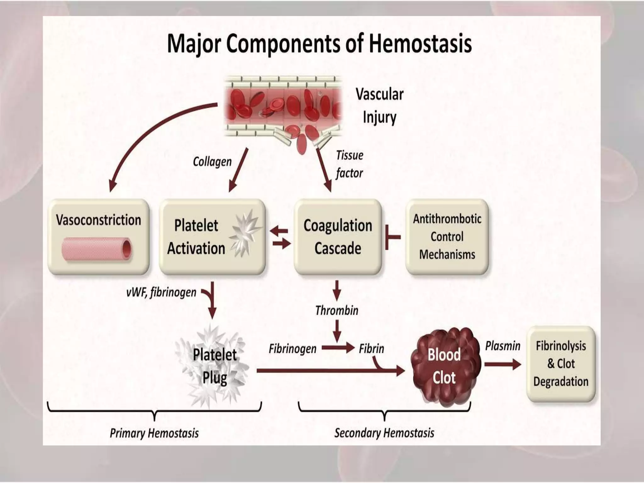
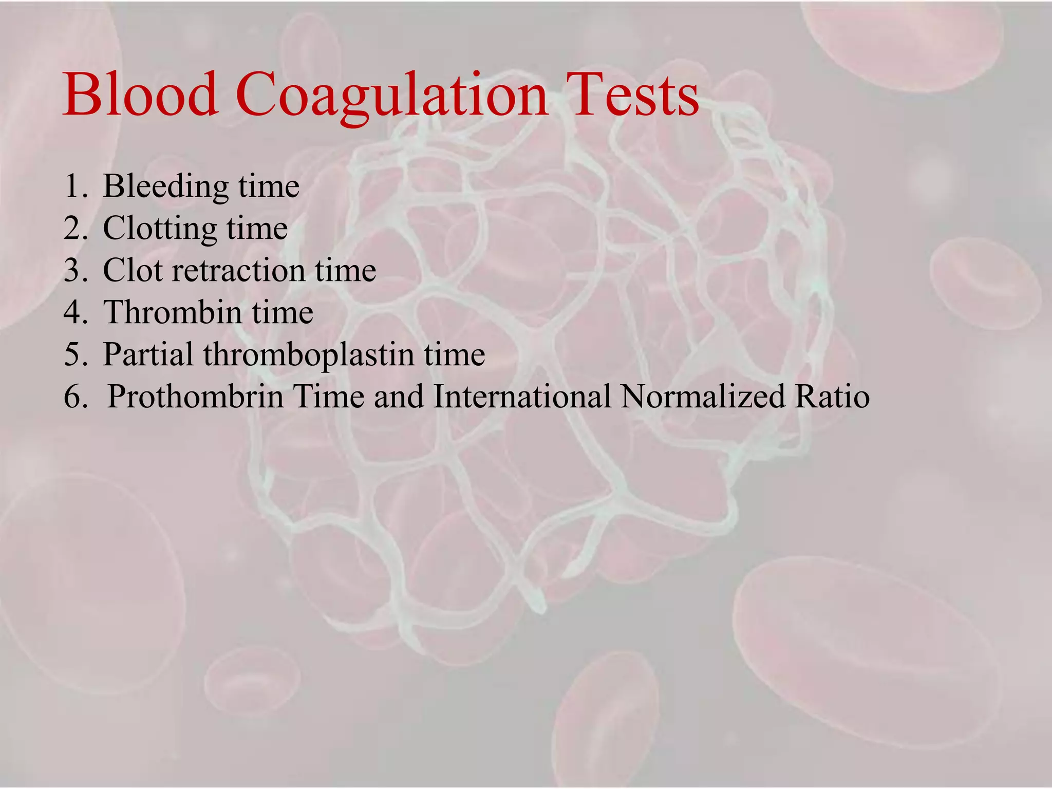
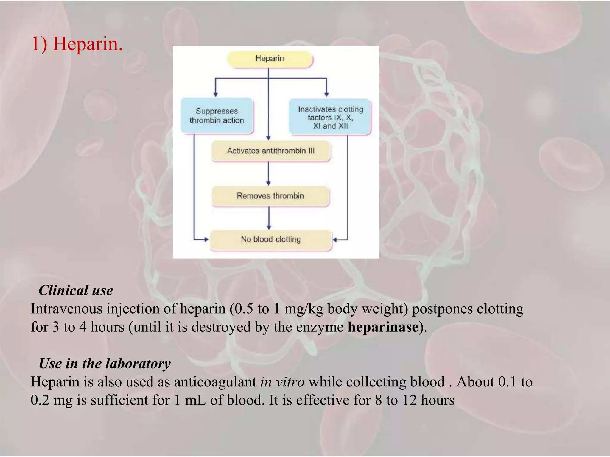
Nazia Yaseen
I MDS
Contents
• Events in haemostasis
• Classification
• Blood coagulation tests
• Hematinics
• Coagulants
• Local hemostatics
• Techniques to maintain hemostasis
• Anticoagulants
• Conclusion
Hemostasis
Haemostasis is the physiologic system of competent blood
vessels, endothelial cells, platelets and numerous plasma
proteins that act in a finely controlled manner to preserve
blood vessel integrity and prevent pathologic hemorrhage or
thrombosis.
Haemostasis is achieved by :-
 Vascular constriction
 Formation of platelet plug
 Formation of blood clot
 Growth of fibrous tissue into the clot.
Vascular Constriction
The contraction results from:-
 Local myogenic spasms
 Local autacoid factors
 Nervous reflexes
Platelets release, serotonin and throboxane A2 which is
responsible for vasoconstriction of smaller vessels.
Formation of the Platelet Plug
 Mechanism of platelet plug
• Platelet adhesion
• Platelet activation
• Platelet aggregation
• Formation of temporary haemostatic plug
PLATELETS
 Platelets are enucleate cells
 1-4 micro meters in size
 Normal blood concentration of
platelets is 1.5L-3L/micro liters
 Formed in bone marrow from
megakaryocytes
 T-1/2 is 8-12 days
 Eliminated from the circulation
by tissue macrophages.
Platelet Plug
MECHANISM OF BLOOD COAGULATION
BASIC
THEORY
GENERAL
MECHANISM
GENERAL MECHANISM
In response to rupture of the vessel or damage to
the blood itself-formation of prothrombin activator
Prothrombin activator catalyzes conversion of
prothrombin to thrombin
Thrombin catalyzes fibrinogen into fibrin fibers.
INITIATION OF COAGUALTION
FORMATION OF PROTHROMBIN ACTIVATOR
These mechanisms are set into play by:-
 Trauma to the vascular wall and the adjacent tissues.
 Contact of the blood with damaged endothelial cells.
Prothrombin activator is generally considered to be formed in
these ways-
a) Extrinsic pathway
b) Intrinsic pathway
Intrinsic Pathway
Conversion of Prothombrin to Thombrin and
formation of fibrin
Clot
The clot is composed of a meshwork of fibrin fibers
running in all directions and entrapping blood cells,
platelets and plasma.
Clot Retraction
- Contractile proteins, namely actin, myosin and thrombosthenin
in the cytoplasm of platelets are responsible for clot retraction.
Fibrinolysis
Blood Coagulation Tests
1. Bleeding time
2. Clotting time
3. Clot retraction time
4. Thrombin time
5. Partial thromboplastin time
6. Prothombrin Time and International Normalized Ratio
Bleeding time (BT)
-The interval from oozing of blood after a cut or injury till arrest of bleeding.
-Ivy method: 2- 8 mins
-Duke method: 2- 5 mins
Prolonged Bleeding Time :
1. Thrombocytopenia
2. Bone Marrow aplasia
3. Defective Platelet aggregation
Clotting time (CT):-
-The time interval from oozing of blood after a cut or injury till the
formation of clot.
Normal Clotting Time : 6 – 10 mins
Prolonged Clotting time :
1. Liver Diseases
2. Anticoagulant therapy
3. Vit K deficiency
Clot retraction time:-
- A measure of platelet function.
- Platelet disorders & thrombocytopenia
Thrombin time:-
Blood test that measures the time it takes for a clot to form in the plasma of a
blood sample containing anticoagulant, after an excess of thrombin has been
added
If it is prolonged, a quantitative or qualitative defect is present.
Normal range is 12 to 14 seconds.
Partial thromboplastin time:-
- Performance indicator measuring the efficacy of both the intrinsic and
the common coagulation pathways.
- Also used to monitor the treatment effects with heparin.
- 30-50 seconds
Prothrombin time (PT):-
- Evaluates the extrinsic pathway of coagulation.
- Prothrombin time indicates the total quantity of prothrombin present in
the blood.
- Normal duration of prothrombin time is 10 to 12 seconds
- PT is used to check five blood clotting factors (factors I, II, V, VII, and
X) and to check the efficiency of anti-coagulant therapy.
- Measured in values of international normalized ratio.
- Blood takes longer time to clot if INR is higher.
- In patients taking anticoagulant therapy for atrial fibrillation-2
and 3.
- For patients with heart valve disorders - 3 and 4.
- But, INR greater than 4 indicates that blood is clotting too
slowly and there is a risk of uncontrolled blood clotting.
- International normalized ratio values may be increased because
of the use of medications enhancing the effect of coumarin, a diet
rich in vitamin K and/or compliance reasons.
INR Significance
4 or greater No surgical treatments
3.5 – 4 Emergency minor surgical
procedures
3 – 3.4 Minor surgical procedures
2.5 – 2.9 Multiple extractions, quadrant
flap surgery, SRP
1.5 -2.5 Full mouth extractions,
gingivectomy, multiple quadrant
flap surgery
Disorders of Blood Coagulation
Inherited Acquired
- Hemophilia A, B, C - Liver diseases
- VWD - DIC
Hematinics
- Compounds required in the formation of blood and employed
in the treatment of anemias.
Iron
-Total body Iron : 2.5 – 5 gms
Daily requirement of Iron :
Adult male : 0.5 -1 mg
Adult female : 1 – 2 mg
Pregnancy and lactation : 3 – 5 mg
Dietary sources of Iron :
Liver, egg, yolk, meat, fish, chicken, spinach, dry fruits, wheat
and apple.
Vit B12 and Folic acid
-Coenzymes for several vital metabolic reactions and essential for
DNA synthesis.
Daily requirement
Dietary sources :
Vit B12 – Liver, fish, egg, meat, cheese and pulses
Folic acid – Green veg, liver, yeast, egg and milk
Adults Pregnancy & Lactation
Vitamin B12 1-3 µg 3-5 µg
Folic acid 50-100 µg 200-400 µg
Coagulants
- These are substances which promote coagulation, and are indicated in
haemorrhagic states.
- Also called Hemostatics.
Vitamin K
1. K1 (from plants : Phytonadione fat-soluble) (Phylloquinone)
2. K3 (synthetic) :
- Fat-soluble : Menadione, Acetomenaphthone
- Water-soluble : Menadione sod. bisulfite : Menadione sod. Diphosphate
Miscellaneous
1. Fibrinogen (human)
2. Antihaemophilic factor
3. Desmopressin
4. Adrenochrome monosemicarbazone
5. Rutin, Ethamsylate
Vitamin K
• Plays important role in coagulation process.
• Helps in production of Factors 2, 7, 9, 10 by the liver.
• K1 - food from plant source
• K2 – gut by bacteria
• K3 – synthetic compound used therapeutically
Uses :-
1. Vit K deficiency
2. New borns
3. Oral anticoagulant toxicity
Preparations
- Phytonadione: VITAMIN-K, KVI, K-WIN 10 mg/ml for i.m. injection.
- Menadione: 0.66 mg in GYNAE CVP with vit C 75 mg, ferrous gluconate
67 mg, Cal. lactate 300 mg and citras bioflavonoid 150 mg per cap
Acetomenaphthone: ACETOMENADIONE 5, 10 mg tab; KAPILIN 10 mg
tab.
- Menadione sod. bisulfite: 20 mg, in CADISPER-C with vit C 100 mg,
adrenochrome monosemicarbazone, 1 mg, rutin 60 mg, methylhesperidin 40
mg, Cal. phosphate 100 mg per tab.
- STYPTOCID 10 mg with adrenochrome monosemicarbazone 0.5 mg, rutin
50 mg, vit C 37.5 mg, vit D 200 i.u., Cal. phosphate 260 mg per tab.
Fibrinogen
- Control bleeding in haemophilia, antihaemophilic globulin (AHG) deficiency and
acute afibrinogenemic states.
FIBRINAL 0.5 g/bottle for i.v. infusion.
Antihaemophilic factor
It is concentrated human AHG prepared from pooled human plasma. It is indicated
in haemophilia and AHG deficiency.
Action is short-lasting (1 to 2 days).
Dose: 5–10 U/kg by i.v. infusion, repeated 6–12 hourly.
FIBRINAL-H, ANTIHAEMOPHILIC FACTOR: 150 U or 200 U + fibrinogen
0.5 g/bottle for i.v. infusion.
Desmopressin
It releases factor VIII and von Willebrand’s factor from vascular endothelium
and checks bleeding in haemophilia and von Willebrand’s disease.
Adrenochrome monosemicarbazone
It is believed to reduce capillary fragility, control oozing from raw surfaces
and prevent microvessel bleeding, e.g. epistaxis, haematuria, secondary
haemorrhage from wounds, etc.
Dose: 1–5 mg oral, i.m. STYPTOCHROME 3 mg/2 ml inj., STYPTOCID: 2
mg/ 2 ml inj
Rutin
It is a plant glycoside claimed to reduce capillary bleeding. It has been used in
a dose of 60 mg oral BD– TDS along with vit C which is believed to facilitate
its action. In CADISPER-C 60 mg tab.
Ethamsylate
It reduces capillary bleeding when platelets are adequate; probably exerts
antihyaluronidase action or corrects abnormalities of platelet adhesion, but
does not stabilize fibrin (not an antifibrinolytic). Ethamsylate has been used in
after tooth extraction, but efficacy is unsubstantiated.
Side effects are nausea, rash, headache, and fall in BP (only after i.v.
injection).
Dose: 250–500 mg TDS oral/i.v.; ETHAMSYL, DICYNENE, HEMSYL, K.
STAT 250, 500 mg tabs; 250 mg/2 ml inj
Local Hemostatics(Styptics)
- Fibrin
- Gelatin foam
- Oxidized cellulose
- Thrombin
- Adrenaline
- Astringents – Tannic acid or metallic salts
TECHNIQUES TO MAINTAIN HEMOSTASIS
MECHANICAL
THERMAL
CHEMICAL
Mechanical haemostasis
Direct pressure
Gauze pack
Suture and ligation
Staples
THERMAL TECHNIQUES
• ELECTROCAUTERY
• HAEMOSTATIC SCALPEL
• LASER
CHEMICAL TECHNIQUES
- Pharmacotherapy with vitamin K, desmopressin, tranexamic
acid, aminocaproic acid, protamine and epinephrine.
- Topical haemostats like collagen, cellulose, gelatins and
thrombins.
- Topical sealants and adhesives like fibrin sealants and synthetic
glues
Commonly used styptics in periodontics:-
Snake venom (Botropase inj/spray).
Absorbable gelatin (surgispon).
Absorbable collagen (oraplug).
Collagen dressings (collasorb).
Tranexamic acid.
Fibrin glue (tisseel).
Bone wax (ethicon).
Laser.
Electrocautery.
Anticoagulants
I. Used in vivo
A. Parenteral anticoagulants
Heparin, Low molecular weight heparins, Heparan sulfate
B. Oral anticoagulants
(i) Coumarin derivatives: Bishydroxycoumarin (dicumarol), Warfarin sod,
Acenocoumarol (Nicoumalone), Ethylbiscoumacetate
(ii) Indandione derivative: Phenindione
II. Used in vitro
A. Heparin
B. Calcium complexing agents:
EDTA
Sodium citrate
Sodium oxalate
Sodium edetate
1) Heparin.
Clinical use
Intravenous injection of heparin (0.5 to 1 mg/kg body weight) postpones clotting
for 3 to 4 hours (until it is destroyed by the enzyme heparinase).
Use in the laboratory
Heparin is also used as anticoagulant in vitro while collecting blood . About 0.1 to
0.2 mg is sufficient for 1 mL of blood. It is effective for 8 to 12 hours
2. Coumarin Deriatives
Dicoumoral and warfarin are the commonly used oral anticoagulants ( in vivo).
Warfarin is used to prevent myocardial infarction (heart attack), strokes and
thrombosis.
3. EDTA
i. Commonly administered intravenously, in cases of lead poisoning.
ii. Used as an anticoagulant in the laboratory (in vitro). 0.5 to 2.0 mg of EDTA per
mL of blood is sufficient to preserve the blood for at least 6 hours. On refrigeration,
it can preserve the blood up to 24 hours.
Conclusion
References
• Guyton CA and Hall B Textbook of Medical Physiology
• Essentials of Medical Physiology – A. Sembulingum
• Essentials of Medical Pharmacology – KD Tripathi
• Hemostasis in Dentistry – Richard P.Szumita
• Vassilopoulos P & Palcanis k. Bleeding disorders and
Periodontology; Periodontology 2000 2007;44; 211-223
Hemostasis

Hemostasis

  • 1.
  • 2.
    Contents • Events inhaemostasis • Classification • Blood coagulation tests • Hematinics • Coagulants • Local hemostatics • Techniques to maintain hemostasis • Anticoagulants • Conclusion
  • 3.
    Hemostasis Haemostasis is thephysiologic system of competent blood vessels, endothelial cells, platelets and numerous plasma proteins that act in a finely controlled manner to preserve blood vessel integrity and prevent pathologic hemorrhage or thrombosis. Haemostasis is achieved by :-  Vascular constriction  Formation of platelet plug  Formation of blood clot  Growth of fibrous tissue into the clot.
  • 4.
    Vascular Constriction The contractionresults from:-  Local myogenic spasms  Local autacoid factors  Nervous reflexes Platelets release, serotonin and throboxane A2 which is responsible for vasoconstriction of smaller vessels.
  • 5.
    Formation of thePlatelet Plug  Mechanism of platelet plug • Platelet adhesion • Platelet activation • Platelet aggregation • Formation of temporary haemostatic plug
  • 6.
    PLATELETS  Platelets areenucleate cells  1-4 micro meters in size  Normal blood concentration of platelets is 1.5L-3L/micro liters  Formed in bone marrow from megakaryocytes  T-1/2 is 8-12 days  Eliminated from the circulation by tissue macrophages.
  • 7.
  • 8.
    MECHANISM OF BLOODCOAGULATION BASIC THEORY GENERAL MECHANISM
  • 9.
    GENERAL MECHANISM In responseto rupture of the vessel or damage to the blood itself-formation of prothrombin activator Prothrombin activator catalyzes conversion of prothrombin to thrombin Thrombin catalyzes fibrinogen into fibrin fibers.
  • 10.
    INITIATION OF COAGUALTION FORMATIONOF PROTHROMBIN ACTIVATOR These mechanisms are set into play by:-  Trauma to the vascular wall and the adjacent tissues.  Contact of the blood with damaged endothelial cells. Prothrombin activator is generally considered to be formed in these ways- a) Extrinsic pathway b) Intrinsic pathway
  • 13.
  • 14.
    Conversion of Prothombrinto Thombrin and formation of fibrin
  • 16.
    Clot The clot iscomposed of a meshwork of fibrin fibers running in all directions and entrapping blood cells, platelets and plasma.
  • 17.
    Clot Retraction - Contractileproteins, namely actin, myosin and thrombosthenin in the cytoplasm of platelets are responsible for clot retraction.
  • 18.
  • 21.
    Blood Coagulation Tests 1.Bleeding time 2. Clotting time 3. Clot retraction time 4. Thrombin time 5. Partial thromboplastin time 6. Prothombrin Time and International Normalized Ratio
  • 22.
    Bleeding time (BT) -Theinterval from oozing of blood after a cut or injury till arrest of bleeding. -Ivy method: 2- 8 mins -Duke method: 2- 5 mins Prolonged Bleeding Time : 1. Thrombocytopenia 2. Bone Marrow aplasia 3. Defective Platelet aggregation
  • 23.
    Clotting time (CT):- -Thetime interval from oozing of blood after a cut or injury till the formation of clot. Normal Clotting Time : 6 – 10 mins Prolonged Clotting time : 1. Liver Diseases 2. Anticoagulant therapy 3. Vit K deficiency
  • 24.
    Clot retraction time:- -A measure of platelet function. - Platelet disorders & thrombocytopenia Thrombin time:- Blood test that measures the time it takes for a clot to form in the plasma of a blood sample containing anticoagulant, after an excess of thrombin has been added If it is prolonged, a quantitative or qualitative defect is present. Normal range is 12 to 14 seconds.
  • 25.
    Partial thromboplastin time:- -Performance indicator measuring the efficacy of both the intrinsic and the common coagulation pathways. - Also used to monitor the treatment effects with heparin. - 30-50 seconds Prothrombin time (PT):- - Evaluates the extrinsic pathway of coagulation. - Prothrombin time indicates the total quantity of prothrombin present in the blood. - Normal duration of prothrombin time is 10 to 12 seconds - PT is used to check five blood clotting factors (factors I, II, V, VII, and X) and to check the efficiency of anti-coagulant therapy. - Measured in values of international normalized ratio.
  • 27.
    - Blood takeslonger time to clot if INR is higher. - In patients taking anticoagulant therapy for atrial fibrillation-2 and 3. - For patients with heart valve disorders - 3 and 4. - But, INR greater than 4 indicates that blood is clotting too slowly and there is a risk of uncontrolled blood clotting. - International normalized ratio values may be increased because of the use of medications enhancing the effect of coumarin, a diet rich in vitamin K and/or compliance reasons.
  • 28.
    INR Significance 4 orgreater No surgical treatments 3.5 – 4 Emergency minor surgical procedures 3 – 3.4 Minor surgical procedures 2.5 – 2.9 Multiple extractions, quadrant flap surgery, SRP 1.5 -2.5 Full mouth extractions, gingivectomy, multiple quadrant flap surgery
  • 29.
    Disorders of BloodCoagulation Inherited Acquired - Hemophilia A, B, C - Liver diseases - VWD - DIC
  • 30.
    Hematinics - Compounds requiredin the formation of blood and employed in the treatment of anemias.
  • 31.
    Iron -Total body Iron: 2.5 – 5 gms Daily requirement of Iron : Adult male : 0.5 -1 mg Adult female : 1 – 2 mg Pregnancy and lactation : 3 – 5 mg Dietary sources of Iron : Liver, egg, yolk, meat, fish, chicken, spinach, dry fruits, wheat and apple.
  • 33.
    Vit B12 andFolic acid -Coenzymes for several vital metabolic reactions and essential for DNA synthesis. Daily requirement Dietary sources : Vit B12 – Liver, fish, egg, meat, cheese and pulses Folic acid – Green veg, liver, yeast, egg and milk Adults Pregnancy & Lactation Vitamin B12 1-3 µg 3-5 µg Folic acid 50-100 µg 200-400 µg
  • 34.
    Coagulants - These aresubstances which promote coagulation, and are indicated in haemorrhagic states. - Also called Hemostatics. Vitamin K 1. K1 (from plants : Phytonadione fat-soluble) (Phylloquinone) 2. K3 (synthetic) : - Fat-soluble : Menadione, Acetomenaphthone - Water-soluble : Menadione sod. bisulfite : Menadione sod. Diphosphate Miscellaneous 1. Fibrinogen (human) 2. Antihaemophilic factor 3. Desmopressin 4. Adrenochrome monosemicarbazone 5. Rutin, Ethamsylate
  • 35.
    Vitamin K • Playsimportant role in coagulation process. • Helps in production of Factors 2, 7, 9, 10 by the liver. • K1 - food from plant source • K2 – gut by bacteria • K3 – synthetic compound used therapeutically Uses :- 1. Vit K deficiency 2. New borns 3. Oral anticoagulant toxicity
  • 36.
    Preparations - Phytonadione: VITAMIN-K,KVI, K-WIN 10 mg/ml for i.m. injection. - Menadione: 0.66 mg in GYNAE CVP with vit C 75 mg, ferrous gluconate 67 mg, Cal. lactate 300 mg and citras bioflavonoid 150 mg per cap Acetomenaphthone: ACETOMENADIONE 5, 10 mg tab; KAPILIN 10 mg tab. - Menadione sod. bisulfite: 20 mg, in CADISPER-C with vit C 100 mg, adrenochrome monosemicarbazone, 1 mg, rutin 60 mg, methylhesperidin 40 mg, Cal. phosphate 100 mg per tab. - STYPTOCID 10 mg with adrenochrome monosemicarbazone 0.5 mg, rutin 50 mg, vit C 37.5 mg, vit D 200 i.u., Cal. phosphate 260 mg per tab.
  • 37.
    Fibrinogen - Control bleedingin haemophilia, antihaemophilic globulin (AHG) deficiency and acute afibrinogenemic states. FIBRINAL 0.5 g/bottle for i.v. infusion. Antihaemophilic factor It is concentrated human AHG prepared from pooled human plasma. It is indicated in haemophilia and AHG deficiency. Action is short-lasting (1 to 2 days). Dose: 5–10 U/kg by i.v. infusion, repeated 6–12 hourly. FIBRINAL-H, ANTIHAEMOPHILIC FACTOR: 150 U or 200 U + fibrinogen 0.5 g/bottle for i.v. infusion.
  • 38.
    Desmopressin It releases factorVIII and von Willebrand’s factor from vascular endothelium and checks bleeding in haemophilia and von Willebrand’s disease. Adrenochrome monosemicarbazone It is believed to reduce capillary fragility, control oozing from raw surfaces and prevent microvessel bleeding, e.g. epistaxis, haematuria, secondary haemorrhage from wounds, etc. Dose: 1–5 mg oral, i.m. STYPTOCHROME 3 mg/2 ml inj., STYPTOCID: 2 mg/ 2 ml inj
  • 39.
    Rutin It is aplant glycoside claimed to reduce capillary bleeding. It has been used in a dose of 60 mg oral BD– TDS along with vit C which is believed to facilitate its action. In CADISPER-C 60 mg tab. Ethamsylate It reduces capillary bleeding when platelets are adequate; probably exerts antihyaluronidase action or corrects abnormalities of platelet adhesion, but does not stabilize fibrin (not an antifibrinolytic). Ethamsylate has been used in after tooth extraction, but efficacy is unsubstantiated. Side effects are nausea, rash, headache, and fall in BP (only after i.v. injection). Dose: 250–500 mg TDS oral/i.v.; ETHAMSYL, DICYNENE, HEMSYL, K. STAT 250, 500 mg tabs; 250 mg/2 ml inj
  • 40.
    Local Hemostatics(Styptics) - Fibrin -Gelatin foam - Oxidized cellulose - Thrombin - Adrenaline - Astringents – Tannic acid or metallic salts
  • 41.
    TECHNIQUES TO MAINTAINHEMOSTASIS MECHANICAL THERMAL CHEMICAL
  • 42.
    Mechanical haemostasis Direct pressure Gauzepack Suture and ligation Staples
  • 43.
    THERMAL TECHNIQUES • ELECTROCAUTERY •HAEMOSTATIC SCALPEL • LASER
  • 44.
    CHEMICAL TECHNIQUES - Pharmacotherapywith vitamin K, desmopressin, tranexamic acid, aminocaproic acid, protamine and epinephrine. - Topical haemostats like collagen, cellulose, gelatins and thrombins. - Topical sealants and adhesives like fibrin sealants and synthetic glues
  • 45.
    Commonly used stypticsin periodontics:- Snake venom (Botropase inj/spray). Absorbable gelatin (surgispon). Absorbable collagen (oraplug). Collagen dressings (collasorb). Tranexamic acid. Fibrin glue (tisseel). Bone wax (ethicon). Laser. Electrocautery.
  • 46.
    Anticoagulants I. Used invivo A. Parenteral anticoagulants Heparin, Low molecular weight heparins, Heparan sulfate B. Oral anticoagulants (i) Coumarin derivatives: Bishydroxycoumarin (dicumarol), Warfarin sod, Acenocoumarol (Nicoumalone), Ethylbiscoumacetate (ii) Indandione derivative: Phenindione II. Used in vitro A. Heparin B. Calcium complexing agents: EDTA Sodium citrate Sodium oxalate Sodium edetate
  • 47.
    1) Heparin. Clinical use Intravenousinjection of heparin (0.5 to 1 mg/kg body weight) postpones clotting for 3 to 4 hours (until it is destroyed by the enzyme heparinase). Use in the laboratory Heparin is also used as anticoagulant in vitro while collecting blood . About 0.1 to 0.2 mg is sufficient for 1 mL of blood. It is effective for 8 to 12 hours
  • 48.
    2. Coumarin Deriatives Dicoumoraland warfarin are the commonly used oral anticoagulants ( in vivo). Warfarin is used to prevent myocardial infarction (heart attack), strokes and thrombosis. 3. EDTA i. Commonly administered intravenously, in cases of lead poisoning. ii. Used as an anticoagulant in the laboratory (in vitro). 0.5 to 2.0 mg of EDTA per mL of blood is sufficient to preserve the blood for at least 6 hours. On refrigeration, it can preserve the blood up to 24 hours.
  • 50.
  • 51.
    References • Guyton CAand Hall B Textbook of Medical Physiology • Essentials of Medical Physiology – A. Sembulingum • Essentials of Medical Pharmacology – KD Tripathi • Hemostasis in Dentistry – Richard P.Szumita • Vassilopoulos P & Palcanis k. Bleeding disorders and Periodontology; Periodontology 2000 2007;44; 211-223