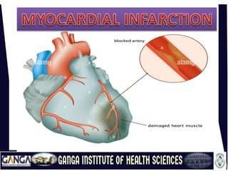
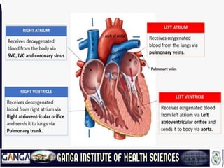
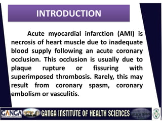
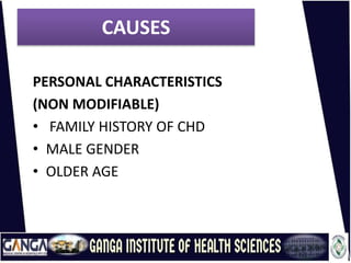
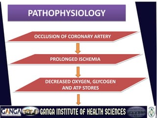
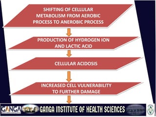
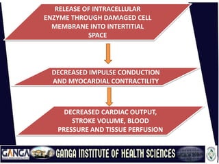
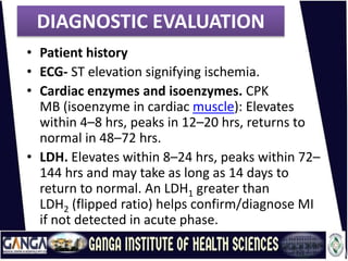
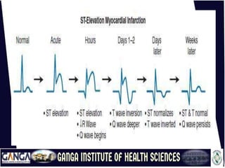
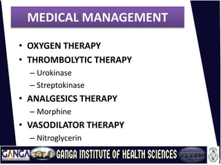
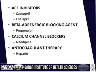
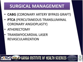
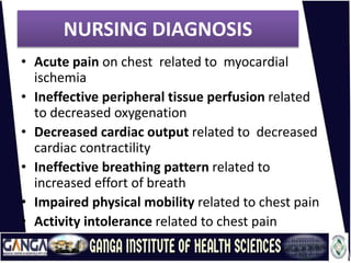
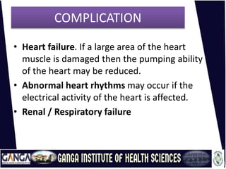
Related with cardio vascular system. Angina is Retrosternal chest pain which if left untreated can cause the higher complications with respect to cardiac health of human body. May be this is simple chest pain but if exceeds can cause major damage # prevention is better than cure :-)
Myocardial infraction or Heart attack are terms used anonymously, but the preferred term is MI.
In an MI an area of the myocardium is permanently destroyed.
MI is usually caused by reduced or decreased blood flow in a coronary artery due to rupture of an atherosclerotic plaque and subsequent occlusion of the artery by a thrombus.
Myocardial infarction (MI) death of the cells of an area of the heart muscle (myocardium) as a result of oxygen deprivation, which in turn is caused by obstruction of the blood supply; commonly referred to as a “heart attack.”
MI refers to the processes by which myocardial tissue is destroyed in regions of the heart that are deprived of an adequate blood supply because of reduced coronary artery blood flow.
Eighty percent to 90% of all acute MI are secondary to thrombus formation. When thrombus develops , perfusion to the myocardium distal to the occlusion is halted, resulting in necrosis.The myocardium receives its blood supply from the two large coronary arteries and their branches.
Occlusion of one or more of these blood vessels (coronary occlusion) is one of the major causes of myocardial infarction.
The occlusion may result from the formation of a clot that develops suddenly when an athermanous plaque ruptures through the sub layers of a blood vessel, or when the narrow, roughened inner lining of a scleroses artery leads to complete thrombosis.
The acute MI process takes time. Cardiac cells can withstand in ischemic conditions for approximately 20 minutes before cellular death begins.
The earliest tissue to become ischemic is the sub endocardium (the innermost layer of tissue in the cardiac muscle)
If ischemia persists, it takes approximately 4 to 6 hours for the entire thickness if the heart muscle to become necrosis.
Francesca Gottschalk - How can education support child empowerment.pptxEduSkills OECD
Francesca Gottschalk from the OECD’s Centre for Educational Research and Innovation presents at the Ask an Expert Webinar: How can education support child empowerment?
Normal Labour/ Stages of Labour/ Mechanism of LabourWasim Ak
Normal labor is also termed spontaneous labor, defined as the natural physiological process through which the fetus, placenta, and membranes are expelled from the uterus through the birth canal at term (37 to 42 weeks
Biological screening of herbal drugs: Introduction and Need for
Phyto-Pharmacological Screening, New Strategies for evaluating
Natural Products, In vitro evaluation techniques for Antioxidants, Antimicrobial and Anticancer drugs. In vivo evaluation techniques
for Anti-inflammatory, Antiulcer, Anticancer, Wound healing, Antidiabetic, Hepatoprotective, Cardio protective, Diuretics and
Antifertility, Toxicity studies as per OECD guidelines
2024.06.01 Introducing a competency framework for languag learning materials ...Sandy Millin
http://sandymillin.wordpress.com/iateflwebinar2024
Published classroom materials form the basis of syllabuses, drive teacher professional development, and have a potentially huge influence on learners, teachers and education systems. All teachers also create their own materials, whether a few sentences on a blackboard, a highly-structured fully-realised online course, or anything in between. Despite this, the knowledge and skills needed to create effective language learning materials are rarely part of teacher training, and are mostly learnt by trial and error.
Knowledge and skills frameworks, generally called competency frameworks, for ELT teachers, trainers and managers have existed for a few years now. However, until I created one for my MA dissertation, there wasn’t one drawing together what we need to know and do to be able to effectively produce language learning materials.
This webinar will introduce you to my framework, highlighting the key competencies I identified from my research. It will also show how anybody involved in language teaching (any language, not just English!), teacher training, managing schools or developing language learning materials can benefit from using the framework.
Unit 8 - Information and Communication Technology (Paper I).pdfThiyagu K
This slides describes the basic concepts of ICT, basics of Email, Emerging Technology and Digital Initiatives in Education. This presentations aligns with the UGC Paper I syllabus.
Acetabularia Information For Class 9 .docxvaibhavrinwa19
Acetabularia acetabulum is a single-celled green alga that in its vegetative state is morphologically differentiated into a basal rhizoid and an axially elongated stalk, which bears whorls of branching hairs. The single diploid nucleus resides in the rhizoid.
5. INTRODUCTION
Acute myocardial infarction (AMI) is
necrosis of heart muscle due to inadequate
blood supply following an acute coronary
occlusion. This occlusion is usually due to
plaque rupture or fissuring with
superimposed thrombosis. Rarely, this may
result from coronary spasm, coronary
embolism or vasculitis.
6. DEFINITION
A Myocardial infarction occurs as a
result of sustained ischemia, causing
irreversible myocardial cell death (necrosis).
A heart attack is when blood vessels
that supply blood to the heart are blocked,
preventing enough oxygen from getting to
the heart. This causes the muscles to die.
7. CAUSES
LIFE STYLE
• Tobacco smoking
• Excessive alcohol
• Physical Inactivity
• High saturated diet and cholesterol
12. SHIFTING OF CELLULAR
METABOLISM FROM AEROBIC
PROCESS TO ANEROBIC PROCESS
PRODUCTION OF HYDROGEN ION
AND LACTIC ACID
CELLULAR ACIDOSIS
INCREASED CELL VULNERABILITY
TO FURTHER DAMAGE
13. RELEASE OF INTRACELLULAR
ENZYME THROUGH DAMAGED CELL
MEMBRANE INTO INTERTITIAL
SPACE
DECREASED IMPULSE CONDUCTION
AND MYOCARDIAL CONTRACTILITY
DECREASED CARDIAC OUTPUT,
STROKE VOLUME, BLOOD
PRESSURE AND TISSUE PERFUSION
14. SYMPTOMS
Chest pain (central chest pain may not be the
main symptom):
• Three quarters of patients present with
characteristic central or epigastric chest pain
radiating to the arms, shoulders, neck, or jaw.
• The pain is described as substernal pressure,
squeezing, aching, burning, or even sharp pain.
• Radiation to the left arm or neck is common.
• Chest pain may be associated with sweating,
nausea, vomiting, dyspnoea, fatigue and/or
palpitations.
15. • Anxiety & Fainting
• Cough
• Light-headedness, dizziness
• Nausea and vomiting
• Palpitations (feeling like your heart is
beating too fast or irregularly)
• Shortness of breath
• Sweating, which may be extreme.
• Pale skin
16. • Diaphoresis (an excessive form of
sweating),
• Light-headedness, and
• Palpitations
• Loss of consciousness
• inadequate blood flow to the brain
• and cardiogenic shock.
• Sudden death
• due to the development of ventricular
fibrillation.
17. DIAGNOSTIC EVALUATION
• Patient history
• ECG- ST elevation signifying ischemia.
• Cardiac enzymes and isoenzymes. CPK
MB (isoenzyme in cardiac muscle): Elevates
within 4–8 hrs, peaks in 12–20 hrs, returns to
normal in 48–72 hrs.
• LDH. Elevates within 8–24 hrs, peaks within 72–
144 hrs and may take as long as 14 days to
return to normal. An LDH1 greater than
LDH2 (flipped ratio) helps confirm/diagnose MI
if not detected in acute phase.
18. • Troponins. Troponin I (cTnI) and troponin T
(cTnT): Levels are elevated at 4–6 hr, peak at
14–18 hr, and return to baseline over 6–7
days. These enzymes have increased
specificity for necrosis and are therefore
useful in diagnosing postoperative MI when
MB-CPK may be elevated related to skeletal
trauma.
• Electrolytes. Imbalances
of sodium and potassium can alter
conduction and compromise contractility.
• WBC. Leukocytosis (10,000–20,000) usually
appears on the second day after MI because
of the inflammatory process.
19. • ESR : Rises on second or third day after MI,
indicating inflammatory response.
• ABGs/pulse oximetry: May indicate hypoxia or
acute/chronic lung disease processes.
• Lipids (total lipids, HDL, LDL, VLDL, total
cholesterol, triglycerides,
phospholipids): Elevations may reflect
arteriosclerosis as a cause for coronary narrowing
or spasm.
• Chest x-ray: May be normal or show an enlarged
cardiac shadow suggestive of HF or
ventricular aneurysm.
• Two-dimensional echocardiogram: May be done to
determine dimensions of chambers,
septal/ventricular wall motion, ejection fraction
(blood flow), and valve configuration/function.
20. • Nuclear imaging studies: Persantine or
Thallium: Evaluates myocardial blood flow
and status of myocardial cells, e.g.,
location/extent of acute/previous MI.
• Coronary angiography: Visualizes
narrowing/occlusion of coronary arteries and
is usually done in conjunction with
measurements of chamber pressures and
assessment of left ventricular function
(ejection fraction). Procedure is not usually
done in acute phase of MI unless angioplasty
or emergency heart surgery is imminent.
21. • Digital subtraction angiography
(DSA). Technique used to visualize status of
arterial bypass grafts and to detect peripheral
artery disease.
• Magnetic resonance imaging (MRI). Allows
visualization of blood flow, cardiac chambers
or intraventricular septum, valves, vascular
lesions, plaque formations, areas of
necrosis/infarction, and blood clots.
• Exercise stress test. Determines cardiovascular
response to activity (often done in conjunction
with thallium imaging in the recovery phase).
22. • Myoglobin: The myoglobin level can
elevate within 1 to 2 hours of acute MI
and peaks within 3 to 15 hours.
• Troponin: (troponin T and troponin I):
• Troponin I levels rise in about 3 hours,
peak at 14 to 18 hours, and remain
elevated for 5 to 7 days.
• Troponin T levels rise in 3 to 5 hours and
remain elevated for 10 to 14 days
27. NURSING DIAGNOSIS
• Acute pain on chest related to myocardial
ischemia
• Ineffective peripheral tissue perfusion related
to decreased oxygenation
• Decreased cardiac output related to decreased
cardiac contractility
• Ineffective breathing pattern related to
increased effort of breath
• Impaired physical mobility related to chest pain
• Activity intolerance related to chest pain
28. COMPLICATION
• Heart failure. If a large area of the heart
muscle is damaged then the pumping ability
of the heart may be reduced.
• Abnormal heart rhythms may occur if the
electrical activity of the heart is affected.
• Renal / Respiratory failure