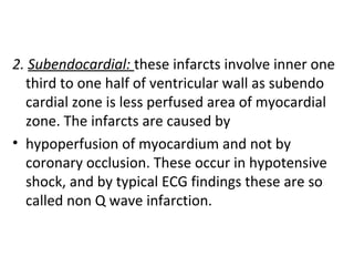
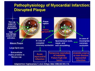
1. A myocardial infarction occurs when blood flow to the heart is blocked, damaging heart muscle.
2. It is caused most often by atherosclerosis and plaque buildup that obstruct coronary arteries.
3. Symptoms include chest pain and other signs of reduced blood supply to the heart. Diagnosis is based on symptoms, electrocardiogram changes, and blood tests showing cardiac enzyme levels.
Acute coronary syndrome result from a sudden blockage in a coronary artery. this blockage causes unstable angina or heart attack (MI), depending on the location and amount of blockage.
people who experience an ACS usually have chest pressure or ache, shortness of breath and fatigue.
People who think they are experiencing ACS should call for emergency help.
Doctors use ECG and blood test (troponin level) to determine whether a person is experiencing an ACS.
Treatment varies depending on the type of syndrome but usually include attempts to increase blood flow to affected area.
will help you in understanding myocardial infarction in more detail with its management and therapy with complications and with graphical knowledge you can understand it better and some laboratry test are also included in it .
Cardiomyopathy (KAR-de-o-mi-OP-ah-thee) refers to diseases of the heart muscle. These diseases have many causes, signs and symptoms, and treatments.
In cardiomyopathy, the heart muscle becomes enlarged, thick, or rigid. In rare cases, the muscle tissue in the heart is replaced with scar tissue.
As cardiomyopathy worsens, the heart becomes weaker. It's less able to pump blood through the body and maintain a normal electrical rhythm. This can lead toheart failure or irregular heartbeats called arrhythmias (ah-RITH-me-ahs). In turn, heart failure can cause fluid to build up in the lungs, ankles, feet, legs, or abdomen.
The weakening of the heart also can cause other complications, such as heart valve problems.
OverviewThe main types of cardiomyopathy are:
Dilated cardiomyopathy
Hypertrophic (hi-per-TROF-ik) cardiomyopathy
Restrictive cardiomyopathy
Arrhythmogenic (ah-rith-mo-JEN-ik) right ventricular dysplasia
(dis-PLA-ze-ah)
Other types of cardiomyopathy sometimes are referred to as "unclassified cardiomyopathy."
Cardiomyopathy can be acquired or inherited. "Acquired" means you aren't born with the disease, but you develop it due to another disease, condition, or factor. "Inherited" means your parents passed the gene for the disease on to you. Many times, the cause of cardiomyopathy isn't known.
Cardiomyopathy can affect people of all ages. However, people in certain age groups are more likely to have certain types of cardiomyopathy. This article focuses on cardiomyopathy in adults.
OutlookSome people who have cardiomyopathy have no signs or symptoms and need no treatment. For other people, the disease develops quickly, symptoms are severe, and serious complications occur.
Treatments for cardiomyopathy include lifestyle changes, medicines, surgery, implanted devices to correct arrhythmias, and a nonsurgical procedure. These treatments can control symptoms, reduce complications, and stop the disease from getting worse.
National Heart Lung and Blood Institute
Coronary artery disease (CAD) also known as atherosclerotic heart disease, atherosclerotic cardiovascular disease, coronary heart disease, or ischemic heart disease (IHD), is the most common type of heart disease and cause of heart attacks. The disease is caused by plaque building up along the inner walls of the arteries of the heart, which narrows the lumen of arteries and reduces blood flow to the heart.
cerebrovascular disease , denotes any abnormality of the brain caused by a, pathologic process, involving , blood vessels.
The three basic processes are
thrombotic occlusion of vessels,
(2) embolic occlusion of vessels,
(3) vascular rupture.
Cerebrovascular diseases include the three major categories:
1.Thrombosis, 2.Embolism, and 3.Hemorrhage.
. Vasculitis, major cause of thrombosis
The prostate is an exocrine gland of the male mammalian reproductive system
It is a walnut-sized gland that forms part of the male reproductive system and is located in front of the rectum and just below the urinary bladder
Function is to store and secrete a clear, slightly alkaline fluid that constitutes 10-30% of the volume of the seminal fluid that along with the spermatozoa, constitutes semen
A healthy human prostate measures (4cm-vertical, by 3cm-horizontal, 2cm ant-post ).
It surrounds the urethra just below the urinary bladder. It has anterior, median, posterior and two lateral lobes
It’s work is regulated by androgens which are responsible for male sex characteristics
Generalised disease of the prostate due to hormonal derangement which leads to non malignant enlargement of the gland (increase in the number of epithelial cells and stromal tissue)to cause compression of the urethra leading to symptoms (LUTS
micro teaching on communication m.sc nursing.pdfAnurag Sharma
Microteaching is a unique model of practice teaching. It is a viable instrument for the. desired change in the teaching behavior or the behavior potential which, in specified types of real. classroom situations, tends to facilitate the achievement of specified types of objectives.
263778731218 Abortion Clinic /Pills In Harare ,sisternakatoto
263778731218 Abortion Clinic /Pills In Harare ,ABORTION WOMEN’S CLINIC +27730423979 IN women clinic we believe that every woman should be able to make choices in her pregnancy. Our job is to provide compassionate care, safety,affordable and confidential services. That’s why we have won the trust from all generations of women all over the world. we use non surgical method(Abortion pills) to terminate…Dr.LISA +27730423979women Clinic is committed to providing the highest quality of obstetrical and gynecological care to women of all ages. Our dedicated staff aim to treat each patient and her health concerns with compassion and respect.Our dedicated group ABORTION WOMEN’S CLINIC +27730423979 IN women clinic we believe that every woman should be able to make choices in her pregnancy. Our job is to provide compassionate care, safety,affordable and confidential services. That’s why we have won the trust from all generations of women all over the world. we use non surgical method(Abortion pills) to terminate…Dr.LISA +27730423979women Clinic is committed to providing the highest quality of obstetrical and gynecological care to women of all ages. Our dedicated staff aim to treat each patient and her health concerns with compassion and respect.Our dedicated group of receptionists, nurses, and physicians have worked together as a teamof receptionists, nurses, and physicians have worked together as a team wwww.lisywomensclinic.co.za/
Lung Cancer: Artificial Intelligence, Synergetics, Complex System Analysis, S...Oleg Kshivets
RESULTS: Overall life span (LS) was 2252.1±1742.5 days and cumulative 5-year survival (5YS) reached 73.2%, 10 years – 64.8%, 20 years – 42.5%. 513 LCP lived more than 5 years (LS=3124.6±1525.6 days), 148 LCP – more than 10 years (LS=5054.4±1504.1 days).199 LCP died because of LC (LS=562.7±374.5 days). 5YS of LCP after bi/lobectomies was significantly superior in comparison with LCP after pneumonectomies (78.1% vs.63.7%, P=0.00001 by log-rank test). AT significantly improved 5YS (66.3% vs. 34.8%) (P=0.00000 by log-rank test) only for LCP with N1-2. Cox modeling displayed that 5YS of LCP significantly depended on: phase transition (PT) early-invasive LC in terms of synergetics, PT N0—N12, cell ratio factors (ratio between cancer cells- CC and blood cells subpopulations), G1-3, histology, glucose, AT, blood cell circuit, prothrombin index, heparin tolerance, recalcification time (P=0.000-0.038). Neural networks, genetic algorithm selection and bootstrap simulation revealed relationships between 5YS and PT early-invasive LC (rank=1), PT N0—N12 (rank=2), thrombocytes/CC (3), erythrocytes/CC (4), eosinophils/CC (5), healthy cells/CC (6), lymphocytes/CC (7), segmented neutrophils/CC (8), stick neutrophils/CC (9), monocytes/CC (10); leucocytes/CC (11). Correct prediction of 5YS was 100% by neural networks computing (area under ROC curve=1.0; error=0.0).
CONCLUSIONS: 5YS of LCP after radical procedures significantly depended on: 1) PT early-invasive cancer; 2) PT N0--N12; 3) cell ratio factors; 4) blood cell circuit; 5) biochemical factors; 6) hemostasis system; 7) AT; 8) LC characteristics; 9) LC cell dynamics; 10) surgery type: lobectomy/pneumonectomy; 11) anthropometric data. Optimal diagnosis and treatment strategies for LC are: 1) screening and early detection of LC; 2) availability of experienced thoracic surgeons because of complexity of radical procedures; 3) aggressive en block surgery and adequate lymph node dissection for completeness; 4) precise prediction; 5) adjuvant chemoimmunoradiotherapy for LCP with unfavorable prognosis.
Knee anatomy and clinical tests 2024.pdfvimalpl1234
This includes all relevant anatomy and clinical tests compiled from standard textbooks, Campbell,netter etc..It is comprehensive and best suited for orthopaedicians and orthopaedic residents.
ARTIFICIAL INTELLIGENCE IN HEALTHCARE.pdfAnujkumaranit
Artificial intelligence (AI) refers to the simulation of human intelligence processes by machines, especially computer systems. It encompasses tasks such as learning, reasoning, problem-solving, perception, and language understanding. AI technologies are revolutionizing various fields, from healthcare to finance, by enabling machines to perform tasks that typically require human intelligence.
- Video recording of this lecture in English language: https://youtu.be/lK81BzxMqdo
- Video recording of this lecture in Arabic language: https://youtu.be/Ve4P0COk9OI
- Link to download the book free: https://nephrotube.blogspot.com/p/nephrotube-nephrology-books.html
- Link to NephroTube website: www.NephroTube.com
- Link to NephroTube social media accounts: https://nephrotube.blogspot.com/p/join-nephrotube-on-social-media.html
Report Back from SGO 2024: What’s the Latest in Cervical Cancer?bkling
Are you curious about what’s new in cervical cancer research or unsure what the findings mean? Join Dr. Emily Ko, a gynecologic oncologist at Penn Medicine, to learn about the latest updates from the Society of Gynecologic Oncology (SGO) 2024 Annual Meeting on Women’s Cancer. Dr. Ko will discuss what the research presented at the conference means for you and answer your questions about the new developments.
HOT NEW PRODUCT! BIG SALES FAST SHIPPING NOW FROM CHINA!! EU KU DB BK substit...GL Anaacs
Contact us if you are interested:
Email / Skype : kefaya1771@gmail.com
Threema: PXHY5PDH
New BATCH Ku !!! MUCH IN DEMAND FAST SALE EVERY BATCH HAPPY GOOD EFFECT BIG BATCH !
Contact me on Threema or skype to start big business!!
Hot-sale products:
NEW HOT EUTYLONE WHITE CRYSTAL!!
5cl-adba precursor (semi finished )
5cl-adba raw materials
ADBB precursor (semi finished )
ADBB raw materials
APVP powder
5fadb/4f-adb
Jwh018 / Jwh210
Eutylone crystal
Protonitazene (hydrochloride) CAS: 119276-01-6
Flubrotizolam CAS: 57801-95-3
Metonitazene CAS: 14680-51-4
Payment terms: Western Union,MoneyGram,Bitcoin or USDT.
Deliver Time: Usually 7-15days
Shipping method: FedEx, TNT, DHL,UPS etc.Our deliveries are 100% safe, fast, reliable and discreet.
Samples will be sent for your evaluation!If you are interested in, please contact me, let's talk details.
We specializes in exporting high quality Research chemical, medical intermediate, Pharmaceutical chemicals and so on. Products are exported to USA, Canada, France, Korea, Japan,Russia, Southeast Asia and other countries.
Explore natural remedies for syphilis treatment in Singapore. Discover alternative therapies, herbal remedies, and lifestyle changes that may complement conventional treatments. Learn about holistic approaches to managing syphilis symptoms and supporting overall health.
These simplified slides by Dr. Sidra Arshad present an overview of the non-respiratory functions of the respiratory tract.
Learning objectives:
1. Enlist the non-respiratory functions of the respiratory tract
2. Briefly explain how these functions are carried out
3. Discuss the significance of dead space
4. Differentiate between minute ventilation and alveolar ventilation
5. Describe the cough and sneeze reflexes
Study Resources:
1. Chapter 39, Guyton and Hall Textbook of Medical Physiology, 14th edition
2. Chapter 34, Ganong’s Review of Medical Physiology, 26th edition
3. Chapter 17, Human Physiology by Lauralee Sherwood, 9th edition
4. Non-respiratory functions of the lungs https://academic.oup.com/bjaed/article/13/3/98/278874
New Drug Discovery and Development .....NEHA GUPTA
The "New Drug Discovery and Development" process involves the identification, design, testing, and manufacturing of novel pharmaceutical compounds with the aim of introducing new and improved treatments for various medical conditions. This comprehensive endeavor encompasses various stages, including target identification, preclinical studies, clinical trials, regulatory approval, and post-market surveillance. It involves multidisciplinary collaboration among scientists, researchers, clinicians, regulatory experts, and pharmaceutical companies to bring innovative therapies to market and address unmet medical needs.
Flu Vaccine Alert in Bangalore Karnatakaaddon Scans
As flu season approaches, health officials in Bangalore, Karnataka, are urging residents to get their flu vaccinations. The seasonal flu, while common, can lead to severe health complications, particularly for vulnerable populations such as young children, the elderly, and those with underlying health conditions.
Dr. Vidisha Kumari, a leading epidemiologist in Bangalore, emphasizes the importance of getting vaccinated. "The flu vaccine is our best defense against the influenza virus. It not only protects individuals but also helps prevent the spread of the virus in our communities," he says.
This year, the flu season is expected to coincide with a potential increase in other respiratory illnesses. The Karnataka Health Department has launched an awareness campaign highlighting the significance of flu vaccinations. They have set up multiple vaccination centers across Bangalore, making it convenient for residents to receive their shots.
To encourage widespread vaccination, the government is also collaborating with local schools, workplaces, and community centers to facilitate vaccination drives. Special attention is being given to ensuring that the vaccine is accessible to all, including marginalized communities who may have limited access to healthcare.
Residents are reminded that the flu vaccine is safe and effective. Common side effects are mild and may include soreness at the injection site, mild fever, or muscle aches. These side effects are generally short-lived and far less severe than the flu itself.
Healthcare providers are also stressing the importance of continuing COVID-19 precautions. Wearing masks, practicing good hand hygiene, and maintaining social distancing are still crucial, especially in crowded places.
Protect yourself and your loved ones by getting vaccinated. Together, we can help keep Bangalore healthy and safe this flu season. For more information on vaccination centers and schedules, residents can visit the Karnataka Health Department’s official website or follow their social media pages.
Stay informed, stay safe, and get your flu shot today!
Pulmonary Thromboembolism - etilogy, types, medical- Surgical and nursing man...VarunMahajani
Disruption of blood supply to lung alveoli due to blockage of one or more pulmonary blood vessels is called as Pulmonary thromboembolism. In this presentation we will discuss its causes, types and its management in depth.
4. Myocardial infarction
A heart attack or acute myocardial infarction (MI) occurs when one of the arteries
that supplies the heart muscle becomes blocked. Blockage may be caused by
spasm of the artery or by atherosclerosis with acute clot formation. The blockage
results in damaged tissue and a permanent loss of contraction of this portion of the
heart muscle.
5. • Myocardial infarction (MI) is the irreversible
necrosis of heart muscle secondary to prolonged
ischemia. This usually results from an imbalance
of oxygen supply and demand.
6. CLASSIFICATION
1. Transmural: :(Q –wave infarction) : most
infartcts are transmural involve the full
thickness of ventricular wall in the
distribution of single coronary artery.
These infracts are caused by
• atherosclerosis,
• acute plaque changes
• less commonly thromboemboli or
vasospasm.
7. 2. Subendocardial: these infarcts involve inner one
third to one half of ventricular wall as subendo
cardial zone is less perfused area of myocardial
zone. The infarcts are caused by
• hypoperfusion of myocardium and not by
coronary occlusion. These occur in hypotensive
shock, and by typical ECG findings these are so
called non Q wave infarction.
9. Occlusion is typically seen in the proximal 2 cm of the
left anterior descending and left circumflex arteries
and in the proximal and distal thirds of the right
coronary artery.
o Rupture of the lipid-rich atheromatous plaque,
intraplaque hemorrhage, andintraluminal thrombus
are three pathological hallmarks most commonly
recognized in the infarct-related coronary artery at
the site of acutemyocardial infarction.
o Role of the platelet-derived mediators (e.g. TXA2,
serotonin, ADP, & PDGF that promote thrombosis
and vasoconstriction occur.
10. o Diminished availability of those natural
endogenous substances that inhibitplatelet
aggregation, such as EDRF, tissue plasminogen
activator, and PGI2.
o Vasospasm is stimulated by mediators release
from platelets.
o Tissue factor activates the coagulation pathway.
o Thrombus occlude the lumen of vessel.
o Ischemia without detection of coronary
thrombosis due to vaculitis
11.
12.
13. These serial sections of a coronary artery demonstrate grossly the
appearance of lumenal narrowing with atherosclerosis.
14. • Grossly :Before 6 to 12 hours: No visible lesion is
seen.
• By 18 to 24 hours: Infarct area becomes pale to
cyanotic & swollen.
• In the first week: The infarct area becomes
progressively more sharply defined, yellow and
softened.
• By the 7to 10 days, circumference of the infarct
area becomes hyperemic, and progressively
expands.
• By the 6 weeks, fibrous scar is well established.
15. myocardial infarction
(2) of the tip of the
anterior wall of
the heart (an
apical infarct)
after occlusion (1)
of a branch of the
left coronary
artery(LCA, right
coronar
17. This is an acute myocardial infarction in the septum. After several days,
there is a yellowish center with necrosis and inflammation surrounded
by a hyperemic border.
18. This is an acute myocardial infarction of the anterior left ventricular free wall and
septum in cross section. Note that the infarction is nearly transmural. There is a
yellowish center with necrosis and inflammation surrounded by a hyperemic borde
19. When the infarction is 3 to 5 days old, the necrosis and inflammation are most
extensive, and the myocardium is the softest, so that transmural infarctions may
be complicated by rupture. A papillary muscle may rupture as well to produce
sudden valvular insufficiency. Rupture through the septum results in a left-to-right
shunt and right heart failure.
21. Time from Onset Gross Morphologic Finding
18 - 24 Hours Pallor of myocardium
24 - 72 Hours Pallor with some hyperemia
3 - 7 Days
Hyperemic border with central
yellowing
10 - 21 Days
Maximally yellow and soft
with vascular margins
7 weeks White fibrosis
Gross morphologic changes evolve over time as follows:
22. This is normal myocardium. There are cross striations and central nuclei. Pale pink
intercalated disks are also present.
23. Microscopic features:
• Within 1 hour of ischemic injury, there is
intercellular edema and “wavy fibers” may be
present at the periphery of the infarct. These
are noncontrctile dead fibers, stretched by the
adjacent viable contracting myocytes.
• In 12 to 72 hours, there is infiltration of
neutrophils with progressive coagulative
necrosis of myocytes. Dead myocytes become
hypereosinophilic with loss of nuclei.
•
24.
25. This is an early acute myocardial infarction. (<iday) Note the
prominent pink contraction bands.
26. 1-2 daysThis is an early acute myocardial infarction. There is increasing loss of
cross striations, and some contraction bands are also seen, and the nuclei are
undergoing karyolysis. Some neutrophils are beginning to infiltrate the
myocardium.
27. 1-2days This is an acute myocardial infarction. There is loss of cross
striations, and the nuclei are not present. There is extensive hemorrhage
here at the border of the infarction, which accounts for the grossly
apparent hyperemic border.
28. 3-4 days This is an acute myocardial infarction of several days'
duration. There is a more extensive neutrophilic infiltrate along
with the prominent necrosis and hemorrhage.
29. • Between 3 and 7 days after onset, dead myocytes begin to
disintegrate and are removed by macrophages and enzyme
proteolysis. There is proliferation of fibroblasts with formation
of granulation tissue, which progressively replaces necrotic
tissue.
• After 6 weeks, healing is complete by fibrosis.
• Contraction band necrosis: Contraction band necrosis,
characterized by hypereosinophilic transverse bands of
precipitated myofibrils in dead myocytes is usually seen at the
edge of an infarct or with reperfusion (e.g. with thrombolytic
therapy).
• Reperfusion of an infarct: Reperfusion of an infarct is also
associated with more hemorrhage, less acute inflammation, less
limitation of the acute inflammation to the periphery in the first
few days, reactive stromal cells, more macrophage infiltration
earlier and a more patchy distribution of necrosis, especially
around the periphery.
30. 2-3 wks Toward the end of the first week, healing of the infarction
becomes more prominent, with capillaries, fibroblasts, and macrophages
filled with hemosiderin. The granulation tissue seen here is most prominent
from 2 to 3 weeks following onset of infarction.
31. weeks –years After a couple of weeks, healing is well under way, and there
is more extensive collagen deposition.
32. wks –yrs The remote myocardial infarction is evidenced by a
collagenous scar seen here in a subendocardial location.
33. Symptoms of a possible heart attack include chest pain and pain that radiates
down the shoulder and arm. Some people (the elderly, people with diabetes, and
women) may have little or no chest pain. Or, they may experience unusual
symptoms (shortness of breath, fatigue, weakness).
Women are more likely than men to have symptoms of nausea, vomiting, back or
jaw pain, and shortness of breath with chest pain.
34. • Clinical features: Chest pain- 20-30%
does not cause chest pain, common in
patients with diabetes mellitus,
hypertension, & in elderly patients.
• 2. Nausea, diaphoresis and dyspnea.
35. • Fate of the patient: hospitalized patients
(where angiography, echocardiography
and perfusion scintigraphy are available)
usual fate are:
• i) About 25 % of patients dye of
cardiogenic shock or fatal arrythmia.
• ii) Patients who survive the acute phase
may develop:
36. • Congestive heart failure
• - Cardiac arrythmia
• - Left ventricular failure with pulmonary edema.
• - Rupture of ventricular wall, interventricular
septum and papillary muscle
• - Thromboembolism.
• iii) 10-20% patients recover with no
complication.
• iv) Early restoration of blood flow by
thrombolysis or balloon angioplasty provides
better prognosis.
•
38. • DIAGNOSIS:
• It is based on
• Symptoms
• electrocardiographic change
• serum elevation of myocardial enzymes
(creatine kinase-MB isoenzyme) or other
proteins (troponin I, troponin T or myoglobin),
that leak out of dead cells.
39. • The classic ECG findings: ST segment
elevation, followed by T wave inversion and Q
waves, are associated with transmural
infarction. ST segment depression and T wave
inversion are associated with subendocardial
infarction.
40.
41.
42.
43. • Complications: It depends on the size , location
duration of the lesion.
With in minutes to 3 days of onset:
1. Arrythmias :75-95% i) ventricular fibrillation ; ii)
block of A-V bundles and its branches causing
acute heart failure.
2. Cardiogenic shock 10-15%(usually in large
infarct) causing acute heart failure.
3. Thrombotic complication- 15-40% mural
thrombus over infarct area or Atrial thrombus,
causing embolism to brain, kidney etc.
4. Rupture of heart.
44. • 3-14 days:
Large infarct: There is softening of dead muscle
(myomalacia cordis) leading to rupture &
death.
Site of rupture is ventricular wall, papillary
muscle & interventricular septum.
5. Acute fibrinous or hemorrhagic pericarditis -
over infarct area.
After weeks or months:
6. Chronic heart failure
7. Cardiac aneurysm, which may rupture
producing hemopericardium and death.
48. Left ventricular aneurysm
containing mural thrombus
A complication of infarction is aneurysm
formation, which is the bulge seen here in
the left ventricular wall. Note the very thin
white wall of the aneurysm toward the apex.
49. The myocytes here are hypertrophied, marked by the large, dark nuclei, and
there is interstitial fibrosis. This is an example of cardiomyopathy. In this case,
long-standing, severe occlusive atherosclerosis led to "ischemic" cardiomyopathy.
50. • 6- Dressler’s syndrome
• Is complication of transmural MI
• -an autoimmune disorder resulting from
damage of the myocardium
• -antibodies developed against protein release
from necrotic myocardial cells
• -autoimmune pericarditis, pericardial friction
rub and pleurisy.
52. 1) Stenting
• a stent is introduced into a blood vessel on a balloon
catheter and advanced into the blocked area of the
artery
• the balloon is then inflated and causes the stent to
expand until it fits the inner wall of the vessel,
conforming to contours as needed
• the balloon is then deflated and drawn back
•The stent stays in place permanently, holding the
vessel open and improving the flow of blood.
53. 2) Angioplasty
• a balloon catheter is passed through the guiding catheter to the
area near the narrowing. A guide wire inside the balloon catheter is
then advanced through the artery until the tip is beyond the
narrowing.
• the angioplasty catheter is moved over the guide wire until the
balloon is within the narrowed segment.
• balloon is inflated, compressing the plaque against the artery wall
• once plaque has been compressed and the artery has been
sufficiently opened, the balloon catheter will be deflated and
removed.
54. 3) Bypass surgery
• healthy blood vessel is removed from leg, arm or chest
• blood vessel is used to create new blood flow path in
your heart
• the “bypass graft” enables blood to reach your heart
by flowing
around (bypassing) the
blocked portion of the
diseased artery. The
increased blood flow
reduces angina and
the risk of heart attack.
55. •Get regular medical checkups.
•Control your blood pressure.
•Check your cholesterol.
•Don’t smoke.
•Exercise regularly.
•Maintain a healthy weight.
•Eat a heart-healthy diet.
•Manage stress.