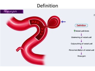
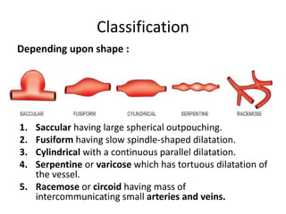
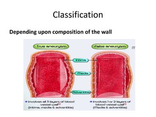
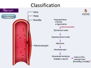
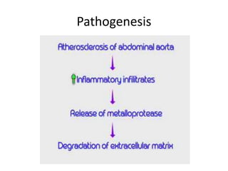
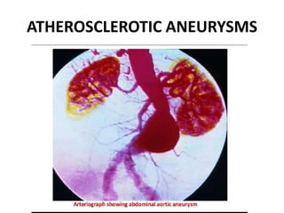
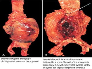
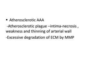
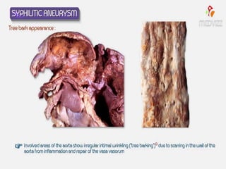
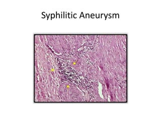
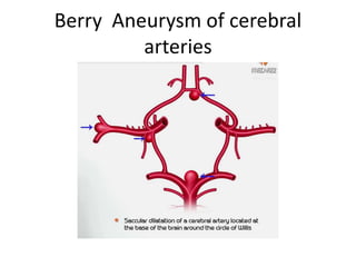
ANEURYSMS
ANEURYSM
 Definition
 Classification
 Pathogenesis
 Complication
Definition-
 Aneurysm is a localized abnormal permanent
dilation of a blood vessel or heart that may be
congenital or acquired
Definition
Classification
 Origin
 Location
 Gross appearance
 Composition of vessel wall
 Etiology
CLASSIFICATION
Depending upon location
-Arterial and venous, with arterial being more
common.
-Capillaries
-The heart, including coronary artery aneurysms,
ventricular aneurysms, aneurysm of sinus of
Valsalva, and aneurysms following cardiac surgery.
-The aorta, namely aortic aneurysms including
thoracic aortic aneurysms and abdominal aortic
aneurysms
Classification
-The brain, including cerebral aneurysms, berry
aneurysms
-The legs, including the popliteal arteries
-The kidney, including renal artery aneurysm
Classification
1. Saccular having large spherical outpouching.
2. Fusiform having slow spindle-shaped dilatation.
3. Cylindrical with a continuous parallel dilatation.
4. Serpentine or varicose which has tortuous dilatation of
the vessel.
5. Racemose or circoid having mass of
intercommunicating small arteries and veins.
Depending upon shape :
 Discrete outpouching of blood vessel
 Ranging from 5-20 cms in diameter
 Often contain thrombus
 Circumferential dilation of blood vessels
 20 cms in diameter
 Most commonly involved Aortic arch ,
Abdominal aorta , iliac arteries
Composition of wall
 True Aneurysm
 False Aneurysm
 Arterial dissection
Classification
Depending upon composition of the wall
Classification
Depending upon composition of the wall
Trauma to vessel wall
Classification
Composition of the wall of
aneurysm
 True Aneurysm- all layers
 False Aneurysm-traumatic rupture or
perforating injury
 Arterial dissection-intimal defect
Classification
Depending upon pathogenic mechanisms
-Atherosclerotic aneurysms are the most
common type
-Syphilitic (luteic) aneurysms found in the
tertiary stage of the syphilis
- Dissecting aneurysms(Dissecting hematoma)
in which the blood enters the separated or
dissected wall of the vessel
Classification
- Mycotic aneurysms resulting from weakening
of the arterial wall by microbial infection.
-Berry aneurysms which are small dilatations
especially affecting the circle of Willis in the
base of the brain.
Pathology
Predisposing factors
 Atherosclerosis especially in aortic aneurysms
 Hypertension especially in ascending aortic
aneurysms
 Trauma
 Vasculitis
 Congenital defects
 Fibromuscular dysplasia
 Berry aneurysms typically in the circle of
willis
 Infections -
–Syphilic aneurysms
–Mycotic aneurysms
–From embolization of a septic embolus
- complication of infective endocarditis
–As an extension of an adjacent
suppurative process
–By circulating organisms directly
infecting the arterial wall.
 Pathogenesis -Occur when structure or function
of the connective tissue within the vascular wall
is compromised.
 Poor intrinsic quality of the vascular wall
connective tissue.
 Imbalance in vascular wall collagen
degradation and synthesis caused by
inflammation and associated proteases.
 Weakening of vascular wall due to
 loss of smooth muscle cells
 synthesis of non-collagenous /non-elastic
extracellular matrix.
 Poor intrinsic quality of the vascular wall
connective tissue
–defective synthesis of fibrillin leads to
aberrant TGF-β activity and weakening of
elastic tissue in the aorta, this may result in
progressive dilation in Marfan syndrome.
–mutations in TGF-β receptors lead to
defective synthesis of elastin and collagens I
and II in Loeys-Dietz syndrome.
–Weak vessel walls due to defective type III
collagen synthesis are also a hallmark of
the vascular forms of Ehlers-Danlos
syndrome.
–altered collagen cross-linking associated
with Vitamin C deficiency (scurvy)
Imbalance in vascular wall collagen degradation
& synthesis
 Increased matrix metalloprotease (MMP)
expression, especially by macrophages in
atherosclerotic plaque or in vasculitis degrade
virtually all components of the extracellular
matrix in the arterial wall (collagens, elastin,
proteoglycans, laminin, fibronectin).
 Decreased expression of tissue inhibitors of
metalloproteases (TIMPs)
Weakening of vascular wall
 Ischemia of the inner media occurs when there
is atherosclerotic thickening of the intima.
 Systemic hypertension causes significant
narrowing of arterioles of the vasa vasorum.
 Medial ischemia may lead to “degenerative
changes” of the aorta.
 Smooth muscle cell loss/change in synthetic
phenotype leads to scarring (and loss of elastic
fibers), inadequate extracellular matrix
synthesis, and production of increasing
amounts of amorphous ground substance
(glycosaminoglycan).
Pathology
ABDOMINAL AORTIC ANEURYSMS
 Most common form of aortic aneurysms.
 Common in men, smokers
 More commonly seen in males over the age of
50 years.
 Most common sites abdominal aorta, thoracic
aorta, iliac arteries and other large systemic
arteries.
Pathogenesis
ATHEROSCLEROTIC ANEURYSMS
External view, gross photograph
of a large aortic aneurysm that ruptured
Opened view, with location of rupture tract
indicated by a probe. The wall of the aneurysm is
exceedingly thin, with lumen filled by large uantity
of layered but largely unorganized thrombus
 Atherosclerotic AAA
-Atherosclerotic plague –intima-necrosis ,
weakness and thinning of arterial wall
-Excessive degradation of ECM by MMP
Inflammatory AAA
 Periaortic fibrosis
 C reactive protein
 Lymphoplasmacytic infiltrate
 Immunoglobulin IgG4 related disease
 Mycotic Abdominal Aortic aneurysm-
salmonella gastroenteritis
Morphology
 Site-below the renal arteries ,above the
bifurcation of aorta
 Saccular/fusiform
 15-25cms
 Intimal surface-atherosclerosis
 Lumen-thrombus
COMPLICATIONS :
1. Rupture
-Most serious and fatal complication.
-Depends upon the size and duration of the
aneurysm and the blood pressure.
-Rupture of abdominal aneurysm may occur
either into the peritoneum or into the
retroperitoneum resulting in sudden and
massive bleeding.
-A ruptured aneurysm is more likely to get
infected.
2. Compression
-The atherosclerotic aneurysm may press upon some
adjacent structures such as compression of ureter and
erosion on the vertebral bodies.
3. Arterial occlusion
-Atherosclerotic aneurysms of the abdominal aorta may
occlude the inferior mesenteric artery, or there may be
development of occlusive thrombosis.
-Thromboembolism is rather common in abdominal
aneurysms.
Thoracic aortic aneurysm
• HT
• Symptoms-respiratory failure , difficulty in
swallowing, cough
SYPHILITIC ANEURYSM
• Luminal narrowing and
obliteration, scarring of the
vessel wall, and a dense
surrounding rim of
lymphocytes and plasma
cells that may extend into
the media
• The aorta loses its elastic
recoil with destruction of
the media and becomes
dilated, producing an
aneurysm.
• Valvular insufficiency and
massive volume overload
lead to hypertrophy of the
left ventricle.
• The greatly enlarged hearts
are sometimes called "cor
bovinum" (cow's heart).
Syphilitic Aneurysm
COMPLICATIONS
1. Rupture
-Causes massive and fatal haemorrhage into
the pleural cavity, pericardial sac, trachea and
oesophagus .
2. Compression
• The aneurysm may press on the adjacent
tissues and cause symptoms :
on trachea causing dyspnoea,
on oesophagus causing dysphagia,
on recurrent laryngeal nerve leading to
hoarseness
3. Cardiac dysfunction
-When the aortic root and valve are involved,
syphilitic aneurysm produces aortic
incompetence and cardiac failure.
-Narrowing of the coronary ostia may further
aggravate cardiac disease
Berry Aneurysm of cerebral
arteries
MYCOTIC ANEURYSM
 Aneurysm that is secondary to weakening of
vessel wall due to infection
 Staphylococcus aureus
 S. epidermis, Salmonella, Streptococcus
pneumonia
 Fungal arterial infections
 Candida , Cryptococcus , Aspergillus
 Immunosupression (Diabets Mellitus)
 Femoral Artery most common site of mycotic
aneurysm
 Abdominal Aorta- second most common site
 Intracranial arteries
 Coronary arteries
Dissecting aneurysm of the aorta
(Aortic dissection)
• Refers to a dissecting haematoma in which the
blood enters the separated (dissected) wall of
the vessel and spreads for varying distance
longitudinally.
• The most common site is the aorta and is an
acute catastrophic aortic disease.
• Men,50-70 yrs
• In women, dissecting aneurysms may occur
during pregnancy.
Dissecting aneurysm of the aorta
(Aortic dissection)
The aortic wall has split (dissected) at the level of the media producing an outer false
lumen running parallel to the central true lumen which is narrowed.
Dissecting aneurysm of the aorta
(Aortic dissection)
PATHOGENESIS
1. Hypertension in about 90% cases of
dissecting aneurysm.
2. 40-60 yrs
3. Marfan’s syndrome.
4. Iatrogenic trauma during cardiac
catheterization or coronary bypass surgery
5. Pregnancy, for some unknown reasons
• Ascending aorta
• Transverse/oblique
• Tunica media
• Extension
Dissecting aneurysm of the aorta (Aortic dissection)
CLASSIFICATION
Dissecting aneurysm of the aorta (Aortic dissection)
HISTOLOGY
i. Focal separation of the
fibromuscular and elastic
tissue of the media.
ii. Numerous cystic spaces in
the media containing
basophilic ground
substance.
iii.Fragmentation of the
elastic tissue.
COMPLICATIONS
Rupture
-Haemorrhage occurs into the pericardium; less
frequently it may rupture into thoracic cavity,
abdominal cavity or retroperitoneum.
• Cardiac disease
- cardiac tamponade.
3. Ischemia
-Obstruction of the branches of aorta by
dissection results in ischaemia of the tissue
supplied.
-There may be renal infarction, cerebral
ischaemia and infarction of the spinal cord.
Arteriosclerosis
• Small arteries and arterioles
• Cause ischemic injury
• Arterial wall thickening and loss of elasticity
Variants
• Hyaline arteriosclerosis
• Hyperplastic arteriosclerosis
Hyaline Arteriosclerosis
• Arterioles with benign HT
• Thickening of wall by hyaline material
Hyperplastic Arteriosclerosis
• Onion skin laminated thickening of arteriolar
walls
• Necrotizing arteriolitis
THANK YOU

aneurysm pathology pptx from.robbins sss

  • 1.
  • 2.
  • 3.
    Definition-  Aneurysm isa localized abnormal permanent dilation of a blood vessel or heart that may be congenital or acquired
  • 4.
  • 5.
    Classification  Origin  Location Gross appearance  Composition of vessel wall  Etiology
  • 6.
    CLASSIFICATION Depending upon location -Arterialand venous, with arterial being more common. -Capillaries -The heart, including coronary artery aneurysms, ventricular aneurysms, aneurysm of sinus of Valsalva, and aneurysms following cardiac surgery. -The aorta, namely aortic aneurysms including thoracic aortic aneurysms and abdominal aortic aneurysms
  • 7.
    Classification -The brain, includingcerebral aneurysms, berry aneurysms -The legs, including the popliteal arteries -The kidney, including renal artery aneurysm
  • 8.
    Classification 1. Saccular havinglarge spherical outpouching. 2. Fusiform having slow spindle-shaped dilatation. 3. Cylindrical with a continuous parallel dilatation. 4. Serpentine or varicose which has tortuous dilatation of the vessel. 5. Racemose or circoid having mass of intercommunicating small arteries and veins. Depending upon shape :
  • 10.
     Discrete outpouchingof blood vessel  Ranging from 5-20 cms in diameter  Often contain thrombus
  • 12.
     Circumferential dilationof blood vessels  20 cms in diameter  Most commonly involved Aortic arch , Abdominal aorta , iliac arteries
  • 13.
    Composition of wall True Aneurysm  False Aneurysm  Arterial dissection
  • 14.
  • 15.
    Classification Depending upon compositionof the wall Trauma to vessel wall
  • 16.
  • 17.
    Composition of thewall of aneurysm  True Aneurysm- all layers  False Aneurysm-traumatic rupture or perforating injury  Arterial dissection-intimal defect
  • 18.
    Classification Depending upon pathogenicmechanisms -Atherosclerotic aneurysms are the most common type -Syphilitic (luteic) aneurysms found in the tertiary stage of the syphilis - Dissecting aneurysms(Dissecting hematoma) in which the blood enters the separated or dissected wall of the vessel
  • 19.
    Classification - Mycotic aneurysmsresulting from weakening of the arterial wall by microbial infection. -Berry aneurysms which are small dilatations especially affecting the circle of Willis in the base of the brain.
  • 20.
    Pathology Predisposing factors  Atherosclerosisespecially in aortic aneurysms  Hypertension especially in ascending aortic aneurysms  Trauma  Vasculitis  Congenital defects  Fibromuscular dysplasia  Berry aneurysms typically in the circle of willis
  • 21.
     Infections - –Syphilicaneurysms –Mycotic aneurysms –From embolization of a septic embolus - complication of infective endocarditis –As an extension of an adjacent suppurative process –By circulating organisms directly infecting the arterial wall.
  • 22.
     Pathogenesis -Occurwhen structure or function of the connective tissue within the vascular wall is compromised.  Poor intrinsic quality of the vascular wall connective tissue.  Imbalance in vascular wall collagen degradation and synthesis caused by inflammation and associated proteases.  Weakening of vascular wall due to  loss of smooth muscle cells  synthesis of non-collagenous /non-elastic extracellular matrix.
  • 23.
     Poor intrinsicquality of the vascular wall connective tissue –defective synthesis of fibrillin leads to aberrant TGF-β activity and weakening of elastic tissue in the aorta, this may result in progressive dilation in Marfan syndrome. –mutations in TGF-β receptors lead to defective synthesis of elastin and collagens I and II in Loeys-Dietz syndrome.
  • 24.
    –Weak vessel wallsdue to defective type III collagen synthesis are also a hallmark of the vascular forms of Ehlers-Danlos syndrome. –altered collagen cross-linking associated with Vitamin C deficiency (scurvy)
  • 25.
    Imbalance in vascularwall collagen degradation & synthesis  Increased matrix metalloprotease (MMP) expression, especially by macrophages in atherosclerotic plaque or in vasculitis degrade virtually all components of the extracellular matrix in the arterial wall (collagens, elastin, proteoglycans, laminin, fibronectin).  Decreased expression of tissue inhibitors of metalloproteases (TIMPs)
  • 26.
    Weakening of vascularwall  Ischemia of the inner media occurs when there is atherosclerotic thickening of the intima.  Systemic hypertension causes significant narrowing of arterioles of the vasa vasorum.  Medial ischemia may lead to “degenerative changes” of the aorta.  Smooth muscle cell loss/change in synthetic phenotype leads to scarring (and loss of elastic fibers), inadequate extracellular matrix synthesis, and production of increasing amounts of amorphous ground substance (glycosaminoglycan).
  • 27.
  • 28.
    ABDOMINAL AORTIC ANEURYSMS Most common form of aortic aneurysms.  Common in men, smokers  More commonly seen in males over the age of 50 years.  Most common sites abdominal aorta, thoracic aorta, iliac arteries and other large systemic arteries.
  • 29.
  • 30.
  • 31.
    External view, grossphotograph of a large aortic aneurysm that ruptured Opened view, with location of rupture tract indicated by a probe. The wall of the aneurysm is exceedingly thin, with lumen filled by large uantity of layered but largely unorganized thrombus
  • 32.
     Atherosclerotic AAA -Atheroscleroticplague –intima-necrosis , weakness and thinning of arterial wall -Excessive degradation of ECM by MMP
  • 33.
    Inflammatory AAA  Periaorticfibrosis  C reactive protein  Lymphoplasmacytic infiltrate
  • 34.
     Immunoglobulin IgG4related disease  Mycotic Abdominal Aortic aneurysm- salmonella gastroenteritis
  • 35.
    Morphology  Site-below therenal arteries ,above the bifurcation of aorta  Saccular/fusiform  15-25cms  Intimal surface-atherosclerosis  Lumen-thrombus
  • 36.
    COMPLICATIONS : 1. Rupture -Mostserious and fatal complication. -Depends upon the size and duration of the aneurysm and the blood pressure. -Rupture of abdominal aneurysm may occur either into the peritoneum or into the retroperitoneum resulting in sudden and massive bleeding. -A ruptured aneurysm is more likely to get infected.
  • 37.
    2. Compression -The atheroscleroticaneurysm may press upon some adjacent structures such as compression of ureter and erosion on the vertebral bodies. 3. Arterial occlusion -Atherosclerotic aneurysms of the abdominal aorta may occlude the inferior mesenteric artery, or there may be development of occlusive thrombosis. -Thromboembolism is rather common in abdominal aneurysms.
  • 38.
    Thoracic aortic aneurysm •HT • Symptoms-respiratory failure , difficulty in swallowing, cough
  • 39.
  • 40.
    • Luminal narrowingand obliteration, scarring of the vessel wall, and a dense surrounding rim of lymphocytes and plasma cells that may extend into the media • The aorta loses its elastic recoil with destruction of the media and becomes dilated, producing an aneurysm. • Valvular insufficiency and massive volume overload lead to hypertrophy of the left ventricle. • The greatly enlarged hearts are sometimes called "cor bovinum" (cow's heart).
  • 42.
  • 43.
    COMPLICATIONS 1. Rupture -Causes massiveand fatal haemorrhage into the pleural cavity, pericardial sac, trachea and oesophagus . 2. Compression • The aneurysm may press on the adjacent tissues and cause symptoms : on trachea causing dyspnoea, on oesophagus causing dysphagia, on recurrent laryngeal nerve leading to hoarseness
  • 44.
    3. Cardiac dysfunction -Whenthe aortic root and valve are involved, syphilitic aneurysm produces aortic incompetence and cardiac failure. -Narrowing of the coronary ostia may further aggravate cardiac disease
  • 45.
    Berry Aneurysm ofcerebral arteries
  • 47.
    MYCOTIC ANEURYSM  Aneurysmthat is secondary to weakening of vessel wall due to infection  Staphylococcus aureus  S. epidermis, Salmonella, Streptococcus pneumonia  Fungal arterial infections  Candida , Cryptococcus , Aspergillus  Immunosupression (Diabets Mellitus)
  • 49.
     Femoral Arterymost common site of mycotic aneurysm  Abdominal Aorta- second most common site  Intracranial arteries  Coronary arteries
  • 50.
    Dissecting aneurysm ofthe aorta (Aortic dissection) • Refers to a dissecting haematoma in which the blood enters the separated (dissected) wall of the vessel and spreads for varying distance longitudinally. • The most common site is the aorta and is an acute catastrophic aortic disease. • Men,50-70 yrs • In women, dissecting aneurysms may occur during pregnancy.
  • 51.
    Dissecting aneurysm ofthe aorta (Aortic dissection) The aortic wall has split (dissected) at the level of the media producing an outer false lumen running parallel to the central true lumen which is narrowed.
  • 52.
    Dissecting aneurysm ofthe aorta (Aortic dissection) PATHOGENESIS 1. Hypertension in about 90% cases of dissecting aneurysm. 2. 40-60 yrs 3. Marfan’s syndrome. 4. Iatrogenic trauma during cardiac catheterization or coronary bypass surgery 5. Pregnancy, for some unknown reasons
  • 53.
    • Ascending aorta •Transverse/oblique • Tunica media • Extension
  • 54.
    Dissecting aneurysm ofthe aorta (Aortic dissection) CLASSIFICATION
  • 55.
    Dissecting aneurysm ofthe aorta (Aortic dissection) HISTOLOGY i. Focal separation of the fibromuscular and elastic tissue of the media. ii. Numerous cystic spaces in the media containing basophilic ground substance. iii.Fragmentation of the elastic tissue.
  • 56.
    COMPLICATIONS Rupture -Haemorrhage occurs intothe pericardium; less frequently it may rupture into thoracic cavity, abdominal cavity or retroperitoneum. • Cardiac disease - cardiac tamponade.
  • 57.
    3. Ischemia -Obstruction ofthe branches of aorta by dissection results in ischaemia of the tissue supplied. -There may be renal infarction, cerebral ischaemia and infarction of the spinal cord.
  • 58.
    Arteriosclerosis • Small arteriesand arterioles • Cause ischemic injury • Arterial wall thickening and loss of elasticity
  • 59.
    Variants • Hyaline arteriosclerosis •Hyperplastic arteriosclerosis
  • 60.
    Hyaline Arteriosclerosis • Arterioleswith benign HT • Thickening of wall by hyaline material
  • 61.
    Hyperplastic Arteriosclerosis • Onionskin laminated thickening of arteriolar walls • Necrotizing arteriolitis
  • 62.