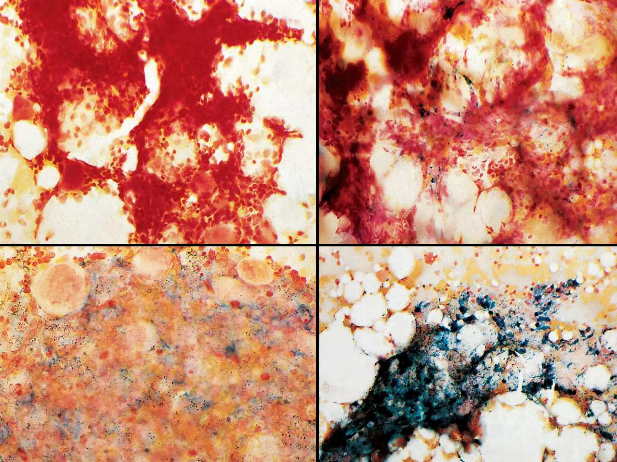
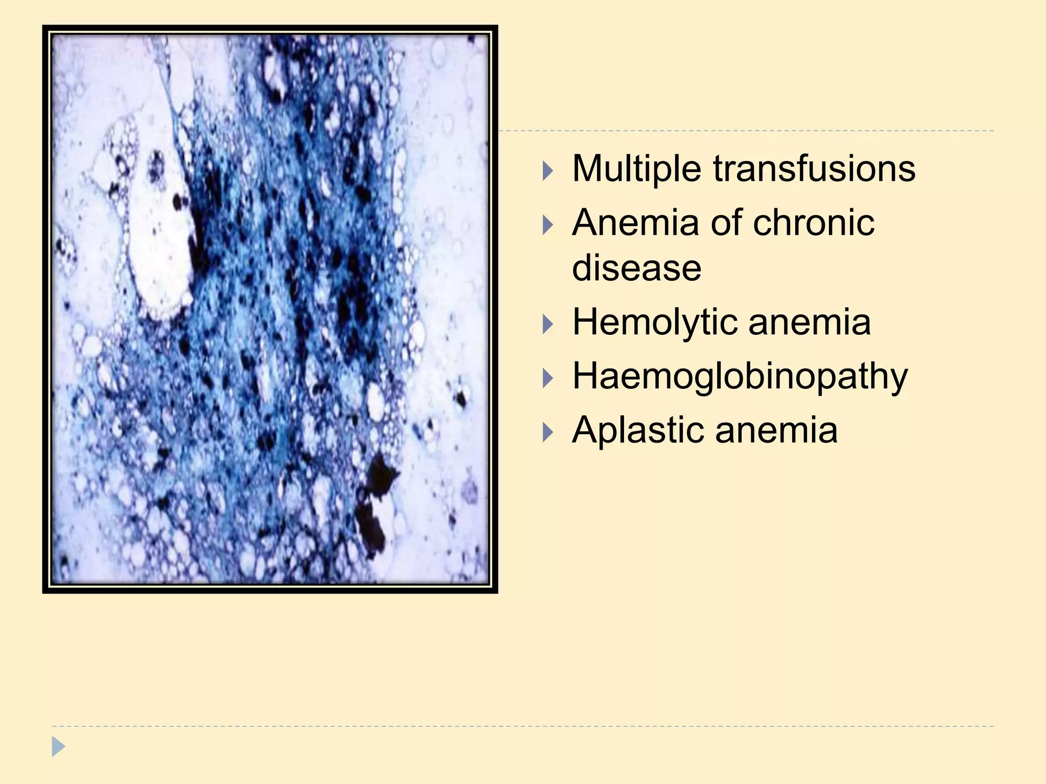
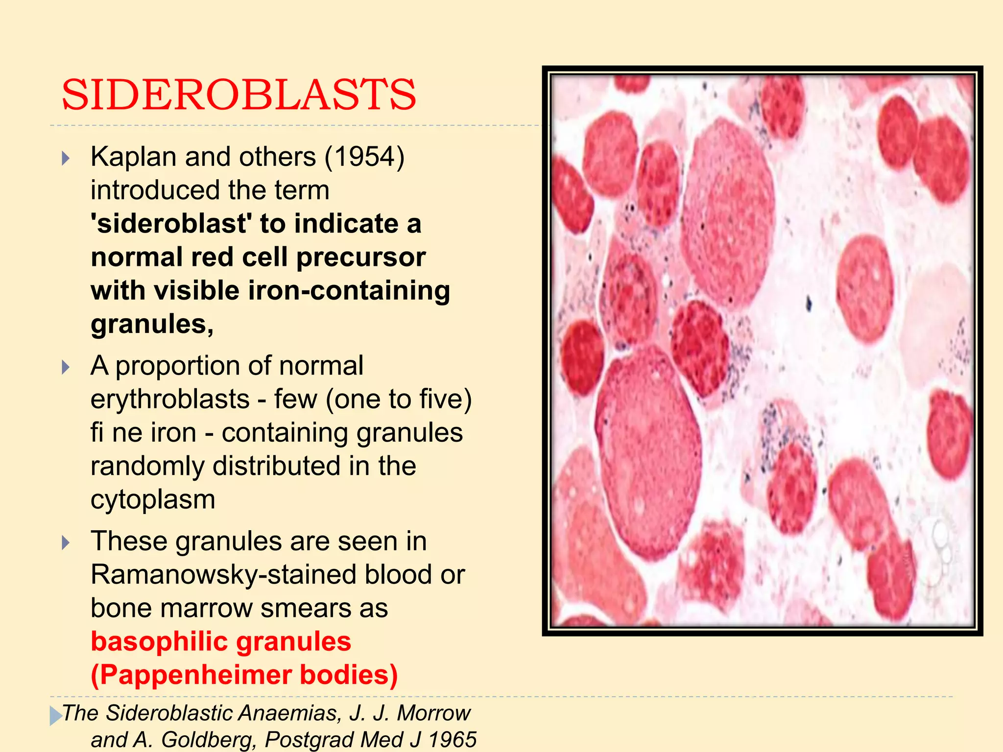
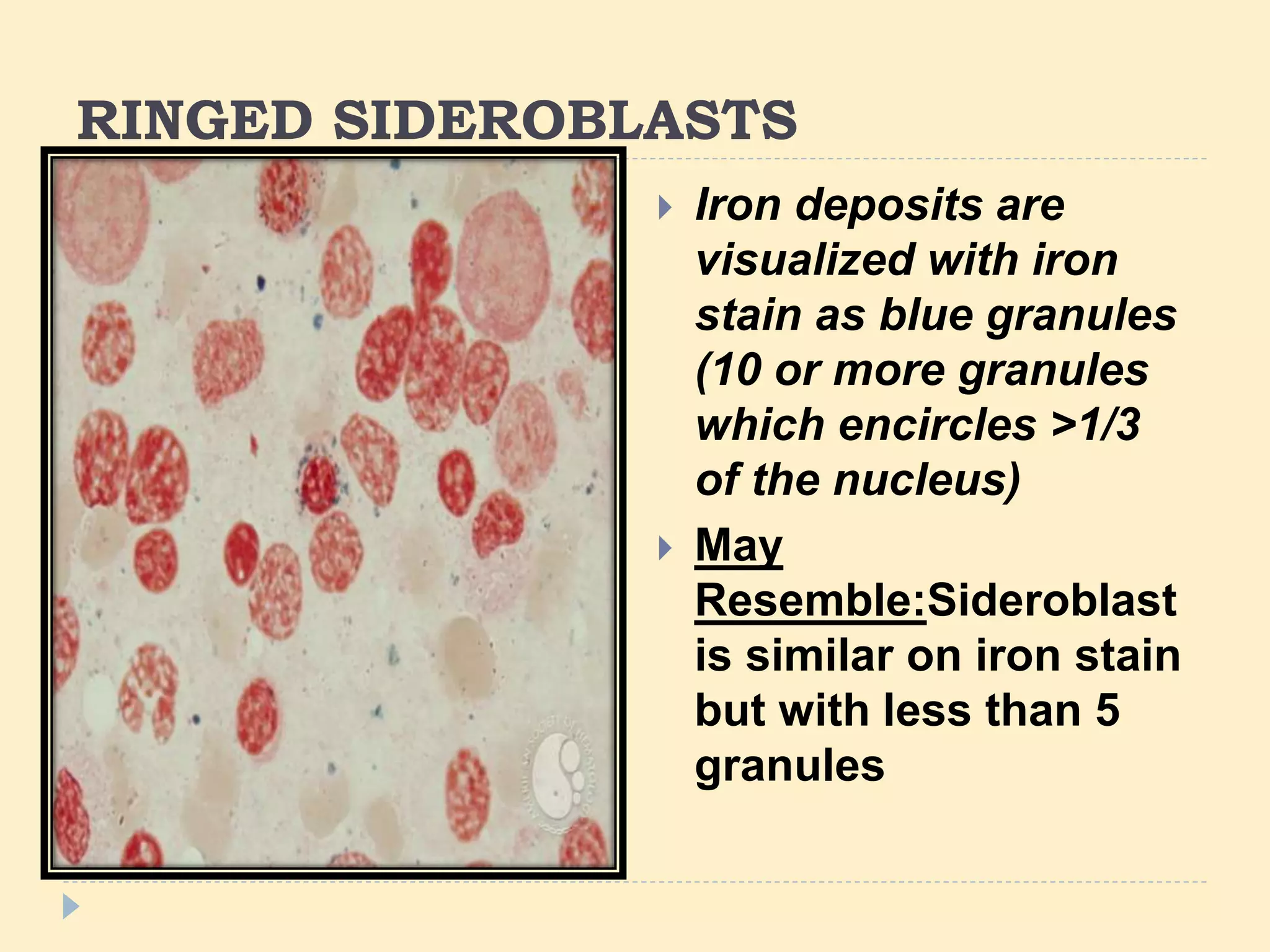
SPECIAL STAINS IN BONE
MARROW EXAMINATION
 Bennett" has divided cytochemical stains into three
groups:
 (i) cytochemical stains of diagnostic significance -
peroxidase, naphthol AS-D acetate esterase without
and with fluoride, leucocyte alkaline phosphatase;
 (it) specialized cytochemical stains - leucocyte acid
phosphatase, naphthol AS-D chloro-acetate
esterase, toluidine blue
 (iii) cytochemical stains of limited value - ,B-
glucuronidase. -acetyl-,B-glucosaminidase, periodic
acid-Schiff, Oil red 0, Sudan black B.
 The Present Status of Cytochemistry in the Diagnosis of
Haematological Malignancy, W. G. STAPLES, E. P.GETAZ, S. Afr. med.
l., 51, 838 (1977).
"Perls' Prussian blue
 Prussian blue - Berlin in the early 1700s
 The original stain formula, known historically (1867)
as "Perls' Prussian blue" after its inventor, German
pathologist Max Perls
 Separate solutions of potassium ferrocyanide and
acid to stain tissue (these are now used combined,
just before staining).
 The formula is also known as Perls Prussian blue
and (incorrectly) as Perl's Prussian blue.
(Perls's Prussian Blue reaction)
 The majority of non-haem iron is stored as
haemosiderin (a ferric iron-protein complex), a
small amount as ferritin and the remainder is
contained in myoglobin and transferrin.
 Ferritin - normally in all cells in the body,
 Haemosiderin - macrophages in the bone marrow,
liver (Kupffer cells) and spleen.
 Most intracellular iron is in macrophages, a small
amount in erythroblasts (sideroblasts).
 A Perls ’ or Prussian blue stain demonstrates
haemosiderin in bone marrow macrophages and
within erythroblasts.
 Assessment of both the amount of iron in reticulo -
endothelial stores and the availability of iron to
developing erythroblasts.
 Detection of increased or decreased proportion of
sideroblasts and abnormal sideroblasts.
Principle
 Ferric iron in haemosiderin is released from protein
by treatment with dilute hydrochloric acid.
 The free iron reacts with a dilute solution of
potassium ferrocyanide to produce an insoluble blue
compound, ferric ferrocyanide (Prussian Blue).
 By contrast, ferritin, which is a water-soluble non-
haem compound of iron with the protein apoferritin, is
not detectable by Perls’ reaction.
 Any contrasting, non-acidic counterstain may be used but
Neutral Red, safranin or eosin are the most popular.
MATERIALS
 Fixative
Methanol as for MGG
 4% HCL
4ml HCL + 96% distilled water
 4% potassium ferrocyanide
4g potassium ferrocyanide + 1000ml distilled water
 Nuclear fast red
Store at room temperature
PROCEDURE
Prepare working solution by adding two parts of 4%
HCL and 4% Potassium ferrocyanide 2-3ml a slide
 Flood the slide with working solution and allow to
stand for 15-20 mins
 Wash well with distilled water for 5 mins
 Counter stain with nuclear fast red in a coplin jar for
5-7mins
 Wash in tap water
 Air dry and observe marrow fragment and erythroid
precursor
 Positive- bright green/bright blue
 Cytoplasm of stain pink
 A bone marrow film or squash preparation - both
intracellular and extracellular iron, the latter being
derived from crushed macrophages.
 Assessment of iron stores mainly on intracellular
iron
INTERPRETATION
 A BM smear with increased iron stores - positive
control.
 Low power to assess storage iron
 40 or × 50 objective to detect abnormally
prominent siderotic granulation
 100 objective to assess whether siderotic
granulation is reduced, normal or increased
Gale E, Torrance J, Bothwell T. The quantitative estimation of total iron
stores in human bone marrow. J Clin Invest 1963;42:1076-82.
Intensive histological grading method
 Intensive histological grading method—the
fragments, macrophages and erythroblasts.
 Iron assessed in the fragments and macrophages -
iron stores
 Iron in the erythroblast - utilisable iron.
 100 erythroblasts were examined and the
percentage containing iron granules in their
cytoplasm (ie, sideroblasts) were enumerated.
Improved method for assessing iron stores in the bone marrow, K S Phiri et al,
J Clin Pathol. 2009 August; 62(8): 685–689.
s
 Multiple transfusions
 Anemia of chronic
disease
 Hemolytic anemia
 Haemoglobinopathy
 Aplastic anemia
SIDEROBLASTS
 Kaplan and others (1954)
introduced the term
'sideroblast' to indicate a
normal red cell precursor
with visible iron-containing
granules,
 A proportion of normal
erythroblasts - few (one to five)
fi ne iron - containing granules
randomly distributed in the
cytoplasm
 These granules are seen in
Ramanowsky-stained blood or
bone marrow smears as
basophilic granules
(Pappenheimer bodies)
The Sideroblastic Anaemias, J. J. Morrow
and A. Goldberg, Postgrad Med J 1965
INTRACELLULAR IRON
 The total number of sideroblasts (normal, reduced
or increased) should be reported and the
frequency and location (cytoplasmic or
perinuclear) of siderotic granules (Bowman, 1961;
Cartwright & Deiss, 1975) should be noted,
 At least 100 erythroblasts should be evaluated for
the percentage of ring sideroblasts, if present
 ICSH guidelines for the standardization of bone marrow specimens and reports
S.-H. LEE*, W. N. ERBER†, A. PORWIT‡, M. TOMONAGA§, L. C. PETERSON–
FOR THE INTERNATIONAL COUNCIL FOR STANDARDIZATION IN
HEMATOLOGY INTERNATIONAL JOURNAL OF LABORATORY
HEMATOLOGY
 The percentage of sideroblasts is increased -
haemolytic anaemias , megaloblastic anaemias
and in haemochromatosis and haemosiderosis,
in proportion to the degree of saturation of transferrin
(i.e. to the amount of iron available).
 A disproportionate increase in the percentage of
sideroblasts - synthesis of haemoglobin is
impaired, in which case the siderotic granules are
both more numerous and larger than normal
RINGED SIDEROBLASTS
 Iron deposits are
visualized with iron
stain as blue granules
(10 or more granules
which encircles >1/3
of the nucleus)
 May
Resemble:Sideroblast
is similar on iron stain
but with less than 5
granules
Ring sideroblasts -
 Refractory anaemia with ring sideroblasts and
refractory cytopenia with multilineage dysplasia and
ring sideroblasts,
 Other categories of MDS.
 Primary myelofibrosis
 Acute myeloid leukaemia (AML), particularly
erythroleukaemia
 WHO categories of therapy related AML and AML
with multilineage myelodysplasia.
 States of iron overload such as
hemochromatosis (sometimes)
 Zinc toxicity induced copper deficiency
 Plasma cells contain haemosiderin inclusions,
which are irregular in shape and relatively large.
 MGG stain - greenish black
 Iron overload (for example in haemochromatosis and
transfusional siderosis) and in chronic alcoholism
VARIANTS
 Films that have previously been stained by
Romanowsky dyes, even after years of storage.
 Stand in methanol overnight to remove most of the
Romanowsky stain.
 The film should be checked before carrying out
Perls’ reaction to ensure that there is no residual
blue staining that could obscure Prussian-blue
staining.
 Rapid method- demonstrating siderotic granules by
staining with 1% bromochlorphenol blue for 1 min.
Iron-containing granules stain dark purple
 Sundberg and Bromann - films were stained first by
a Romanowsky dye (Wright’s stain) and then
overstained by the acid-ferrocyanide method.
 Hayhoe and Quaglino described a method for
combined periodic acid–Schiff (PAS) and iron
staining.
 This may be helpful in the investigation of abnormal
erythropoiesis in which the erythroblasts give a
positive PAS reaction
 Aspirate films are more sensitive than trephine
biopsy sections for the detection of haemosiderin
when the biopsy specimens are decalcified in formic
acid.
 Decalcification leads to an unquantifiable loss of
iron.
 Decalcification - ethylenediaminetetraacetic acid,
which is a less powerful decalcifying agent.
 Distributed irregularly within bone marrow
macrophages - minimum of seven fragments
before concluding that storage iron is absent or
reduced.
 If less than seven particles are present and no iron is
seen, the sample should be reported as lacking
stainable iron, but the assessment should be
stated to be unreliable
How should stainable iron in bone marrow films be assessed? D A
Hughes, S E Stuart-Smith, B J Bain
J Clin Pathol 2004;57:1038–1040. doi: 10.1136/jcp.2003.015834
LEUCOCYTE CYTOCHEMISTRY
 To identify diagnostically useful enzymes or other
substances in the cytoplasm of haemopoietic cells.
 To characterize the blast cells in acute leukaemia
as myeloid (leading to a diagnosis of AML unless
there is also evidence of lymphoid differentiation)
 To diagnosis a mixed-phenotype acute leukaemia,
 To identify granulocytic and monocytic
components in AML
 To identify unusual lineages occasionally involved
in clonal myeloid disorders (e.g. basophils and
mast cells)
 To detect cytoplasmic abnormalities and enzyme
deficiencies in myeloid disorders (e.g. MPO
neutrophils in myelodysplasia or acute leukaemia,
NAP -deficient neutrophils in CML)
 To identify Auer rods in MDS
Periodic Acid – Schiff [PAS] Reaction
 Periodic acid oxidizes the 1-2 glycol groups
in sugars, to produce dialdehydes
 The oxidation condition has to be sufficiently
regulated so as to not oxidize the aldehydes further.
 Aldehydes + Schiff reagent = purple-magenta
color.
PAS
 A variety of intracellular compounds react with the
PAS reagents, but in the blood and marrow cells
glycogen is the most abundant compound
 Intensity of colour - number of aldehyde groups
liberated
 For blood smears, the recommended fixative
is methanol.
 Glutaraldehyde- not recommended - free aldehyde
groups may be available to react with the Schiff
reagent, which may result in false positive staining.
 Fixative- Methanol
 1% periodic acid
10g HIO4.2H2O+ 1000ml Distilled water
 Schiff’s reagent
 Counter stain
Aqeous haematoxylin
PROCEDURE
 Fix films 20 min in methanol
 Rinse gently under tap water
 If required expose to diastase
 Flood slides with 1% PAS , allow to stand for 5-7min
 Rinse under running tap water, 5-7min
 Immerse in Schiff’s reagent for 15 min
 Rinse in running tap water
 Counter stain- Aqeous haematoxylin for 30 seconds
 QC control- high neutrophil count
 PAS stain can be used in old but well-preserved
smear and smear stained with Wright’s stain.
 Smear stained with Wright’s stain should be
decolorized with ethanol before performing PAS
staining.
 Microscopic examination should be performed
immediately after PAS staining, since positive
reaction will turn weaker after one week.
 Neutrophil precursors contain many mitochondria ,
respiration is the main source of metabolic energy.
 As they mature, mitochondria decrease in number
 Glycolysis is now the predominant mechanism for
energy production and the neutrophil can be
sustained anaerobically to operate in hypoxic
infected tissues and exudates
 Glycogen particles accumulate in the
metamyelocyte and in band and mature
neutrophils which represent the non-dividing non-
secretory stages.'·
Granulocyte
 Myeloblasts and
promyelocyte diffuse
faint positive
 Myelocyte- some coarse
and some fine granules
 Neutrophils-
numerous granules
packed tightly
 Eosinophils- diffuse
intergranular cytoplasm
with large unstained
granules
 Normal erythroid
precursors- variable
 Normoblasts may be
positive in
sideroblastic anemia,
iron deficiency,
thallassemia, and
severe hemolytic
anemias
 -
 It was found that
megakaryocytes are rich in
glycogen which is not only
confined to the intensely PAS-
positive granules and inclusion
bodies, but also makes up a
good part of the diffuse
cytoplasmic staining.
 Glycogen content and PAS
staining pattern of human
megakaryocytesDr. F. V.
Shammas*, A. Engeset
Scandinavian Journal of
Haematology , September
1986
 Monocytes - weakly positive reaction. Monocytes
have active aerobic glycolysis
 Macrophages - strong positive reaction.
 Lymphocytes- smaller content
 Ewings sarcoma and alveolar
rhabdomyosarcoma cells are PAS positive
AML
 Usually negative, but may
show a weak diffuse
reaction with
superimposed fine
granular positivity. AML
M1 and M2, Auer rods-
weak positive
 a cytoplasmic ‘blush’ – a
fine diffuse or dust-like
positivity-AML M3
 Negative to focal or
granular positivity, to
strongly positive staining.-
AML M7
 Acute basophilc lekemia-
positive
PAS IN AML
 Positive in acute erythroid leukemia
 Pronormoblasts and other stages of normoblasts
positive
 Nucleated RBCs PAS positive in MDS
PAS IN ALL
 95% of cases show positive blocks or granules
of bright red PAS-positive material.
 This may be present in very few blasts (<1%) or the
majority.
 The critical difference from granular or block
positivity in other leukaemic cells is the glass-clear
background cytoplasm in lymphoblasts.
 Myeloblasts, monoblasts, leukaemic erythroblasts
and megakaryoblasts all show some degree of
diffuse cytoplasmic positivity and occasionally block
positivity is seen.
PAS IN ALL
 ALL –varied staining
pattern
 Lymphoblasts- coarse
block, fine diffuse or
both or negative
 Negative staining does
not rule out ALL
 Burkitt’s lymphome –
negative
 Some authors suggest that strong PAS-positivity
indicates a good prognosis (Laurie, 1968;Vowels and
Willoughby, 1973; Feldges et al., 1974; Ascari et al.,
1975) but others claim that it has no such
significance (Bennett and Henderson 1969; Berrebi et
al., 1973; Humphrey et al., 1974; Shaw et al., 1977).
 There are varying reports of the relation of P.A.S.
positivity, to the duration of first remission and
survival.
Kurz, R. and Haas, H.: Value of the combined cytological and cytochemical
classification in the management of acute childhood leukaemia. Acta.
Haematol. 52: 1-7, 1974
Feldges, A. J., Aur, R. J. A., Verzosa, M. S. and Daniel, S.: Periodic acid
Schiff reaction-a useful index of duration of complete remission in acute
childhood lymphocytic leukaemia. Acta. Haematol. 52: 8-13, 1974.
Myeloperoxidase (MPO)
 Primary and secondary granules of neutrophils and
their precursors
 Eosinophil granules
 Azurophilic granules of monocytes.
 The MPO in eosinophil granules is cyanide
resistant, whereas that in neutrophils and
monocytes is cyanide sensitive.
 MPO has a heme pigment, which causes its green
color in secretions rich in neutrophils, such as pus
and some forms of mucus
Peroxidase stain :
 Principle:
 In the presence of peroxidase, H2O2 is split
liberating O2 that oxidizes benzidine or 4-chlor-1-
naphthol
Myeloperoxidase (MPO)
Bluish-black granules red brown precipitate
The reaction product is blue/ black/ red brown.
 Dissolve 1,4 chloro 1 naphthol in 15ml ethanol
 Add 45ml distilled water
 10 drops of tris(hydromethylaminomethane)- Hcl
buffer and H2O2 each
 Fix in leucognost for 1 min
 Wash and place in staining solution for 10 min
 Wash and stain with Harris Haematoxylin
 Wash under tap water and dry in air
 Staining can be enhanced by immersing the slides in
copper sulphate or nitrate, but this is generally not
required in normal diagnostic practice
SUBSTRATES
 The most common substrates used are benzidine
derivatives (Graham, 1918; Goodpasture, 1919).
 Benzidine dihydrochloride (BDC) according to
Kaplow's (1965) technique.
 Alternative non-benzidine-based techniques use 4-
chloro-1-naphthol (4CN) or 3-amino-9-
ethylcarbazole.
 The former gives very crisp staining but is soluble in
some mounting media and immersion oil
 The latter shows some diffusibility and does not stain
as strongly as DAB.
 MPO is not inhibited by heparin, oxalate or EDTA
anticoagulants.
 Films should be made within 12 h of blood collection.
 Staining is satisfactory on slides kept at room
temperature for at least a week.
 QC control slide- High neutrophil count
NORMAL MARROW
 The most primitive myeloblasts are negative-
granular positivity appearing progressively as they
mature toward the promyelocyte stage- positivity
may be localized to the Golgi region.
 Promyelocytes and myelocytes are the most
strongly.
 Metamyelocytes and neutrophils - fewer positive
(secondary) granules.
 Neutrophils and eosinophils - primary granules
 Eosinophils – also secondary granules.
 Monocyte lineage- peroxidase activity detectable at
the promonocyte stage, fewer scattered granules
than neutrophils and their precursors.
 MPO activity - basophil granules- not
demonstrable in mature basophils by the DAB
reaction described earlier.
 Erythroid positive when treated with methanol-
staining reagent- Hb-
pseudoperoxidase/Lepehne reaction
 Some individuals have congenital deficiency of
neutrophil MPO.
 All stages of the neutrophil lineage, from the
myeloblast onward, are negative.
 In these individuals, the eosinophils stain normally.
MPO in AML
 AML with MDS- MPO may be aberrant, because
patients may develop an acquired MPO deficiency
as part of the dysplastic process.
 AML 1, 2- >3% positive
 Auer rods- MPO +
 AML M3- MPO- strongly positive, reaction
product covering cytoplasm and nucleus
 Monoblasts- MPO negative, Promonocytes-
scattered positivity
 AML M7- Consistently negative for MPO and SBB
s
 AML without maturation
 AML with maturation
 Pox type 1- upto 5% Peroxidase positive blasts- AML
without maturation
 Pox type 2- 5-65%- AML without maturation/ Acute
myelomonocytic leukemia
 Pox type 3- >65% - AML with maturation, Acute
promyelocytic leukemia
 Percentage of MPO-positive blast cells has an impact
on the clinical outcome of AML.
 MPO-negative M0 subtype with immature features
showed a disappointing clinical outcome, higher
resistance rate in patients
 MPO-positive rate of blast cells is associated with drug-
resistance mechanisms.
 The percentage of myeloperoxidase-positive blast cells is a strong
independent prognostic factor in acute myeloid leukemia, even in the
patients with normal karyotype
T Matsuo1, K Kuriyama1, Y Miyazaki1, S Yoshida1, M Tomonaga1, N Emi3,
T Kobayashi4, S Miyawaki5, T Matsushima6, K Shinagawa7, S Honda2 and
R Ohno8for the Japan Adult Leukemia Study Group
 Leukemia (2003) 17, 1538–1543. doi:10.1038/sj.leu.2403010
SUDAN BLACK
 More sensitive than MPO in early precursors
 More reliable and stable
 Principle: Sudan black B dye is fat soluble, then it
stains fat particles (Steroles, phospholipids and
neutral fats) in the primary and secondery granules
of myelocytic and lysosomes in monocytic cells.
 Fix air dried smears in formalin vapor
 Wash in running water and air dry
 Immerse in working stain solution ( 60 ml of 0.3
Sudan Black B in 100 ml absolute alcohol+ 40 ml of
crystalline phenol in absolute alcohol and disodium
hydrogen phosphate)
 Transfer to staining rack- dip in 70% alcohol and
rinse
 Counter stain with nuclear fast red
 Wash in running water
 SBB stains the granules of neutrophils (both the
primary and the specific granules) , specific granules
of eosinophils
 The staining of eosinophil granules may be
peripheral with the central core remaining
unstained.
 Promyelocytes- few sudanophilic granules, mature
polymorphonuclear neutrophils - large numbers of
sudanophilic granules.
 Monoblasts - either negative or few small SBB
positive granules.
 Promonocytes and monocytes - variable number of
fine positively staining granules.
 Hereditary neutrophil, eosinophil and monocyte
peroxidase deficiencies- granules of cells of the
deficient lineages are SBB-negative.
 Basophils - generally not positive - may show bright
red/purple metachromatic staining of the granules.
ALL
 Rare cases (1–2%) of acute lymphoblastic
leukaemia (ALL) show non-granular smudgy
positivity not seen with MPO staining.
 Very rarely a stronger reaction - lymphoblasts of ALL
or in lymphoma cells of T or B lineage
 AML: Acute Myeloid
Leukemia(M1, M2 and
M3)
 Auer rods
 AMML: Acute
Myelomonocytic
Leukemia
 Erythroleukemia: pos in
myeloblasts, neg in
normoblasts
ESTERASES
 Leucocyte esterases - enzymes that hydrolyse acyl
or chloroacyl esters of a-naphthol or naphthol AS. Li
et al
 Nine isenzymes of esterases in leucocytes
 - Napthol AS-D Chloroacetate (1,2,7,9)
- Alpha-naphtyhl acetate
- Alpha-naphthyl butyrate (3,4,5,6)
 Specific- myelocyte
 Non specific- other cells
 Debris digesting action of the neutrophil.
SPECIFIC ESTERASE
 A specific esterase (capable of liberating naphthol
from a naphthol AS-D chloroacetate substrate)
present in the non specific granules - granulocytes
and mast cells
 Chloracetate esterase has an optimum pH between
7.0 and 7.6 and is insensitive to fluoride inhibition
Specific esterase or chloroacetate
 Principle:
Interpretation:
 Myeloid cells (+ve)
 Mast cells
 Monocyte and basophile (–ve) to weak (+ve)
 Other cells {lymph – plasma –megakaryocyte – nrbc } (-
ve)
A naphthol compound is released which combines with a
diazonium salt to produce a brightly coloured compound at the
site of enzyme activity
 The incubation time is important- most haemopoietic
cells show some scattered granular staining if the
incubation is prolonged.
 The substrate used is naphthol AS- D chloro-acetate
and the coupler is garnet GBC diazo salt
 Haematoxylin is used as a counterstain.
 Positivity is represented by a reddish coloration.
 The reaction is resistant to sodium fluoride
inhibition.
 Aminocaproate esterase is found in mast cells but
cytochemically has no practical value.
CAE in ACUTE MYELOID LEUKEMIA
 CAE - more often
positive in M2 than in
M1 AML and reactions
are stronger.
 Auer rods- usually weak
or negative except in
M2 AML associated
with t(8;21) and AML
M3 in which Auer rods
are often positive for
CAE
Non Specific Esterase:
{with fluoride inhibition}
 Specificity - sodium fluoride as an inhibitor
(NASDA-F).
 Sodium fluoride, inhibits the activity in
monocytes, megakaryocytes, platelets, and
plasma cells but not that in lymphocytes
Nonspecific esterases
 Monocytes and precursors
 macrophages, megakaryocytes and platelets
 Mature T cells and T-all (cytoplasmic dot)
 Normal granulocytes are negative- MDS or AML
may give positive reactions of varying intensity.
 Carcinomas, megaloblastic erythrocytes,
cytoplasm focally in AML-M7
 Air dried smears in Leucognost fixing mixture for 1-3
mins
 Wash with distilled water for 1 min
 Place in staining solution(60 ml of phosphate buffer
in distilled water+1 naphthyl acetate in
acetone+pararosaniline Hcl+ nitrite solution)and
incubate in dark for 1-2 hr
 Wash in distilled water for 10 sec
 Stain with haematoxylin – 2min
 Wash in tap water – 2 min
 Air dry and examine
 QC Control-slide with high platelet count
a-Naphthyl Acetate Esterase
(ANAE)
 The reaction product is
diffuse red/brown in
colour
 Sensitive but not
specific (picks up T-
lymphocytes
(punctate staining
pattern) as well as
monocytes (diffuse
staining pattern)
 The T lymphocytic
pattern is resistant to
NaF , staining in
monocytes is NaF
sensitive and favoured
 The reaction product is
brown and granular
 More specific, although
slightly less sensitive
 Differential staining with
the different esterases -
megakaryoblasts, which
do not stain with the α-
naphthyl butyrate, but
stain with the α-naphthyl
acetate substrate
α-Naphthyl Butyrate Esterases
 Peroxidase type- < 20% EST- positive blasts- AML
 Pox EST mixed type- 25-50% pos blasts- AML M5
 Esterase type- > 50% EST positive blasts- AML M5
USES OF NON SPECIFIC ESTERASES
 Helps diagnosis in AML of M5 and AML M5
 ALL of T-ALL and in CLL T-CLL (POS) from B-
CLL (NEG)
 Erythroblasts are usually ANAE NEG, however
they may react in megaloblastic anaemia and
erythroleukaemia
 AML M7- They are alpha naphthyl butyrate
esterase negative
 Variable alpha naphythyl acetate esterase activity -
scattered clumps or granules inhibited by flouride
(vs diffuse cytoplasmic positivity)
Monocytes, monoblasts and
promonocytes- NSE positive
 Alpha-naphthyl acetate esterase as substrate and
fast blue B as coupler- histiocytes in lymph node
imprints in true histiocytic lymphoma.
 Histiocytic lymphoma is extremely rare and can only
be identified positively by this stain.'
 By this means we can differentiate between the
histiocyte and the transformed lymphocyte or
immunoblast
Double esterase- Sequential Combined
Esterase Stain Using ANAE and CAE
 Double esterase combines two substrates:
naphthol AS-D chloracetate that reacts with
primary granules in the neutrophilic series and -
naphthyl butyrate that stains material in the
endoplasmic reticulum of cells in the monocytic
lineage.
 The combined methods have the advantage of
demonstrating pathological double staining of
individual cells.
 Identification of monocytic and granulocytic
component in AML M4
 Step 1- Fix in buffered formalin acetone
 Add 10 ml of non specific buffer+ 0.5 ml butrate
 50 microl pararosaniline+ 50 microl sodium nitate
 Stand for 1 min
 50microl from 3 to 2
 Add5 to slide and incubate in dark room for 45 min
 Step 2- 10 ml specific esterase buffer to 0.5ml
chloracetate substrate+ 5 ml of fast blue BB salt
 Wash and air dry
 Counter and counter stain with haematoxyline
 The ANAE - brown
reaction product
 CAE - granular bright
blue product
 Staining patterns are
identical to those seen
with the two stains used
separately.
 Avoids the need to
compare results from
separate slides and
reveals aberrant
staining patterns.
 In myelomonocytic
leukaemias – cells
staining with both
esterases may be
present.
 In MDS and AML with
dysplastic granulocytes,
double staining of
individual cells
 Non-clonal dysplastic
states such as
megaloblastic anaemia
Acid phosphatase ( with tartrate
resistance)
 Principle: The acid phosphatase hydrolyzes
naphthol AS-BI phosphoric acid.
 The hydrolyzed substrate + dye - colored complex is
insoluble, it precipitates out at the site of enzyme
activity.
 Tartaric acid when added to the incubation
mixture, will not inhibit the enzyme fraction
found in hairy cell leukemia.

ACID PHOSPHATASE
 Enzymes which hyrolysze phosphate esters at acid
Ph
 Seven isoenzymes(0,1,2,3,3b,4 and 5)
 All haematopoetic cells
 Most intense - macrophages and osteoclasts
 Moderate staining - plasma cells, megakaryocytes,
and monocytes.
 Weak reactions- neutrophils, bands,
metamyelocytes, myelocytes, and promyelocytes.
 Main diagnostic use - diagnosis of T-cell ALL and
hairy cell leukaemia
ISOENZYMES POSITIVE CELLS
2 AND 4 NEUTROPHILS AND PROSTATE
EPITHELIAL CELLS
4 MONOCYTES
3
3b
LYMPHOCYTES AND PLATELETS
PRIMITIVE BLASTS
5 HCL
EPITHELIOID CELLS
GAUCHER CELLS
OSTEOCLASTS
 Fixative- Formalin vapour for 10 mins
 Bromathine fast quarnet GBC salt
 Substrate- 10mg Naphthol AS-BI
 Counter stain- Haematoxylin
 Tartaric acid
 Working solution A- 40ml stock substrate+ 20 mg
Bromathine fast quartnet GBC salt.
 B- 266 mg crystalline tartaric acid+40 ml stock
solution A
 Air dry for several hours
 Fix in formalin vapour
 Incubate in working solutions A and B FOR 1 HOUR
 Rinse in tap water, air dry
 Counter stain with haematoxylin
 Rinse in tap water and mount slide
 QC control- high neutrophil count
 Increased acid phosphatase activity - lymphocytes
from patients with macroglobulinemia, atypical
lymphocytes from infectious mononucleosis(<40
granules)
 Lymphoblasts from patients with T-cell
leukemia.(golgi region)
 Tartarate-resistant acid phosphatase reaction
diffusely prominent in the cytoplasm of
neoplastic cells is highly characteristic of hairy-
cell leukemia.
 Not all Hairy Cells are Tartrate resistant and not all
other cells are Tartrate sensitive
a. Infectious mononucleosis has increased acid
phosphatase activity with some of the atypical
lymphs Tartrate resistant.
b. Gaucher cells are acid phosphatase positive,
Tartrate resistant.
c. Sezary cells are acid phosphatase positive,
Tartrate resistant.
d. Occasionally, some CLL clones are Tartrate
resistant.
REACTION
 The naphthol--ASBI phosphoric acid-fast garnet
GBC method is sensitive, cytochemical
demonstration of acid phosphatase and TRAP
activity in cytologic preparations.
 The naphthol--ASBI phosphoric acid--
pararosaniline method is highly specific -
histochemical demonstration of acid phosphatase
TRAP in tissue sections.
 The reaction product is red with a mixture of
granular and diffuse positivity
 The test is considered positive when two or more
cells are found with 4+ activity.
Positive staining - disease
 Osteoclastic giant cells in tumors
 Acute basophilic leukemia,
 T-ALL (focal paranuclear),
 T cell CLL
 AML-M6b,
 T cell large granular lymphocytic leukemia, T cell
prolymphocytic leukemia
Toluidine blue
 Basic thiazine
metachromatic dye
 Metachromatic property
 Red violet – positive
 It is a member of the
thiazine group and is
partially soluble in both
water and alcohol.
Toluidine blue
 Enumeration of basophils and mast cells.
 It binds strongly to the granules in these cells
 Particularly useful in pathological states in which the
cells may not be easily identifiable on
Romanowsky stains.
 In AML and in CML and other myeloproliferative
neoplasms, basophils may be dysplastic and poorly
granular, as may the mast cells in systemic
mastocytosis.
 Nuclei stain blue and cells with abundant RNA may
show a blue tint to the cytoplasm.
 Although toluidine blue is said to be specific for
these granules, with >10 min incubation, the
primary granules of promyelocytes are stained
red/purple.
 However, these are smaller and finer than the mast
cell or basophil granules and easily distinguished
 Acute basophilic
leukemia
 CML in blast crisis
 Acute mast cell
leukemia
Leukocyte Alkaline phosphatase (LAP):
 Purpose: Distinguishing the cells of leukemoid
reactions with increase activity from these of (CML)
with decreased activity.
 Secondary granules of neutrophils.
 The substrate naphthol AS-BI phosphate
is hydrolyzed by the enzyme at an alkaline pH.
 This hydrolyzed substrate in combination with
a dye such as fast garnet produces a colored
precipitate at site of the enzyme activity
Leukocyte Alkaline phosphatase (LAP):
 Mature neutrophils, but not eosinophils, have
alkaline phosphatase in specific cytoplasmic
organelles - secretory vesicles or phosphosomes.
 The cytochemical reaction - within 8 hours of
obtaining the blood specimen
 Films can be fixed and stored, in the dark, at room
temperature.
 EDTA)-anticoagulated blood is not ideal as
enzyme activity is inhibited
 Capillary blood preferred
 The films should be made within 10–20 minutes of
obtaining the blood, but even then there is some loss
of activity
 N,N-dimethylformamide may dissolve some types of
plastic; therefore a glass tube should be used to
dissolve the substrate. .
 Once spread, the blood film should be stained within
6 h.
CONTROLS
 Low, normal and high controls should be stained in
parallel with the patient’s sample.
 A low control - patient with chronic granulocytic
leukaemia (CGL), or can be prepared by immersing
an appropriately fixed film of normal blood in boiling
water for 1 minute.
 A high control - be obtained from a patient with
infection or from a pregnant or postpartum woman or
from a woman taking oral contraceptives.
 Fixative- con formalin methanol
 Fix and refrigerate for 90 seconds
 Wash with distilled water and air dry
 Fix the substrate solution(10mg naphthol acid
phosphate+10mg fast garnet+10ml working
propanediol solution) onto the slides
 Allow to stand for 15 min at 20 degrees
 Rinse and wash with distilled water
 Counter stain with nuclear fast red
LAP
 Intracellular metabolic activity
 Ruby red/ blue purple colour
100 consecutive neutrophils
 0 Colourless
 1Diffuse positivity- occasional granules
 2 Diffuse positive moderate amouns of granules
 3 Strong- numerous granules
 4 Very strong- dark confluent granules
 The reaction is scored
from 0 to 4 depending
on the number of
stained granules and
the intensity of the stain.
 The number of cells is
multiplied by the score
and added up with a
normal range being
from 40 to 100
Leukocyte Alkaline phosphatase
(LAP)
Positive LAP reaction
Negative LAP reaction
LAP decreased in:
LAP elevated in:
CML.
Leukemoid reaction.
Paroxymal Nocturnal
Hemoglobinuria.
Pregnancy
Sickle cell anemia.
Polycythemia vera.
Hypophosphatasia.
Aplastic anemia.
Multiuple myeloma
Obstructive juindice.
Hodgkins` disease.
Myelofibrosis
Blast crisis-CML
 Normal- 20-100
 CML <13
 Leukemoid reaction>100
 Polycythemia Vera- 100- 200
 Secondary polycythemia : 10- 100
 Cortisol and stress
 Osteoblasts and endothelial cells
 Stimulated by granulocyte colony-stimulating factor
(G-CSF)
 Inhibited by granulocyte-macrophage colony-
stimulating factor (GM-CSF), interleukin 3 (IL3) and
interferon.
 Neonates - very high NAP scores, usually exceeding
200.
 A fall to levels more typical of childhood occurs
between 5 and 10 months of age
 Premature and low birth-weight babies - lower
scores than full-term babies.
 Children have higher NAP scores than adults with a
gradual fall to adult levels occurring before puberty
 Patients with CGL - normal or elevated NAP during
pregnancy, postoperatively (particularly following
splenectomy), during bacterial infection, blast crisis,
when the bone marrow is rendered hypoplastic by
chemotherapy, and following the onset of
transformation..
 In multiple myeloma, the increased NAP score
correlates with disease activity.
TdT
 TdT catalyses the addition of dinucleoside
triphosphate onto the free end of ssDNA without a
template
 Not in normal, mature lymphocytes but is present in
65% of the total thymic population of lymphocytes
 1-3% of all normal BM cells are POS
 T-ALL are TdT POS but B-ALL are NEG
 Minority of patients with acute nonlymphocytic
leukemia (ANLL)
 Red to brown staining/lime green flouresence
 N-acetyl-,B-glucosaminidase - lysosomal enzyme
that hydrolyses ,B-glycosidic bonds and releases N-
acetyl-,B-glucosamine.
 Naphthol AS-Bl-N-acetyl-,B-glycosaminide -
substrate
 Fast garnet GBe - coupler
 Monocytes and Neutrophils
 B-Glucuronidase - lysosomal enzyme.
 Naphthol-AS-BI-B-D glucuronide as substrate
and hexazonium pararosanilin as coupler
 Granulocytes,T lymphocytes
 More sensitive than the PAS reaction in
lymphoblastic leukaemia.
 B-glucuronidase activity is low in ,B-cell chronic
lymphatic leukaemia and high in the T-cell variety.
 There is a close correlation between acid
phosphatase and ,Bglucuronidase activity.
 Oil red 0, a diazo dye, selectively stain neutral or
simple fats - lipid substance, showing up as round
orange-pink areas.
 Both lymphoid precursors and histiocytes exhibit
positivity.
 Positive in ALL L3/Burkitt’s lymphoma
 Atlas of Hematology By Shauna Christine Anderson,
Keila Poulsen
 Dacie and Lewis Practical Haematology
 Barbara Bain The diagnosis of leukemia
 Cytochemical Markers of Differentiation in Acute
Leukemia1Daniel Catovsky, Luigi de Salvo Cardullo,
Maureen O'Brien, Ricardo Morilla, Christine
Costello,David Galton, Kanagabasai Ganeshaguru,
and Victor Hoffbrand
 Standard operating protocol- Dept of clinical
pathology, CMC Vellore
THANK YOU

Special stains in Bone marrow examination

  • 1.
    SPECIAL STAINS INBONE MARROW EXAMINATION
  • 2.
     Bennett" hasdivided cytochemical stains into three groups:  (i) cytochemical stains of diagnostic significance - peroxidase, naphthol AS-D acetate esterase without and with fluoride, leucocyte alkaline phosphatase;  (it) specialized cytochemical stains - leucocyte acid phosphatase, naphthol AS-D chloro-acetate esterase, toluidine blue  (iii) cytochemical stains of limited value - ,B- glucuronidase. -acetyl-,B-glucosaminidase, periodic acid-Schiff, Oil red 0, Sudan black B.  The Present Status of Cytochemistry in the Diagnosis of Haematological Malignancy, W. G. STAPLES, E. P.GETAZ, S. Afr. med. l., 51, 838 (1977).
  • 4.
    "Perls' Prussian blue Prussian blue - Berlin in the early 1700s  The original stain formula, known historically (1867) as "Perls' Prussian blue" after its inventor, German pathologist Max Perls  Separate solutions of potassium ferrocyanide and acid to stain tissue (these are now used combined, just before staining).  The formula is also known as Perls Prussian blue and (incorrectly) as Perl's Prussian blue.
  • 5.
    (Perls's Prussian Bluereaction)  The majority of non-haem iron is stored as haemosiderin (a ferric iron-protein complex), a small amount as ferritin and the remainder is contained in myoglobin and transferrin.  Ferritin - normally in all cells in the body,  Haemosiderin - macrophages in the bone marrow, liver (Kupffer cells) and spleen.  Most intracellular iron is in macrophages, a small amount in erythroblasts (sideroblasts).
  • 6.
     A Perls’ or Prussian blue stain demonstrates haemosiderin in bone marrow macrophages and within erythroblasts.  Assessment of both the amount of iron in reticulo - endothelial stores and the availability of iron to developing erythroblasts.  Detection of increased or decreased proportion of sideroblasts and abnormal sideroblasts.
  • 7.
    Principle  Ferric ironin haemosiderin is released from protein by treatment with dilute hydrochloric acid.  The free iron reacts with a dilute solution of potassium ferrocyanide to produce an insoluble blue compound, ferric ferrocyanide (Prussian Blue).  By contrast, ferritin, which is a water-soluble non- haem compound of iron with the protein apoferritin, is not detectable by Perls’ reaction.  Any contrasting, non-acidic counterstain may be used but Neutral Red, safranin or eosin are the most popular.
  • 8.
    MATERIALS  Fixative Methanol asfor MGG  4% HCL 4ml HCL + 96% distilled water  4% potassium ferrocyanide 4g potassium ferrocyanide + 1000ml distilled water  Nuclear fast red Store at room temperature
  • 9.
    PROCEDURE Prepare working solutionby adding two parts of 4% HCL and 4% Potassium ferrocyanide 2-3ml a slide  Flood the slide with working solution and allow to stand for 15-20 mins  Wash well with distilled water for 5 mins  Counter stain with nuclear fast red in a coplin jar for 5-7mins  Wash in tap water  Air dry and observe marrow fragment and erythroid precursor
  • 10.
     Positive- brightgreen/bright blue  Cytoplasm of stain pink  A bone marrow film or squash preparation - both intracellular and extracellular iron, the latter being derived from crushed macrophages.  Assessment of iron stores mainly on intracellular iron
  • 11.
    INTERPRETATION  A BMsmear with increased iron stores - positive control.  Low power to assess storage iron  40 or × 50 objective to detect abnormally prominent siderotic granulation  100 objective to assess whether siderotic granulation is reduced, normal or increased
  • 12.
    Gale E, TorranceJ, Bothwell T. The quantitative estimation of total iron stores in human bone marrow. J Clin Invest 1963;42:1076-82.
  • 14.
    Intensive histological gradingmethod  Intensive histological grading method—the fragments, macrophages and erythroblasts.  Iron assessed in the fragments and macrophages - iron stores  Iron in the erythroblast - utilisable iron.  100 erythroblasts were examined and the percentage containing iron granules in their cytoplasm (ie, sideroblasts) were enumerated. Improved method for assessing iron stores in the bone marrow, K S Phiri et al, J Clin Pathol. 2009 August; 62(8): 685–689.
  • 15.
    s  Multiple transfusions Anemia of chronic disease  Hemolytic anemia  Haemoglobinopathy  Aplastic anemia
  • 16.
    SIDEROBLASTS  Kaplan andothers (1954) introduced the term 'sideroblast' to indicate a normal red cell precursor with visible iron-containing granules,  A proportion of normal erythroblasts - few (one to five) fi ne iron - containing granules randomly distributed in the cytoplasm  These granules are seen in Ramanowsky-stained blood or bone marrow smears as basophilic granules (Pappenheimer bodies) The Sideroblastic Anaemias, J. J. Morrow and A. Goldberg, Postgrad Med J 1965
  • 17.
    INTRACELLULAR IRON  Thetotal number of sideroblasts (normal, reduced or increased) should be reported and the frequency and location (cytoplasmic or perinuclear) of siderotic granules (Bowman, 1961; Cartwright & Deiss, 1975) should be noted,  At least 100 erythroblasts should be evaluated for the percentage of ring sideroblasts, if present  ICSH guidelines for the standardization of bone marrow specimens and reports S.-H. LEE*, W. N. ERBER†, A. PORWIT‡, M. TOMONAGA§, L. C. PETERSON– FOR THE INTERNATIONAL COUNCIL FOR STANDARDIZATION IN HEMATOLOGY INTERNATIONAL JOURNAL OF LABORATORY HEMATOLOGY
  • 18.
     The percentageof sideroblasts is increased - haemolytic anaemias , megaloblastic anaemias and in haemochromatosis and haemosiderosis, in proportion to the degree of saturation of transferrin (i.e. to the amount of iron available).  A disproportionate increase in the percentage of sideroblasts - synthesis of haemoglobin is impaired, in which case the siderotic granules are both more numerous and larger than normal
  • 19.
    RINGED SIDEROBLASTS  Irondeposits are visualized with iron stain as blue granules (10 or more granules which encircles >1/3 of the nucleus)  May Resemble:Sideroblast is similar on iron stain but with less than 5 granules
  • 20.
    Ring sideroblasts - Refractory anaemia with ring sideroblasts and refractory cytopenia with multilineage dysplasia and ring sideroblasts,  Other categories of MDS.  Primary myelofibrosis  Acute myeloid leukaemia (AML), particularly erythroleukaemia  WHO categories of therapy related AML and AML with multilineage myelodysplasia.  States of iron overload such as hemochromatosis (sometimes)  Zinc toxicity induced copper deficiency
  • 21.
     Plasma cellscontain haemosiderin inclusions, which are irregular in shape and relatively large.  MGG stain - greenish black  Iron overload (for example in haemochromatosis and transfusional siderosis) and in chronic alcoholism
  • 22.
    VARIANTS  Films thathave previously been stained by Romanowsky dyes, even after years of storage.  Stand in methanol overnight to remove most of the Romanowsky stain.  The film should be checked before carrying out Perls’ reaction to ensure that there is no residual blue staining that could obscure Prussian-blue staining.  Rapid method- demonstrating siderotic granules by staining with 1% bromochlorphenol blue for 1 min. Iron-containing granules stain dark purple
  • 23.
     Sundberg andBromann - films were stained first by a Romanowsky dye (Wright’s stain) and then overstained by the acid-ferrocyanide method.  Hayhoe and Quaglino described a method for combined periodic acid–Schiff (PAS) and iron staining.  This may be helpful in the investigation of abnormal erythropoiesis in which the erythroblasts give a positive PAS reaction
  • 24.
     Aspirate filmsare more sensitive than trephine biopsy sections for the detection of haemosiderin when the biopsy specimens are decalcified in formic acid.  Decalcification leads to an unquantifiable loss of iron.  Decalcification - ethylenediaminetetraacetic acid, which is a less powerful decalcifying agent.
  • 25.
     Distributed irregularlywithin bone marrow macrophages - minimum of seven fragments before concluding that storage iron is absent or reduced.  If less than seven particles are present and no iron is seen, the sample should be reported as lacking stainable iron, but the assessment should be stated to be unreliable How should stainable iron in bone marrow films be assessed? D A Hughes, S E Stuart-Smith, B J Bain J Clin Pathol 2004;57:1038–1040. doi: 10.1136/jcp.2003.015834
  • 26.
    LEUCOCYTE CYTOCHEMISTRY  Toidentify diagnostically useful enzymes or other substances in the cytoplasm of haemopoietic cells.  To characterize the blast cells in acute leukaemia as myeloid (leading to a diagnosis of AML unless there is also evidence of lymphoid differentiation)  To diagnosis a mixed-phenotype acute leukaemia,
  • 27.
     To identifygranulocytic and monocytic components in AML  To identify unusual lineages occasionally involved in clonal myeloid disorders (e.g. basophils and mast cells)  To detect cytoplasmic abnormalities and enzyme deficiencies in myeloid disorders (e.g. MPO neutrophils in myelodysplasia or acute leukaemia, NAP -deficient neutrophils in CML)  To identify Auer rods in MDS
  • 28.
    Periodic Acid –Schiff [PAS] Reaction  Periodic acid oxidizes the 1-2 glycol groups in sugars, to produce dialdehydes  The oxidation condition has to be sufficiently regulated so as to not oxidize the aldehydes further.  Aldehydes + Schiff reagent = purple-magenta color.
  • 29.
    PAS  A varietyof intracellular compounds react with the PAS reagents, but in the blood and marrow cells glycogen is the most abundant compound  Intensity of colour - number of aldehyde groups liberated  For blood smears, the recommended fixative is methanol.  Glutaraldehyde- not recommended - free aldehyde groups may be available to react with the Schiff reagent, which may result in false positive staining.
  • 30.
     Fixative- Methanol 1% periodic acid 10g HIO4.2H2O+ 1000ml Distilled water  Schiff’s reagent  Counter stain Aqeous haematoxylin
  • 31.
    PROCEDURE  Fix films20 min in methanol  Rinse gently under tap water  If required expose to diastase  Flood slides with 1% PAS , allow to stand for 5-7min  Rinse under running tap water, 5-7min  Immerse in Schiff’s reagent for 15 min  Rinse in running tap water  Counter stain- Aqeous haematoxylin for 30 seconds  QC control- high neutrophil count
  • 32.
     PAS staincan be used in old but well-preserved smear and smear stained with Wright’s stain.  Smear stained with Wright’s stain should be decolorized with ethanol before performing PAS staining.  Microscopic examination should be performed immediately after PAS staining, since positive reaction will turn weaker after one week.
  • 33.
     Neutrophil precursorscontain many mitochondria , respiration is the main source of metabolic energy.  As they mature, mitochondria decrease in number  Glycolysis is now the predominant mechanism for energy production and the neutrophil can be sustained anaerobically to operate in hypoxic infected tissues and exudates  Glycogen particles accumulate in the metamyelocyte and in band and mature neutrophils which represent the non-dividing non- secretory stages.'·
  • 34.
    Granulocyte  Myeloblasts and promyelocytediffuse faint positive  Myelocyte- some coarse and some fine granules  Neutrophils- numerous granules packed tightly  Eosinophils- diffuse intergranular cytoplasm with large unstained granules
  • 35.
     Normal erythroid precursors-variable  Normoblasts may be positive in sideroblastic anemia, iron deficiency, thallassemia, and severe hemolytic anemias  -
  • 36.
     It wasfound that megakaryocytes are rich in glycogen which is not only confined to the intensely PAS- positive granules and inclusion bodies, but also makes up a good part of the diffuse cytoplasmic staining.  Glycogen content and PAS staining pattern of human megakaryocytesDr. F. V. Shammas*, A. Engeset Scandinavian Journal of Haematology , September 1986
  • 37.
     Monocytes -weakly positive reaction. Monocytes have active aerobic glycolysis  Macrophages - strong positive reaction.  Lymphocytes- smaller content  Ewings sarcoma and alveolar rhabdomyosarcoma cells are PAS positive
  • 38.
    AML  Usually negative,but may show a weak diffuse reaction with superimposed fine granular positivity. AML M1 and M2, Auer rods- weak positive  a cytoplasmic ‘blush’ – a fine diffuse or dust-like positivity-AML M3  Negative to focal or granular positivity, to strongly positive staining.- AML M7  Acute basophilc lekemia- positive
  • 39.
    PAS IN AML Positive in acute erythroid leukemia  Pronormoblasts and other stages of normoblasts positive  Nucleated RBCs PAS positive in MDS
  • 40.
    PAS IN ALL 95% of cases show positive blocks or granules of bright red PAS-positive material.  This may be present in very few blasts (<1%) or the majority.  The critical difference from granular or block positivity in other leukaemic cells is the glass-clear background cytoplasm in lymphoblasts.  Myeloblasts, monoblasts, leukaemic erythroblasts and megakaryoblasts all show some degree of diffuse cytoplasmic positivity and occasionally block positivity is seen.
  • 41.
    PAS IN ALL ALL –varied staining pattern  Lymphoblasts- coarse block, fine diffuse or both or negative  Negative staining does not rule out ALL  Burkitt’s lymphome – negative
  • 42.
     Some authorssuggest that strong PAS-positivity indicates a good prognosis (Laurie, 1968;Vowels and Willoughby, 1973; Feldges et al., 1974; Ascari et al., 1975) but others claim that it has no such significance (Bennett and Henderson 1969; Berrebi et al., 1973; Humphrey et al., 1974; Shaw et al., 1977).  There are varying reports of the relation of P.A.S. positivity, to the duration of first remission and survival. Kurz, R. and Haas, H.: Value of the combined cytological and cytochemical classification in the management of acute childhood leukaemia. Acta. Haematol. 52: 1-7, 1974 Feldges, A. J., Aur, R. J. A., Verzosa, M. S. and Daniel, S.: Periodic acid Schiff reaction-a useful index of duration of complete remission in acute childhood lymphocytic leukaemia. Acta. Haematol. 52: 8-13, 1974.
  • 43.
    Myeloperoxidase (MPO)  Primaryand secondary granules of neutrophils and their precursors  Eosinophil granules  Azurophilic granules of monocytes.  The MPO in eosinophil granules is cyanide resistant, whereas that in neutrophils and monocytes is cyanide sensitive.  MPO has a heme pigment, which causes its green color in secretions rich in neutrophils, such as pus and some forms of mucus
  • 44.
    Peroxidase stain : Principle:  In the presence of peroxidase, H2O2 is split liberating O2 that oxidizes benzidine or 4-chlor-1- naphthol
  • 45.
    Myeloperoxidase (MPO) Bluish-black granulesred brown precipitate The reaction product is blue/ black/ red brown.
  • 46.
     Dissolve 1,4chloro 1 naphthol in 15ml ethanol  Add 45ml distilled water  10 drops of tris(hydromethylaminomethane)- Hcl buffer and H2O2 each  Fix in leucognost for 1 min  Wash and place in staining solution for 10 min  Wash and stain with Harris Haematoxylin  Wash under tap water and dry in air  Staining can be enhanced by immersing the slides in copper sulphate or nitrate, but this is generally not required in normal diagnostic practice
  • 47.
    SUBSTRATES  The mostcommon substrates used are benzidine derivatives (Graham, 1918; Goodpasture, 1919).  Benzidine dihydrochloride (BDC) according to Kaplow's (1965) technique.  Alternative non-benzidine-based techniques use 4- chloro-1-naphthol (4CN) or 3-amino-9- ethylcarbazole.  The former gives very crisp staining but is soluble in some mounting media and immersion oil  The latter shows some diffusibility and does not stain as strongly as DAB.
  • 48.
     MPO isnot inhibited by heparin, oxalate or EDTA anticoagulants.  Films should be made within 12 h of blood collection.  Staining is satisfactory on slides kept at room temperature for at least a week.  QC control slide- High neutrophil count
  • 49.
    NORMAL MARROW  Themost primitive myeloblasts are negative- granular positivity appearing progressively as they mature toward the promyelocyte stage- positivity may be localized to the Golgi region.  Promyelocytes and myelocytes are the most strongly.  Metamyelocytes and neutrophils - fewer positive (secondary) granules.  Neutrophils and eosinophils - primary granules  Eosinophils – also secondary granules.
  • 50.
     Monocyte lineage-peroxidase activity detectable at the promonocyte stage, fewer scattered granules than neutrophils and their precursors.  MPO activity - basophil granules- not demonstrable in mature basophils by the DAB reaction described earlier.  Erythroid positive when treated with methanol- staining reagent- Hb- pseudoperoxidase/Lepehne reaction
  • 51.
     Some individualshave congenital deficiency of neutrophil MPO.  All stages of the neutrophil lineage, from the myeloblast onward, are negative.  In these individuals, the eosinophils stain normally.
  • 52.
    MPO in AML AML with MDS- MPO may be aberrant, because patients may develop an acquired MPO deficiency as part of the dysplastic process.  AML 1, 2- >3% positive  Auer rods- MPO +  AML M3- MPO- strongly positive, reaction product covering cytoplasm and nucleus  Monoblasts- MPO negative, Promonocytes- scattered positivity  AML M7- Consistently negative for MPO and SBB
  • 53.
    s  AML withoutmaturation  AML with maturation
  • 54.
     Pox type1- upto 5% Peroxidase positive blasts- AML without maturation  Pox type 2- 5-65%- AML without maturation/ Acute myelomonocytic leukemia  Pox type 3- >65% - AML with maturation, Acute promyelocytic leukemia
  • 55.
     Percentage ofMPO-positive blast cells has an impact on the clinical outcome of AML.  MPO-negative M0 subtype with immature features showed a disappointing clinical outcome, higher resistance rate in patients  MPO-positive rate of blast cells is associated with drug- resistance mechanisms.  The percentage of myeloperoxidase-positive blast cells is a strong independent prognostic factor in acute myeloid leukemia, even in the patients with normal karyotype T Matsuo1, K Kuriyama1, Y Miyazaki1, S Yoshida1, M Tomonaga1, N Emi3, T Kobayashi4, S Miyawaki5, T Matsushima6, K Shinagawa7, S Honda2 and R Ohno8for the Japan Adult Leukemia Study Group  Leukemia (2003) 17, 1538–1543. doi:10.1038/sj.leu.2403010
  • 56.
    SUDAN BLACK  Moresensitive than MPO in early precursors  More reliable and stable  Principle: Sudan black B dye is fat soluble, then it stains fat particles (Steroles, phospholipids and neutral fats) in the primary and secondery granules of myelocytic and lysosomes in monocytic cells.
  • 57.
     Fix airdried smears in formalin vapor  Wash in running water and air dry  Immerse in working stain solution ( 60 ml of 0.3 Sudan Black B in 100 ml absolute alcohol+ 40 ml of crystalline phenol in absolute alcohol and disodium hydrogen phosphate)  Transfer to staining rack- dip in 70% alcohol and rinse  Counter stain with nuclear fast red  Wash in running water
  • 58.
     SBB stainsthe granules of neutrophils (both the primary and the specific granules) , specific granules of eosinophils  The staining of eosinophil granules may be peripheral with the central core remaining unstained.  Promyelocytes- few sudanophilic granules, mature polymorphonuclear neutrophils - large numbers of sudanophilic granules.
  • 59.
     Monoblasts -either negative or few small SBB positive granules.  Promonocytes and monocytes - variable number of fine positively staining granules.  Hereditary neutrophil, eosinophil and monocyte peroxidase deficiencies- granules of cells of the deficient lineages are SBB-negative.  Basophils - generally not positive - may show bright red/purple metachromatic staining of the granules.
  • 60.
    ALL  Rare cases(1–2%) of acute lymphoblastic leukaemia (ALL) show non-granular smudgy positivity not seen with MPO staining.  Very rarely a stronger reaction - lymphoblasts of ALL or in lymphoma cells of T or B lineage
  • 61.
     AML: AcuteMyeloid Leukemia(M1, M2 and M3)  Auer rods  AMML: Acute Myelomonocytic Leukemia  Erythroleukemia: pos in myeloblasts, neg in normoblasts
  • 62.
    ESTERASES  Leucocyte esterases- enzymes that hydrolyse acyl or chloroacyl esters of a-naphthol or naphthol AS. Li et al  Nine isenzymes of esterases in leucocytes  - Napthol AS-D Chloroacetate (1,2,7,9) - Alpha-naphtyhl acetate - Alpha-naphthyl butyrate (3,4,5,6)  Specific- myelocyte  Non specific- other cells  Debris digesting action of the neutrophil.
  • 63.
    SPECIFIC ESTERASE  Aspecific esterase (capable of liberating naphthol from a naphthol AS-D chloroacetate substrate) present in the non specific granules - granulocytes and mast cells  Chloracetate esterase has an optimum pH between 7.0 and 7.6 and is insensitive to fluoride inhibition
  • 64.
    Specific esterase orchloroacetate  Principle: Interpretation:  Myeloid cells (+ve)  Mast cells  Monocyte and basophile (–ve) to weak (+ve)  Other cells {lymph – plasma –megakaryocyte – nrbc } (- ve) A naphthol compound is released which combines with a diazonium salt to produce a brightly coloured compound at the site of enzyme activity
  • 65.
     The incubationtime is important- most haemopoietic cells show some scattered granular staining if the incubation is prolonged.  The substrate used is naphthol AS- D chloro-acetate and the coupler is garnet GBC diazo salt  Haematoxylin is used as a counterstain.  Positivity is represented by a reddish coloration.  The reaction is resistant to sodium fluoride inhibition.  Aminocaproate esterase is found in mast cells but cytochemically has no practical value.
  • 66.
    CAE in ACUTEMYELOID LEUKEMIA  CAE - more often positive in M2 than in M1 AML and reactions are stronger.  Auer rods- usually weak or negative except in M2 AML associated with t(8;21) and AML M3 in which Auer rods are often positive for CAE
  • 67.
    Non Specific Esterase: {withfluoride inhibition}  Specificity - sodium fluoride as an inhibitor (NASDA-F).  Sodium fluoride, inhibits the activity in monocytes, megakaryocytes, platelets, and plasma cells but not that in lymphocytes
  • 68.
    Nonspecific esterases  Monocytesand precursors  macrophages, megakaryocytes and platelets  Mature T cells and T-all (cytoplasmic dot)  Normal granulocytes are negative- MDS or AML may give positive reactions of varying intensity.  Carcinomas, megaloblastic erythrocytes, cytoplasm focally in AML-M7
  • 69.
     Air driedsmears in Leucognost fixing mixture for 1-3 mins  Wash with distilled water for 1 min  Place in staining solution(60 ml of phosphate buffer in distilled water+1 naphthyl acetate in acetone+pararosaniline Hcl+ nitrite solution)and incubate in dark for 1-2 hr  Wash in distilled water for 10 sec  Stain with haematoxylin – 2min  Wash in tap water – 2 min  Air dry and examine  QC Control-slide with high platelet count
  • 70.
    a-Naphthyl Acetate Esterase (ANAE) The reaction product is diffuse red/brown in colour  Sensitive but not specific (picks up T- lymphocytes (punctate staining pattern) as well as monocytes (diffuse staining pattern)  The T lymphocytic pattern is resistant to NaF , staining in monocytes is NaF sensitive and favoured  The reaction product is brown and granular  More specific, although slightly less sensitive  Differential staining with the different esterases - megakaryoblasts, which do not stain with the α- naphthyl butyrate, but stain with the α-naphthyl acetate substrate α-Naphthyl Butyrate Esterases
  • 71.
     Peroxidase type-< 20% EST- positive blasts- AML  Pox EST mixed type- 25-50% pos blasts- AML M5  Esterase type- > 50% EST positive blasts- AML M5
  • 72.
    USES OF NONSPECIFIC ESTERASES  Helps diagnosis in AML of M5 and AML M5  ALL of T-ALL and in CLL T-CLL (POS) from B- CLL (NEG)  Erythroblasts are usually ANAE NEG, however they may react in megaloblastic anaemia and erythroleukaemia  AML M7- They are alpha naphthyl butyrate esterase negative  Variable alpha naphythyl acetate esterase activity - scattered clumps or granules inhibited by flouride (vs diffuse cytoplasmic positivity)
  • 73.
  • 75.
     Alpha-naphthyl acetateesterase as substrate and fast blue B as coupler- histiocytes in lymph node imprints in true histiocytic lymphoma.  Histiocytic lymphoma is extremely rare and can only be identified positively by this stain.'  By this means we can differentiate between the histiocyte and the transformed lymphocyte or immunoblast
  • 76.
    Double esterase- SequentialCombined Esterase Stain Using ANAE and CAE  Double esterase combines two substrates: naphthol AS-D chloracetate that reacts with primary granules in the neutrophilic series and - naphthyl butyrate that stains material in the endoplasmic reticulum of cells in the monocytic lineage.  The combined methods have the advantage of demonstrating pathological double staining of individual cells.  Identification of monocytic and granulocytic component in AML M4
  • 77.
     Step 1-Fix in buffered formalin acetone  Add 10 ml of non specific buffer+ 0.5 ml butrate  50 microl pararosaniline+ 50 microl sodium nitate  Stand for 1 min  50microl from 3 to 2  Add5 to slide and incubate in dark room for 45 min  Step 2- 10 ml specific esterase buffer to 0.5ml chloracetate substrate+ 5 ml of fast blue BB salt  Wash and air dry  Counter and counter stain with haematoxyline
  • 78.
     The ANAE- brown reaction product  CAE - granular bright blue product  Staining patterns are identical to those seen with the two stains used separately.  Avoids the need to compare results from separate slides and reveals aberrant staining patterns.
  • 79.
     In myelomonocytic leukaemias– cells staining with both esterases may be present.  In MDS and AML with dysplastic granulocytes, double staining of individual cells  Non-clonal dysplastic states such as megaloblastic anaemia
  • 80.
    Acid phosphatase (with tartrate resistance)  Principle: The acid phosphatase hydrolyzes naphthol AS-BI phosphoric acid.  The hydrolyzed substrate + dye - colored complex is insoluble, it precipitates out at the site of enzyme activity.  Tartaric acid when added to the incubation mixture, will not inhibit the enzyme fraction found in hairy cell leukemia. 
  • 81.
    ACID PHOSPHATASE  Enzymeswhich hyrolysze phosphate esters at acid Ph  Seven isoenzymes(0,1,2,3,3b,4 and 5)  All haematopoetic cells  Most intense - macrophages and osteoclasts  Moderate staining - plasma cells, megakaryocytes, and monocytes.  Weak reactions- neutrophils, bands, metamyelocytes, myelocytes, and promyelocytes.  Main diagnostic use - diagnosis of T-cell ALL and hairy cell leukaemia
  • 82.
    ISOENZYMES POSITIVE CELLS 2AND 4 NEUTROPHILS AND PROSTATE EPITHELIAL CELLS 4 MONOCYTES 3 3b LYMPHOCYTES AND PLATELETS PRIMITIVE BLASTS 5 HCL EPITHELIOID CELLS GAUCHER CELLS OSTEOCLASTS
  • 83.
     Fixative- Formalinvapour for 10 mins  Bromathine fast quarnet GBC salt  Substrate- 10mg Naphthol AS-BI  Counter stain- Haematoxylin  Tartaric acid  Working solution A- 40ml stock substrate+ 20 mg Bromathine fast quartnet GBC salt.  B- 266 mg crystalline tartaric acid+40 ml stock solution A
  • 84.
     Air dryfor several hours  Fix in formalin vapour  Incubate in working solutions A and B FOR 1 HOUR  Rinse in tap water, air dry  Counter stain with haematoxylin  Rinse in tap water and mount slide  QC control- high neutrophil count
  • 85.
     Increased acidphosphatase activity - lymphocytes from patients with macroglobulinemia, atypical lymphocytes from infectious mononucleosis(<40 granules)  Lymphoblasts from patients with T-cell leukemia.(golgi region)  Tartarate-resistant acid phosphatase reaction diffusely prominent in the cytoplasm of neoplastic cells is highly characteristic of hairy- cell leukemia.
  • 86.
     Not allHairy Cells are Tartrate resistant and not all other cells are Tartrate sensitive a. Infectious mononucleosis has increased acid phosphatase activity with some of the atypical lymphs Tartrate resistant. b. Gaucher cells are acid phosphatase positive, Tartrate resistant. c. Sezary cells are acid phosphatase positive, Tartrate resistant. d. Occasionally, some CLL clones are Tartrate resistant.
  • 87.
    REACTION  The naphthol--ASBIphosphoric acid-fast garnet GBC method is sensitive, cytochemical demonstration of acid phosphatase and TRAP activity in cytologic preparations.  The naphthol--ASBI phosphoric acid-- pararosaniline method is highly specific - histochemical demonstration of acid phosphatase TRAP in tissue sections.  The reaction product is red with a mixture of granular and diffuse positivity  The test is considered positive when two or more cells are found with 4+ activity.
  • 88.
    Positive staining -disease  Osteoclastic giant cells in tumors  Acute basophilic leukemia,  T-ALL (focal paranuclear),  T cell CLL  AML-M6b,  T cell large granular lymphocytic leukemia, T cell prolymphocytic leukemia
  • 90.
    Toluidine blue  Basicthiazine metachromatic dye  Metachromatic property  Red violet – positive  It is a member of the thiazine group and is partially soluble in both water and alcohol.
  • 91.
    Toluidine blue  Enumerationof basophils and mast cells.  It binds strongly to the granules in these cells  Particularly useful in pathological states in which the cells may not be easily identifiable on Romanowsky stains.  In AML and in CML and other myeloproliferative neoplasms, basophils may be dysplastic and poorly granular, as may the mast cells in systemic mastocytosis.
  • 92.
     Nuclei stainblue and cells with abundant RNA may show a blue tint to the cytoplasm.  Although toluidine blue is said to be specific for these granules, with >10 min incubation, the primary granules of promyelocytes are stained red/purple.  However, these are smaller and finer than the mast cell or basophil granules and easily distinguished
  • 93.
     Acute basophilic leukemia CML in blast crisis  Acute mast cell leukemia
  • 94.
    Leukocyte Alkaline phosphatase(LAP):  Purpose: Distinguishing the cells of leukemoid reactions with increase activity from these of (CML) with decreased activity.  Secondary granules of neutrophils.  The substrate naphthol AS-BI phosphate is hydrolyzed by the enzyme at an alkaline pH.  This hydrolyzed substrate in combination with a dye such as fast garnet produces a colored precipitate at site of the enzyme activity
  • 95.
    Leukocyte Alkaline phosphatase(LAP):  Mature neutrophils, but not eosinophils, have alkaline phosphatase in specific cytoplasmic organelles - secretory vesicles or phosphosomes.  The cytochemical reaction - within 8 hours of obtaining the blood specimen  Films can be fixed and stored, in the dark, at room temperature.  EDTA)-anticoagulated blood is not ideal as enzyme activity is inhibited  Capillary blood preferred
  • 96.
     The filmsshould be made within 10–20 minutes of obtaining the blood, but even then there is some loss of activity  N,N-dimethylformamide may dissolve some types of plastic; therefore a glass tube should be used to dissolve the substrate. .  Once spread, the blood film should be stained within 6 h.
  • 97.
    CONTROLS  Low, normaland high controls should be stained in parallel with the patient’s sample.  A low control - patient with chronic granulocytic leukaemia (CGL), or can be prepared by immersing an appropriately fixed film of normal blood in boiling water for 1 minute.  A high control - be obtained from a patient with infection or from a pregnant or postpartum woman or from a woman taking oral contraceptives.
  • 98.
     Fixative- conformalin methanol  Fix and refrigerate for 90 seconds  Wash with distilled water and air dry  Fix the substrate solution(10mg naphthol acid phosphate+10mg fast garnet+10ml working propanediol solution) onto the slides  Allow to stand for 15 min at 20 degrees  Rinse and wash with distilled water  Counter stain with nuclear fast red
  • 99.
    LAP  Intracellular metabolicactivity  Ruby red/ blue purple colour 100 consecutive neutrophils  0 Colourless  1Diffuse positivity- occasional granules  2 Diffuse positive moderate amouns of granules  3 Strong- numerous granules  4 Very strong- dark confluent granules
  • 100.
     The reactionis scored from 0 to 4 depending on the number of stained granules and the intensity of the stain.  The number of cells is multiplied by the score and added up with a normal range being from 40 to 100
  • 101.
    Leukocyte Alkaline phosphatase (LAP) PositiveLAP reaction Negative LAP reaction
  • 102.
    LAP decreased in: LAPelevated in: CML. Leukemoid reaction. Paroxymal Nocturnal Hemoglobinuria. Pregnancy Sickle cell anemia. Polycythemia vera. Hypophosphatasia. Aplastic anemia. Multiuple myeloma Obstructive juindice. Hodgkins` disease. Myelofibrosis Blast crisis-CML
  • 103.
     Normal- 20-100 CML <13  Leukemoid reaction>100  Polycythemia Vera- 100- 200  Secondary polycythemia : 10- 100  Cortisol and stress  Osteoblasts and endothelial cells
  • 104.
     Stimulated bygranulocyte colony-stimulating factor (G-CSF)  Inhibited by granulocyte-macrophage colony- stimulating factor (GM-CSF), interleukin 3 (IL3) and interferon.  Neonates - very high NAP scores, usually exceeding 200.  A fall to levels more typical of childhood occurs between 5 and 10 months of age  Premature and low birth-weight babies - lower scores than full-term babies.  Children have higher NAP scores than adults with a gradual fall to adult levels occurring before puberty
  • 105.
     Patients withCGL - normal or elevated NAP during pregnancy, postoperatively (particularly following splenectomy), during bacterial infection, blast crisis, when the bone marrow is rendered hypoplastic by chemotherapy, and following the onset of transformation..  In multiple myeloma, the increased NAP score correlates with disease activity.
  • 106.
    TdT  TdT catalysesthe addition of dinucleoside triphosphate onto the free end of ssDNA without a template  Not in normal, mature lymphocytes but is present in 65% of the total thymic population of lymphocytes  1-3% of all normal BM cells are POS  T-ALL are TdT POS but B-ALL are NEG  Minority of patients with acute nonlymphocytic leukemia (ANLL)  Red to brown staining/lime green flouresence
  • 107.
     N-acetyl-,B-glucosaminidase -lysosomal enzyme that hydrolyses ,B-glycosidic bonds and releases N- acetyl-,B-glucosamine.  Naphthol AS-Bl-N-acetyl-,B-glycosaminide - substrate  Fast garnet GBe - coupler  Monocytes and Neutrophils
  • 108.
     B-Glucuronidase -lysosomal enzyme.  Naphthol-AS-BI-B-D glucuronide as substrate and hexazonium pararosanilin as coupler  Granulocytes,T lymphocytes  More sensitive than the PAS reaction in lymphoblastic leukaemia.  B-glucuronidase activity is low in ,B-cell chronic lymphatic leukaemia and high in the T-cell variety.  There is a close correlation between acid phosphatase and ,Bglucuronidase activity.
  • 109.
     Oil red0, a diazo dye, selectively stain neutral or simple fats - lipid substance, showing up as round orange-pink areas.  Both lymphoid precursors and histiocytes exhibit positivity.  Positive in ALL L3/Burkitt’s lymphoma
  • 110.
     Atlas ofHematology By Shauna Christine Anderson, Keila Poulsen  Dacie and Lewis Practical Haematology  Barbara Bain The diagnosis of leukemia  Cytochemical Markers of Differentiation in Acute Leukemia1Daniel Catovsky, Luigi de Salvo Cardullo, Maureen O'Brien, Ricardo Morilla, Christine Costello,David Galton, Kanagabasai Ganeshaguru, and Victor Hoffbrand  Standard operating protocol- Dept of clinical pathology, CMC Vellore
  • 111.