Downloaded 102 times

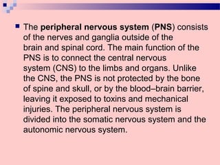
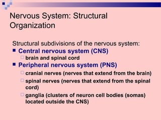

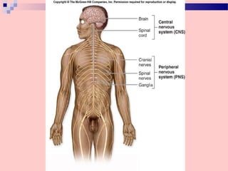
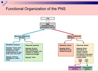
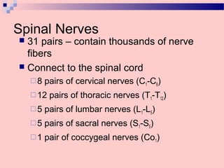
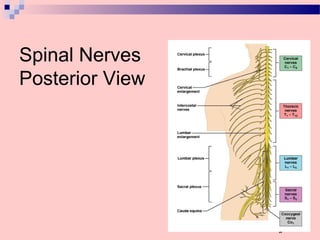


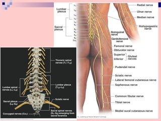
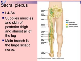

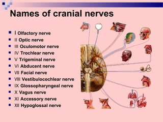
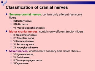

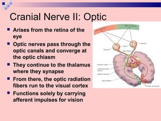
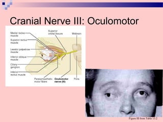
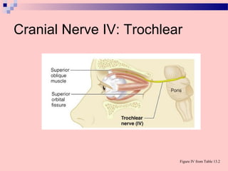
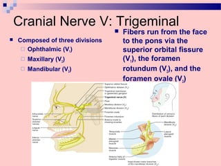

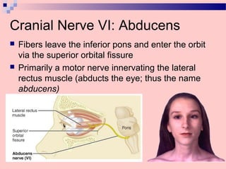

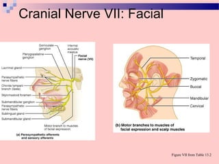

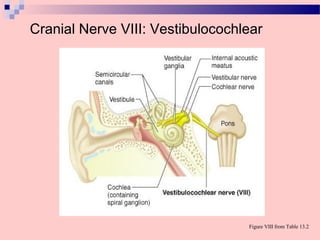
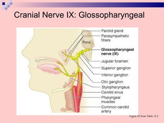
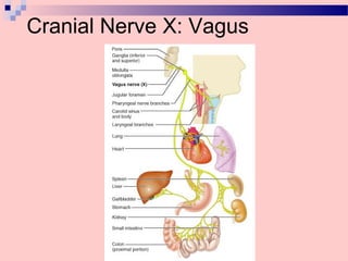


The document provides information on the structural organization and functional roles of the peripheral nervous system (PNS). It discusses how the PNS is divided into 31 pairs of spinal nerves and 12 pairs of cranial nerves. It also summarizes the key characteristics and functions of each of the 12 cranial nerves.


