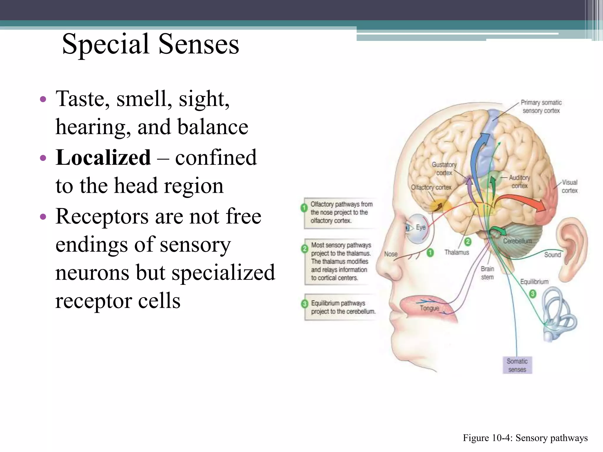
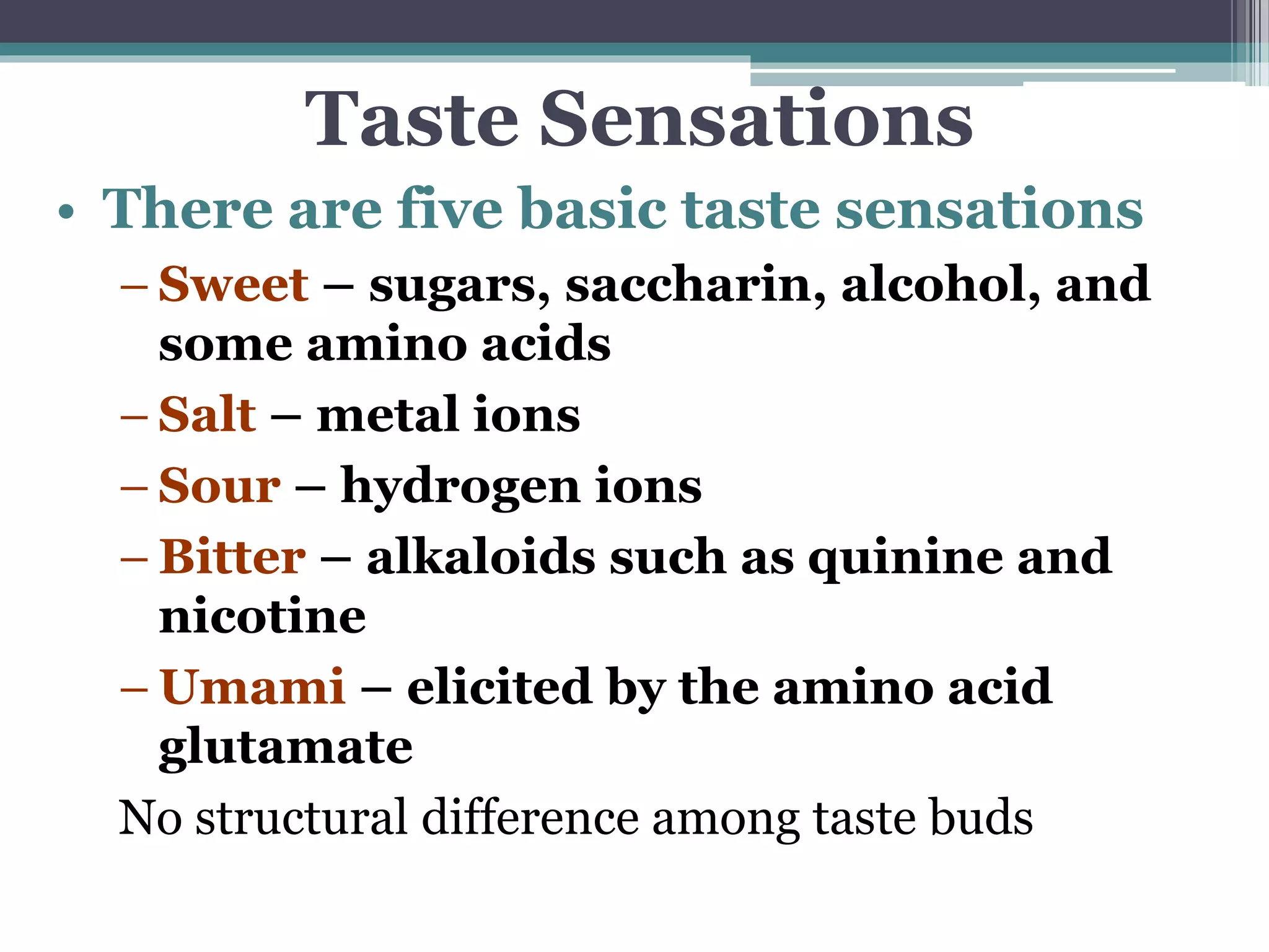
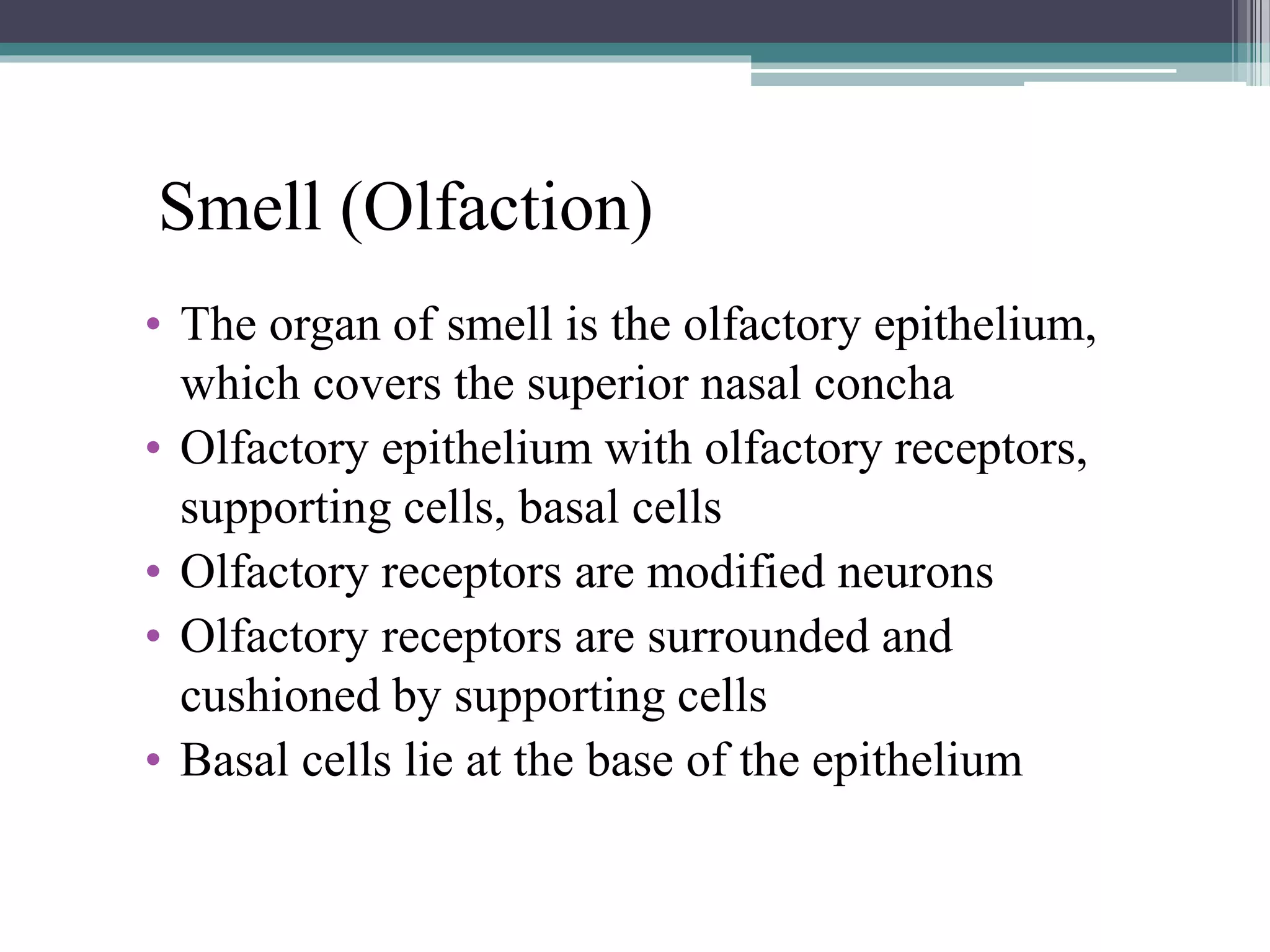
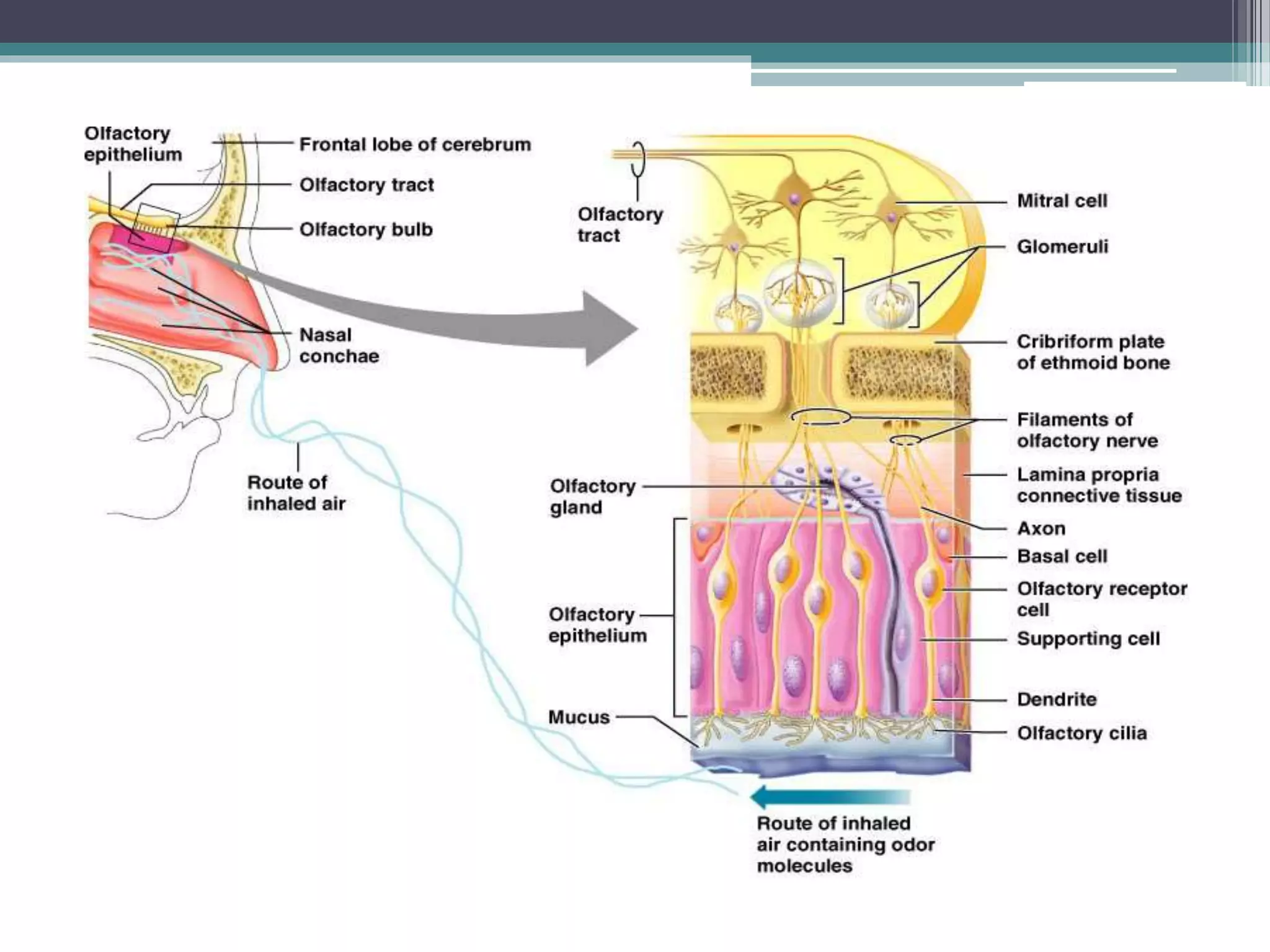
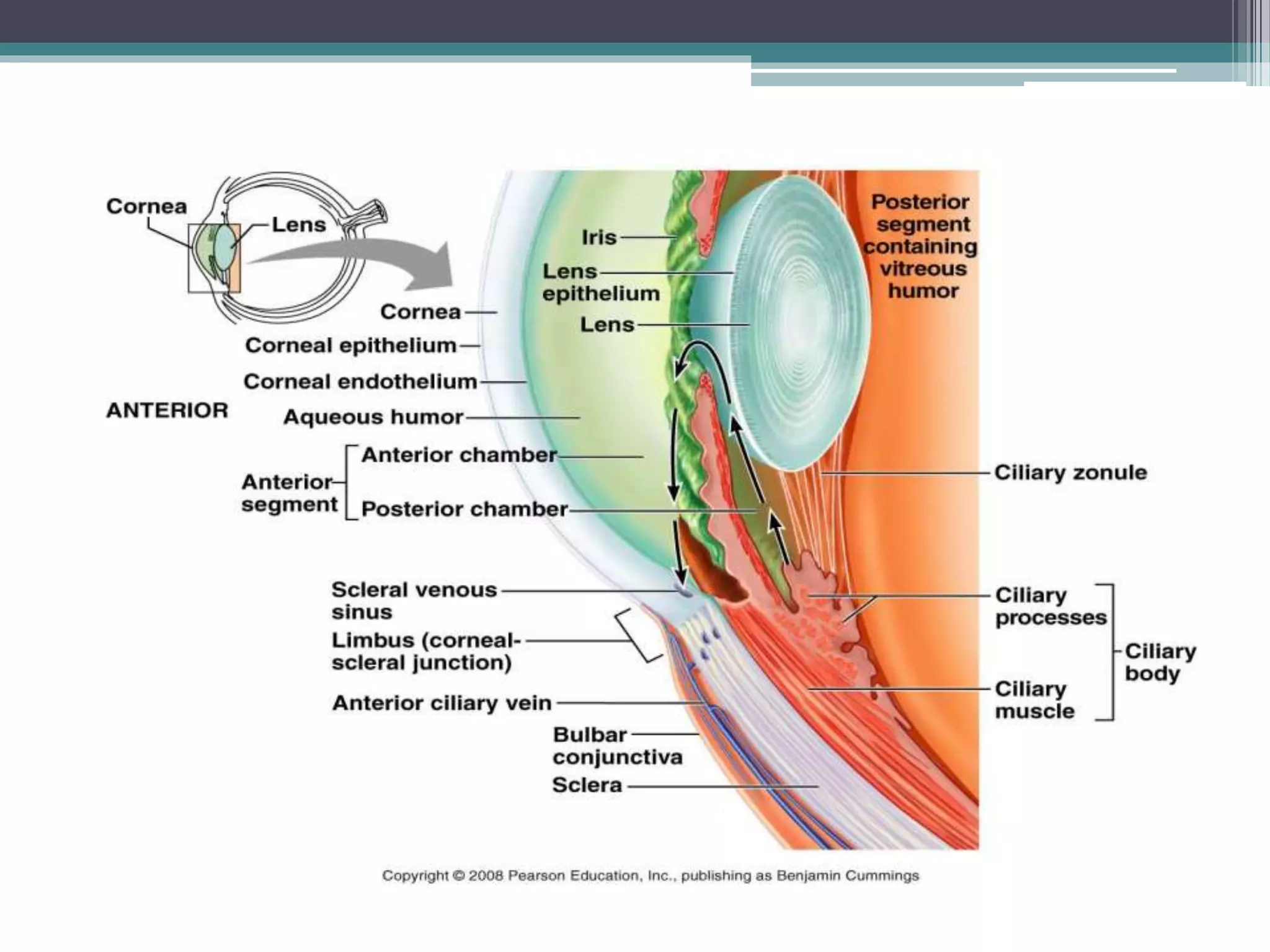
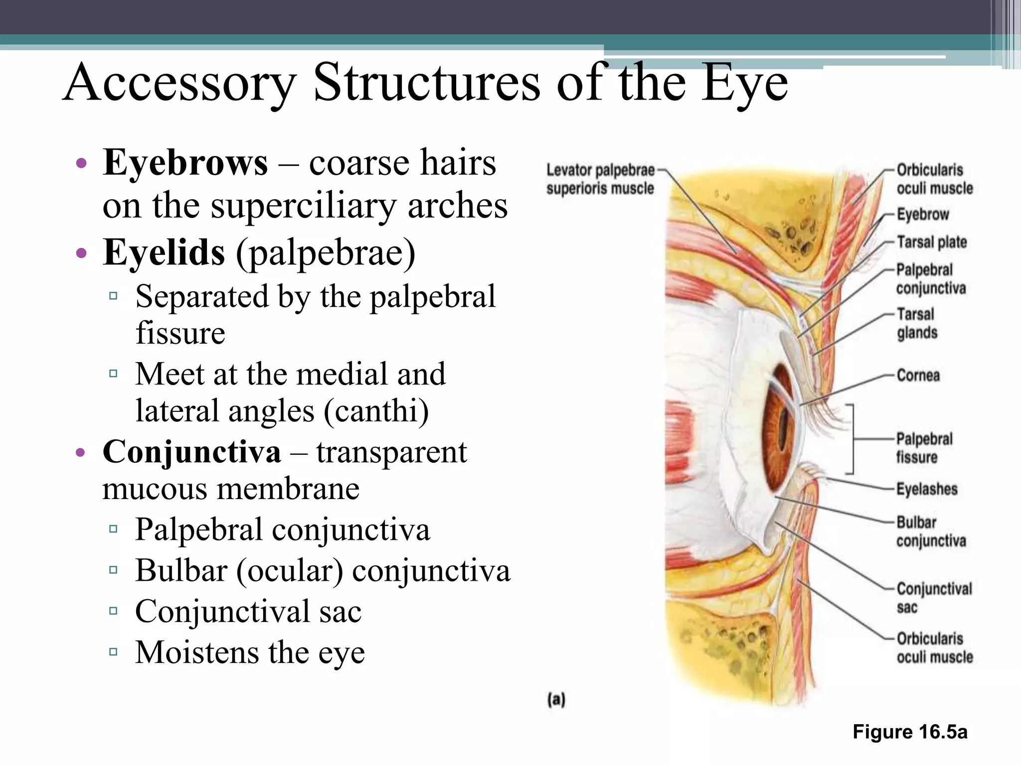
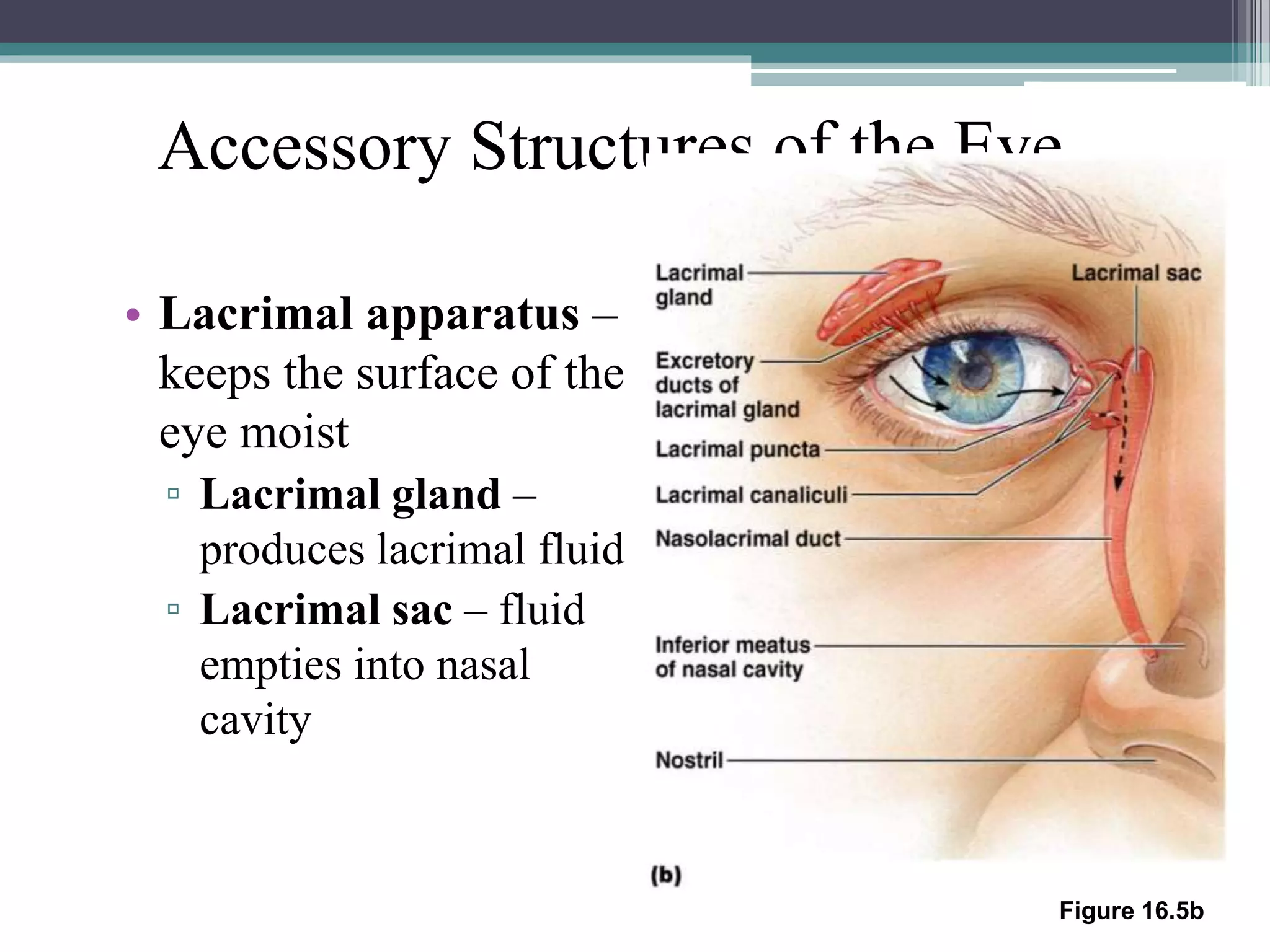
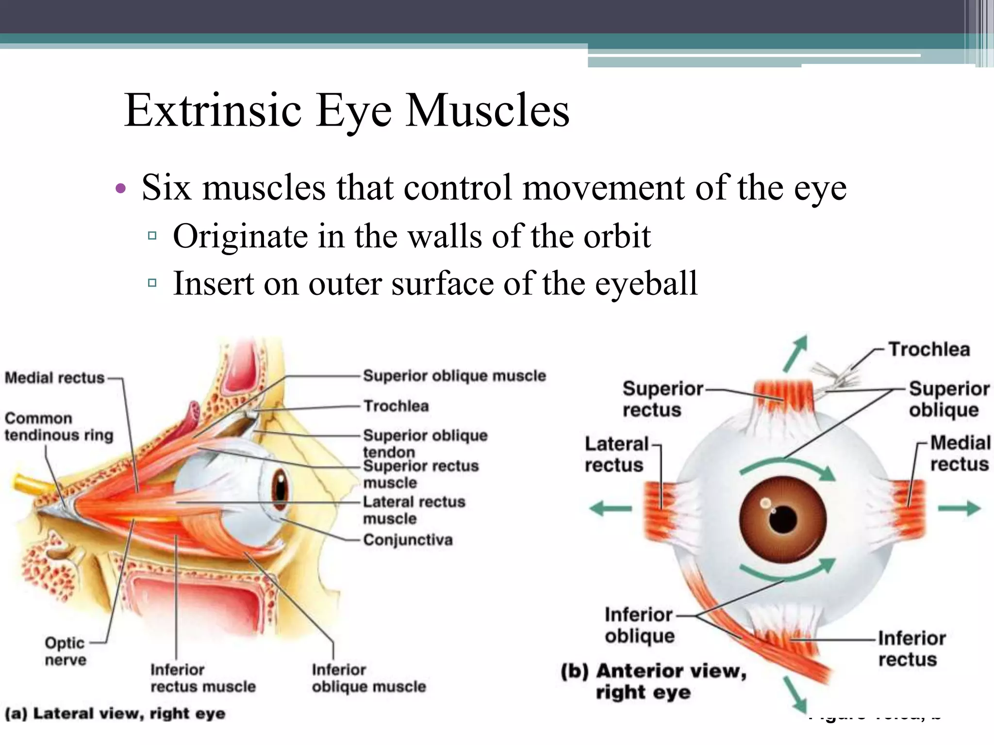
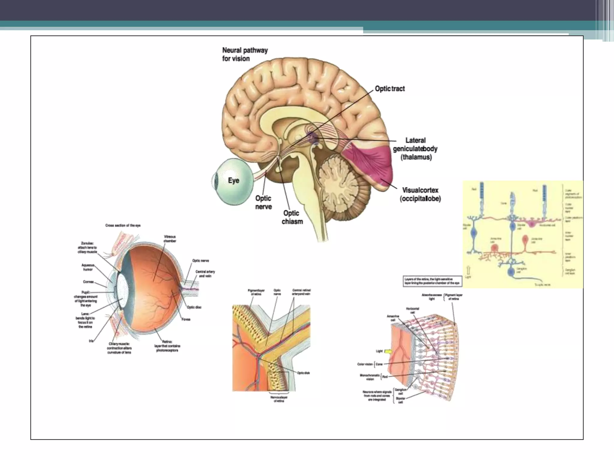
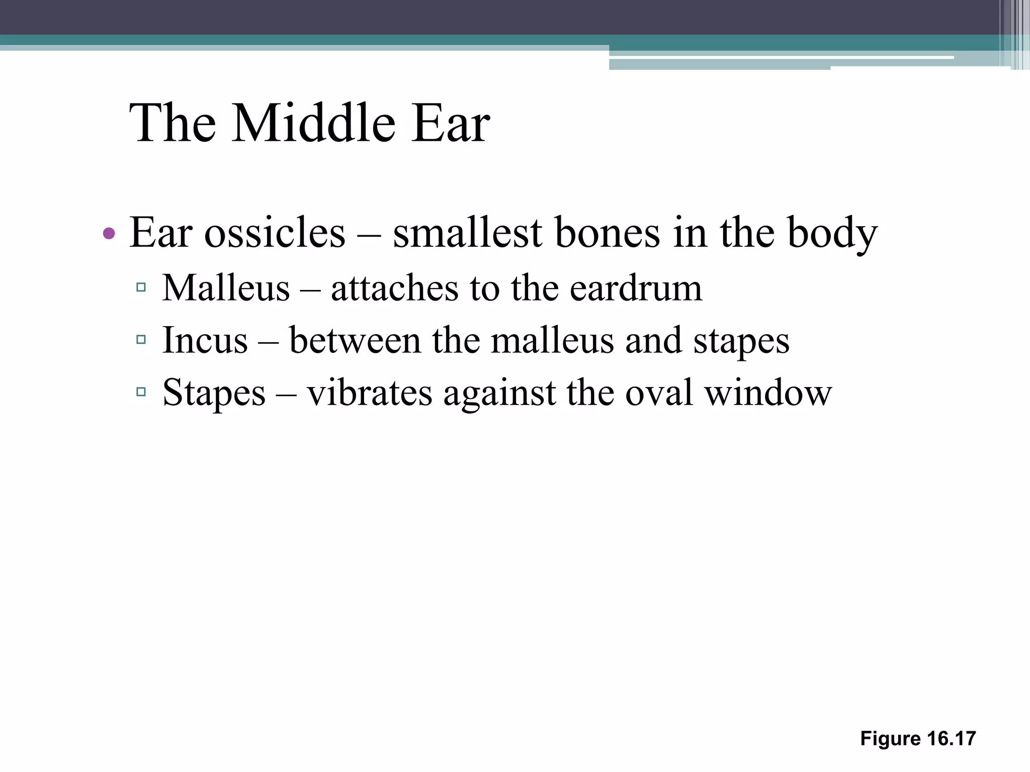
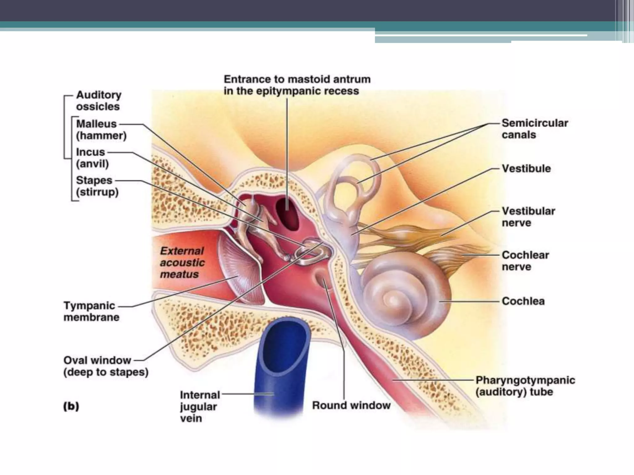
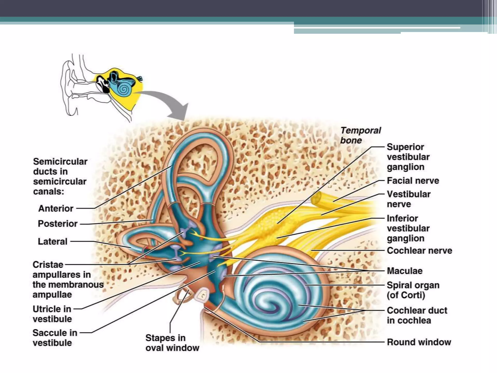
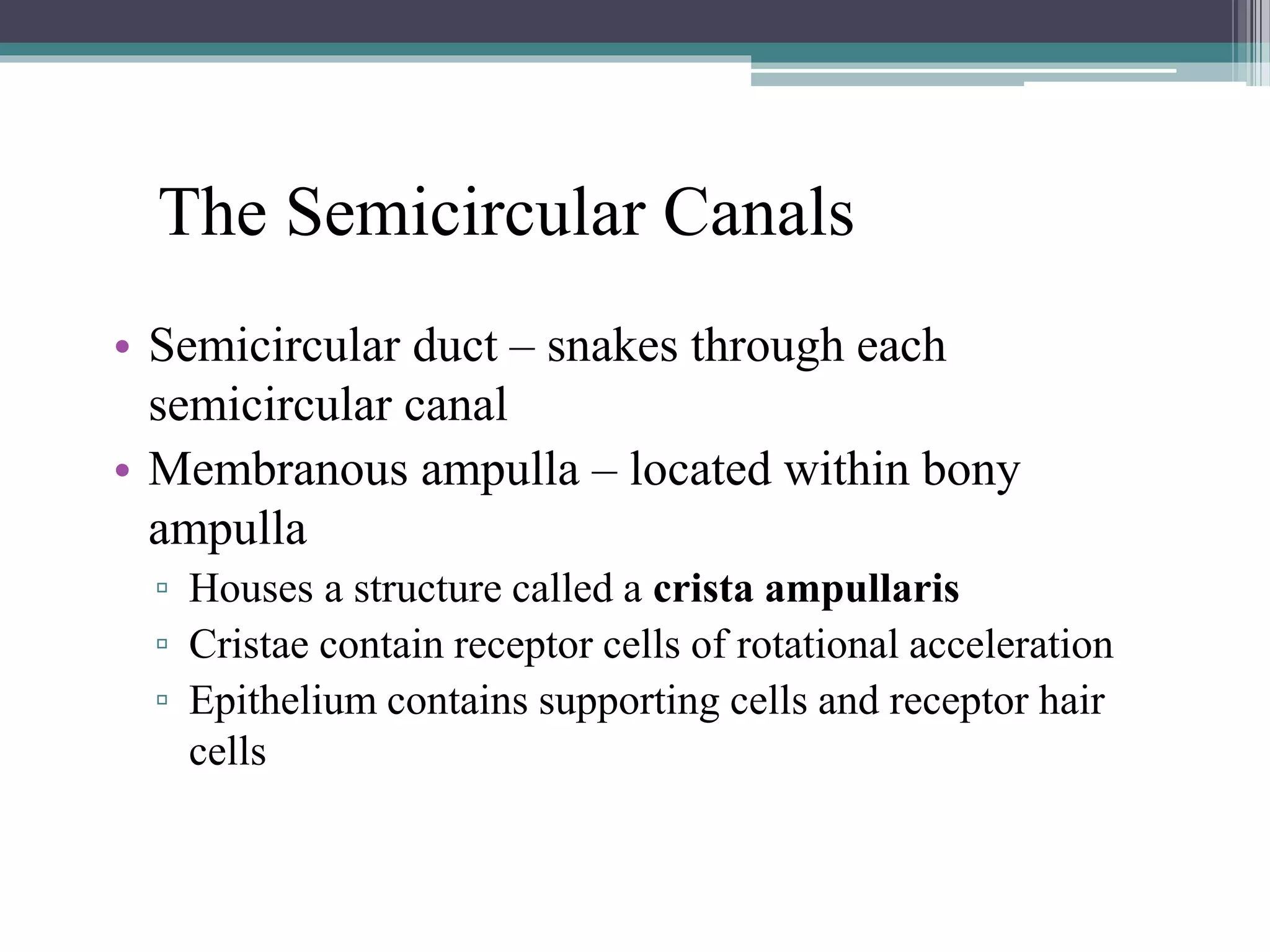
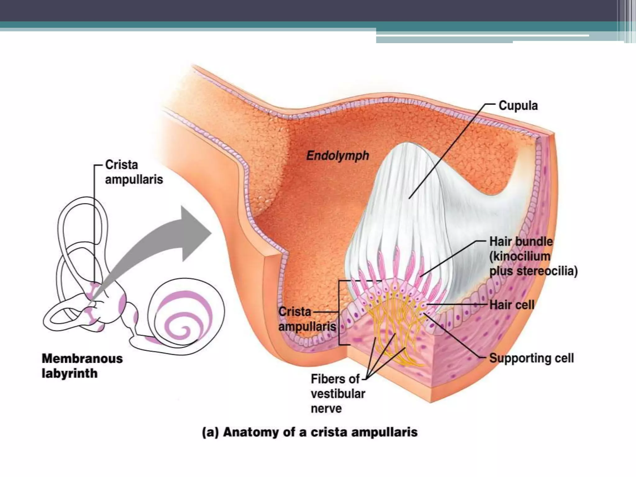
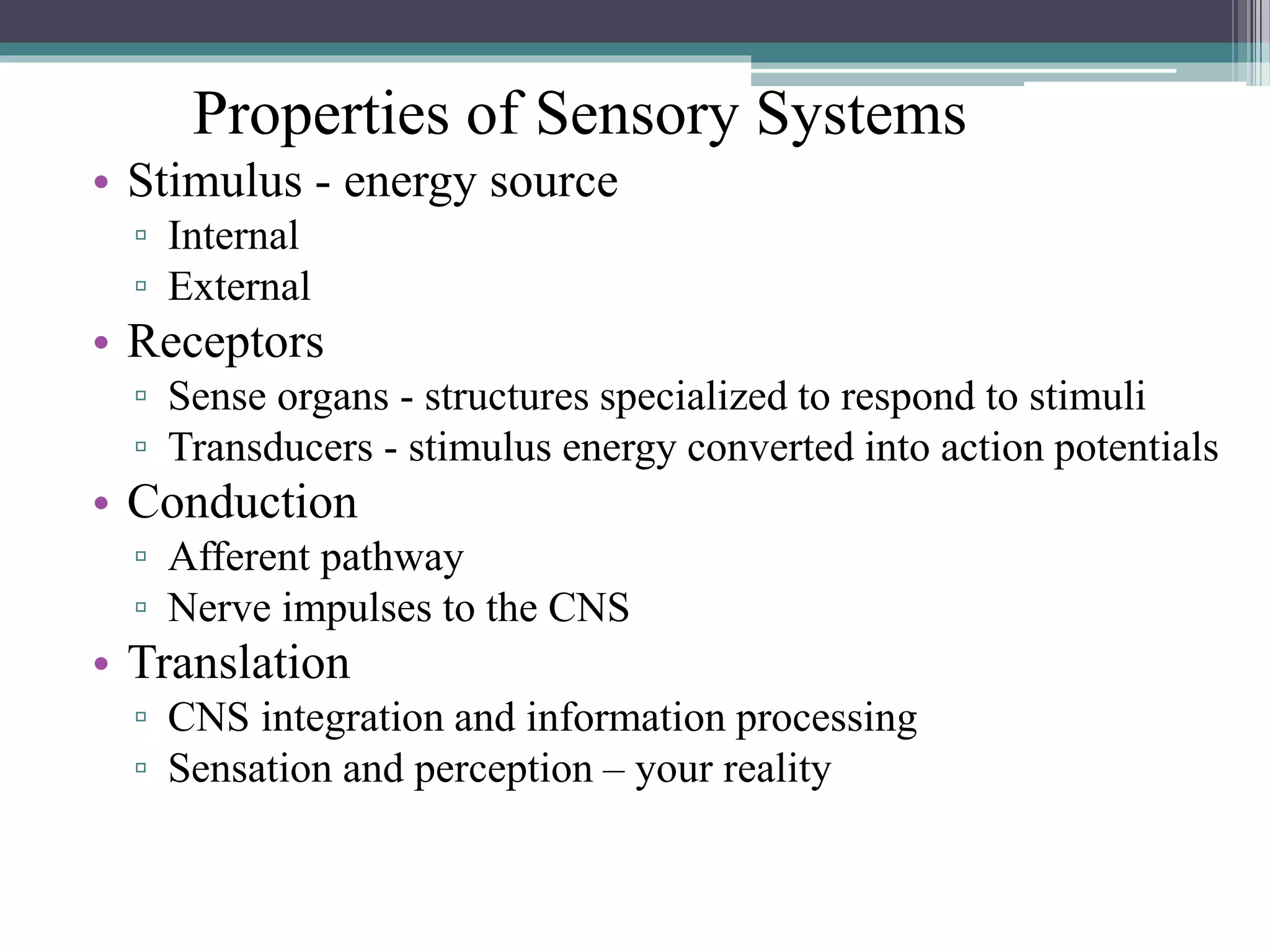
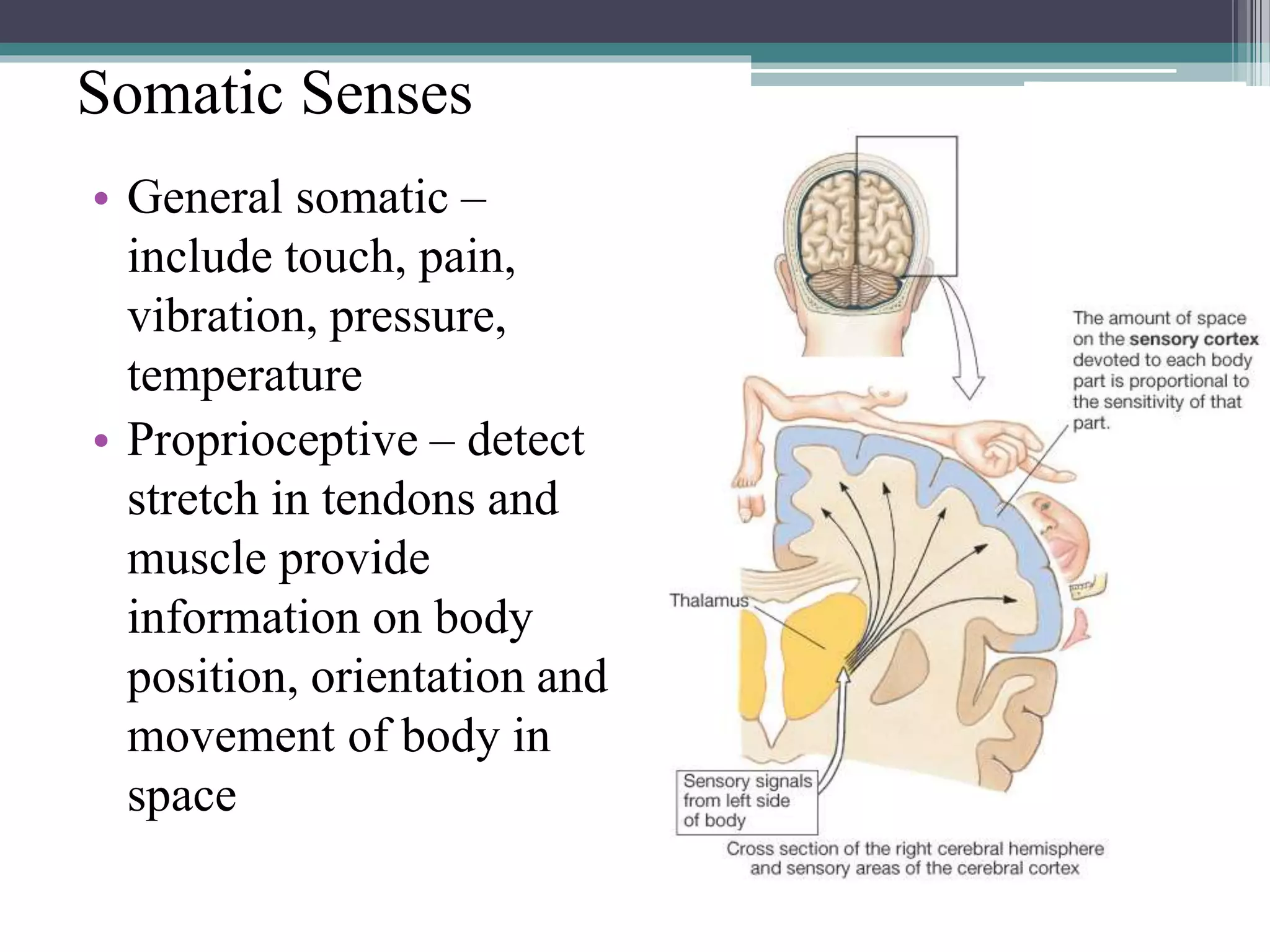
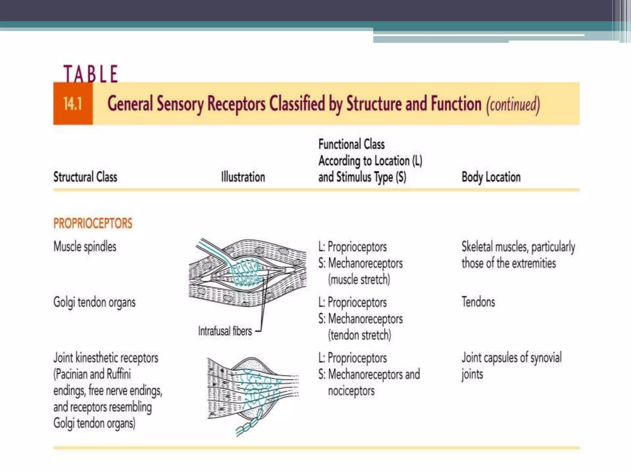
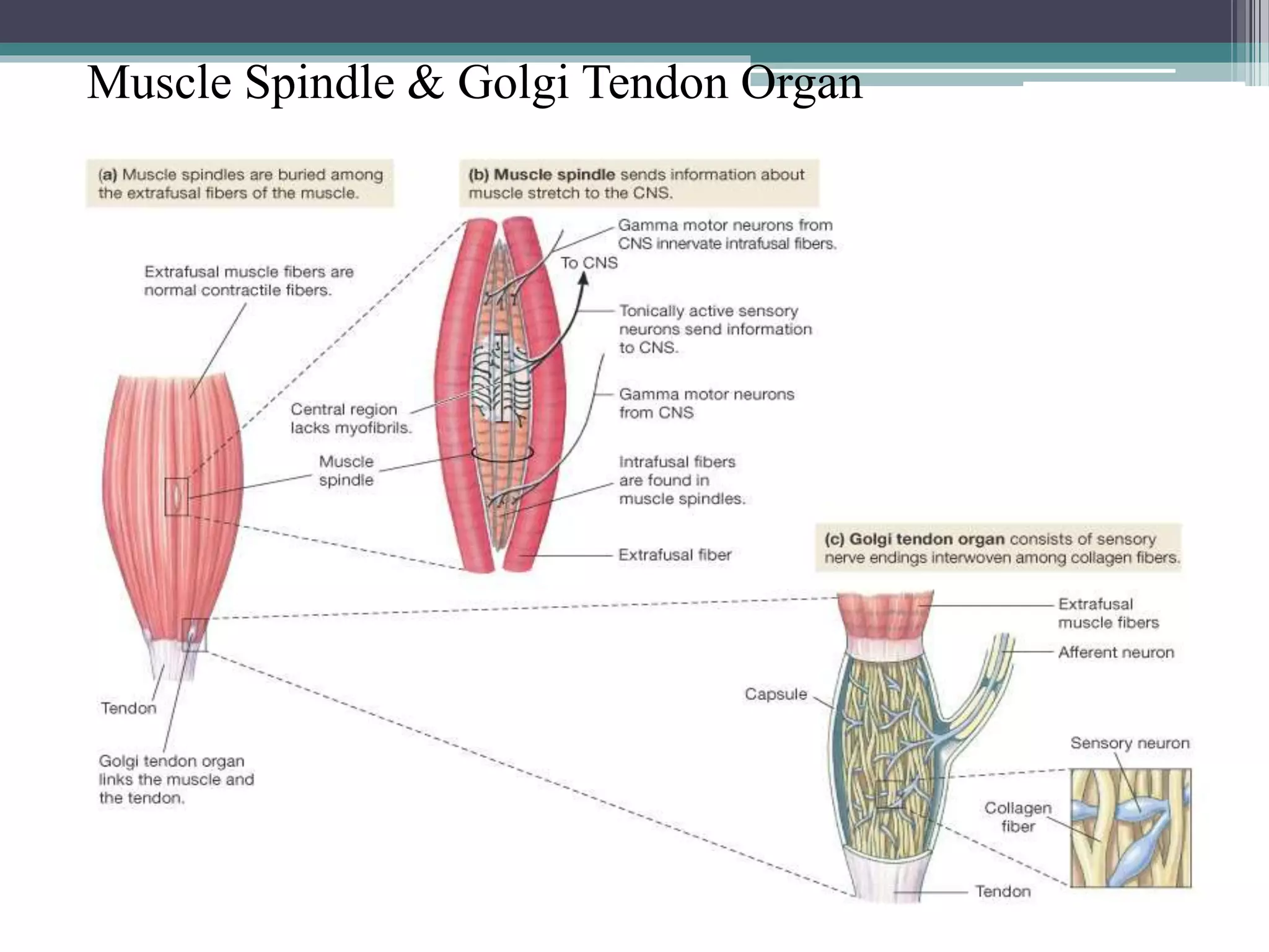
The sense organs
Sense organs
• Besides the eyes, ears, nose and taste buds, millions
of sense organs are located throughout the body in
skin, internal organs and muscles.
• Many receptors allows us to respond to internal and
external stimuli. the receptors are located on the tips
of dendrites of sensory neurons.
• Ability to sense changes in environment is a
requirement for maintaining homeostasis.
• Conversion of a stimulus to a sensation:
▫ Stimuli (light, sound, temperature, etc. are changed into
an electrical signal or nerve impulse.
▫ The signal is then transmitted over a neuron to the
other one.
▫ The signal is interpreted and become consciously
aware of a sensation.
Classification of sense organs
• General sense organs: are microscopic receptors
widely distributed throughout the body in the skin,
muscle, tendons joints and other internal organs.
• They are responsible for pain, touch, temperature and
pressure.
• Special sense organs: such us those
responsible for smell, taste, vision, hearing
and equilibrium are grouped into localized
areas.
• All sense organs must be able to detect
changes in the environment.
Special Senses
• Taste, smell, sight,
hearing, and balance
• Localized – confined
to the head region
• Receptors are not free
endings of sensory
neurons but specialized
receptor cells
Figure 10-4: Sensory pathways
Five Senses
• Vision (Sight)
• Hearing
• Smell
• Taste
• Touch (Palpation)
Taste – Gustation
• Taste receptors
▫ Occur in taste buds
 Most are found on the
surface of the tongue
 Located within tongue
papillae
▫ Two types of papillae
(with taste buds)
 Fungiform papillae
 Circumvallate papillae
Taste Buds
• Taste buds – chemical receptors that generate nervous
impulses resulting in the sense of taste. There are
about 10,000 microscopic taste buds located on the
papillae of the tongue.
Taste Buds
• Collection of 50 –100
epithelial cells
• Contain three major cell
types (similar in all special
senses)
▫ Supporting cells
▫ Gustatory cells
▫ Basal cells
• Contain long microvilli –
extend through a taste pore
Taste Sensations
• There are five basic taste sensations
– Sweet – sugars, saccharin, alcohol, and
some amino acids
– Salt – metal ions
– Sour – hydrogen ions
– Bitter – alkaloids such as quinine and
nicotine
– Umami – elicited by the amino acid
glutamate
No structural difference among taste buds
Gustatory Pathway from Taste Buds
• Taste information
reaches the cerebral
cortex
▫ Primarily through the
facial (VII) and
glossopharyngeal (IX)
nerves
▫ Some taste information
through the vagus nerve
(X)
▫ Sensory neurons synapse
in the medulla
 Located in the solitary
nucleus
Figure 16.2
Smell (Olfaction)
• The organ of smell is the olfactory epithelium,
which covers the superior nasal concha
• Olfactory epithelium with olfactory receptors,
supporting cells, basal cells
• Olfactory receptors are modified neurons
• Olfactory receptors are surrounded and
cushioned by supporting cells
• Basal cells lie at the base of the epithelium
• Surfaces are coated with secretions from olfactory
glands
• Olfactory reception involves detecting dissolved
chemicals as they interact with odorant binding
proteins
Olfactory Receptors
• Bipolar sensory neurons located within olfactory epithelium
▫ Dendrite projects into nasal cavity, terminates in cilia
▫ Axon projects directly up into olfactory bulb of cerebrum
▫ Olfactory bulb projects to olfactory cortex, hippocampus,
and amygdaloid nuclei
Anatomy of the Eyeball
• Function of the eyeball
▫ Protect and support the photoreceptors
▫ Gather, focus, and process light into precise images
• External walls – composed of three tunics (layers)
• Internal cavity – contains fluids (humors)
The Fibrous Layer
• Most external layer of the eyeball
▫ Cornea
 Anterior one-sixth of the fibrous tunic
 Composed of stratified Squamous externally, simple
squamous internally
 Refracts (bends) light
▫ Sclera
 Posterior five-sixths of the tunic
 White, opaque region composed of dense irregular
connective tissue Provides shape and an anchor for eye
muscles,
▫ Scleral venous sinus – allows aqueous humor to drain
The Vascular Layer
• Middle layer consists of choroid, ciliary body,
and iris
• Iris and Pupil
▫ Composed of smooth muscle, melanocytes, and
blood vessels that forms the colored portion of the
eye.
▫ Function: It regulates the amount of light entering
the eye through the pupil.
▫ It is attached to the ciliary body.
▫ Pupil is the opening in center of iris through which
light enters the eye
• Ciliary body
▫ Composed of a ring of muscle called ciliary muscle
and ciliary processes which are folds located at the
posterior surface of ciliary bodies
▫ Suspensory ligaments attach to these processes
▫ Function: secretes the aqueous humor
▫ The suspensory ligaments position the lens so that
light passing through the pupil passes through the
center of the lens of the eye.
The Vascular Layer
• Choroid - vascular layer in the wall of the eye.
▫ Dark brown (pigmented) membrane with melanocytes
that lines most of the internal surface of the sclera. Has
lots of blood vessels
▫ Lines most of the interior of the sclera.
▫ Extends from the ciliary body to the lens.
▫ Corresponds to arachnoid and pia mater
• Functions:
▫ Delivers oxygen and nutrients to the retina.
▫ Absorb light rays so that the light rays are not reflected
within the eye
The Inner Layer (Retina)
• Retina is the innermost layer of the eye lining the
posterior cavity
• The retina contains 2 layers:
▫ Pigmented layer made of a single layer of
melanocytes, absorbs light after it passes through
the neural layer
▫ Neural layer – sheet of nervous tissue, contains
three main types of neurons
 Photoreceptor cells
 Bipolar cells
 Ganglion cells
Photoreceptors
• Two main types
▫ Rod cells
 More sensitive to light
 Allow vision in dim light
 In periphery
▫ Cone cells
 Operate best in bright light
 High-acuity
 Color vision – blue, green, red
cones
 Concentrated in fovea
Regional Specializations of the
Retina
• Ora serrata retinae
▫ Neural layer ends at the posterior margin of the
ciliary body
▫ Pigmented layer covers ciliary body and
posterior surface of the iris
• Macula lutea – contains mostly cones
• Fovea centralis – contains only cones
▫ Region of highest visual acuity
• Optic disc – blind spot
The Lens
• A thick, transparent, biconvex disc
• Held in place by its ciliary zonule
• Lens epithelium – covers anterior surface
of the lens
Internal Chambers and Fluids
Figure 16.8
Internal Chambers and Fluids
• Anterior segment
• Divided into anterior and posterior
chambers
▫ Anterior chamber – between the cornea and iris
▫ Posterior chamber – between the iris and lens
▫ Filled with aqueous humor
 Renewed continuously
 Formed as a blood filtrate
 Supplies nutrients to the lens and cornea
Internal Chambers and Fluids
• The lens and ciliary zonules divide the eye
• Posterior segment (cavity)
▫ Filled with vitreous humor - clear, jelly-
like substance
 Transmits light
 Supports the posterior surface of the lens
 Helps maintain intraocular pressure
Accessory Structures of the Eye
• Eyebrows – coarse hairs
on the superciliary arches
• Eyelids (palpebrae)
▫ Separated by the palpebral
fissure
▫ Meet at the medial and
lateral angles (canthi)
• Conjunctiva – transparent
mucous membrane
▫ Palpebral conjunctiva
▫ Bulbar (ocular) conjunctiva
▫ Conjunctival sac
▫ Moistens the eye
Figure 16.5a
Accessory Structures of the Eye
• Lacrimal apparatus –
keeps the surface of the
eye moist
▫ Lacrimal gland –
produces lacrimal fluid
▫ Lacrimal sac – fluid
empties into nasal
cavity
Figure 16.5b
Extrinsic Eye Muscles
• Six muscles that control movement of the eye
▫ Originate in the walls of the orbit
▫ Insert on outer surface of the eyeball
Figure 16.6a, b
Visual Pathways to the Cerebral
Cortex
• Pathway begins at the retina
▫ Light activates photoreceptors
▫ Photoreceptors signal bipolar cells
▫ Bipolar cells signal ganglion cells
▫ Axons of ganglion cells exit eye as the
optic nerve
Vision Integration / Pathway
• Optic nerve
• Optic chiasm
• Optic tract
• Thalamus
• Visual cortex
• Other pathways include
the midbrain and
diencephalon
Figure 10-29b, c: Neural pathways for vision and the papillary reflex
The Ear: Hearing and Equilibrium
• The ear – receptor organ for hearing and equilibrium
• Composed of three main regions
▫ Outer ear – functions in hearing
▫ Middle ear – functions in hearing
▫ Inner ear – functions in both hearing and equilibrium
The Outer (External) Ear
• Auricle (pinna) - helps direct sounds
• External acoustic meatus
▫ Lined with skin
▫ Contains hairs, sebaceous glands, and
ceruminous glands
• Tympanic membrane
 Forms the boundary between the external and middle
ear
The Middle Ear
• The tympanic cavity
▫ A small, air-filled space
▫ Located within the petrous portion of the
temporal bone
• Medial wall is penetrated by
▫ Oval window
▫ Round window
• Pharyngotympanic tube (auditory or
eustachian tube) - Links the middle ear and
pharynx
Figure 16.17
The Middle Ear
• Ear ossicles – smallest bones in the body
▫ Malleus – attaches to the eardrum
▫ Incus – between the malleus and stapes
▫ Stapes – vibrates against the oval window
The Inner (Internal) Ear
• Inner ear – also called the labyrinth
• Bony labyrinth – a cavity consisting of three parts
▫ Semicircular canals
▫ Vestibule
▫ Cochlea
▫ Bony labyrinth is filled with perilymph
The Membranous Labyrinth
• Membranous labyrinth - series of membrane-walled
sacs and ducts
▫ Fit within the bony labyrinth
▫ Consists of three main parts
 Semicircular ducts
 Utricle and saccule
 Cochlear duct
▫ Filled with a clear fluid – endolymph
Figure 16.18
The Cochlea
• A spiraling chamber in the bony labyrinth
▫ Coils around a pillar of bone – the modiolus
 Spiral lamina – a spiral of bone in the modiolus
 The cochlear nerve runs through the core of the
modiolus
The Cochlea
• The cochlear duct (scala media) – contains
receptors for hearing
▫ Lies between two chambers
 The scala vestibuli
 The scala tympani
▫ The vestibular membrane – the roof of the
cochlear duct
▫ The basilar membrane – the floor of the
cochlear duct
The Cochlea
• The cochlear duct (scala media) – contains
receptors for hearing
▫ Organ of Corti – the receptor epithelium for hearing
▫ Consists of hair cells (receptor cells)
The Role of the Cochlea in Hearing
Figure 16.20
Auditory Pathway from the Organ of
Corti
• The ascending
auditory pathway
• Transmits
information from
cochlear receptors
to the cerebral
cortex
Figure 16.23
The Vestibule
• Utricle and saccule – suspended in perilymph
 Two egg-shaped parts of the membranous labyrinth
 House the macula – a spot of sensory epithelium
• Macula – contains receptor cells
▫ Monitor the position of the head when the head is still
▫ Contains columnar supporting cells
▫ Receptor cells – called hair cells
 Synapse with the vestibular nerve
Anatomy and Function of the Maculae
Figure 16.21b
The Semicircular Canals
• Lie posterior and lateral to the vestibule
• Anterior and posterior semicircular canals lie in
the vertical plane at right angles
• Lateral semicircular canal lies in the horizontal
plane
The Semicircular Canals
• Semicircular duct – snakes through each
semicircular canal
• Membranous ampulla – located within bony
ampulla
▫ Houses a structure called a crista ampullaris
▫ Cristae contain receptor cells of rotational acceleration
▫ Epithelium contains supporting cells and receptor hair
cells
Structure and Function of the Crista Ampullaris
Figure 16.22b
Sensory Systems
• Somatic sensory
▫ General – transmit impulses from skin, skeletal
muscles, and joints
▫ Special senses - hearing, balance, vision
• Visceral sensory
▫ Transmit impulses from visceral organs
▫ Special senses - olfaction (smell), gustation
(taste)
Properties of Sensory Systems
• Stimulus - energy source
▫ Internal
▫ External
• Receptors
▫ Sense organs - structures specialized to respond to stimuli
▫ Transducers - stimulus energy converted into action potentials
• Conduction
▫ Afferent pathway
▫ Nerve impulses to the CNS
• Translation
▫ CNS integration and information processing
▫ Sensation and perception – your reality
Sensory Pathways
• Stimulus as physical energy  sensory
receptor acts as a transducer
• Stimulus > threshold  action potential to
CNS
• Integration in CNS  cerebral cortex or acted
on subconsciously
Classification by Function (Stimuli)
• Mechanoreceptors – respond to touch, pressure,
vibration, stretch, and itch
• Thermoreceptors – sensitive to changes in
temperature
• Photoreceptors – respond to light energy (e.g., retina)
• Chemoreceptors – respond to chemicals (e.g., smell,
taste, changes in blood chemistry)
• Nociceptors – sensitive to pain-causing stimuli
• Osmoreceptors – detect changes in concentration of
solutes, osmotic activity
• Baroreceptors – detect changes in fluid pressure
Classification by Location
• Exteroceptors – sensitive to stimuli arising from
outside the body
▫ Located at or near body surfaces
▫ Include receptors for touch, pressure, pain, and temperature
• Interoceptors – (visceroceptors) receive stimuli from
internal viscera
▫ Monitor a variety of stimuli
• Proprioceptors – monitor degree of stretch
▫ Located in musculoskeletal organs
Classification by Structure
Somatic Senses
• General somatic –
include touch, pain,
vibration, pressure,
temperature
• Proprioceptive – detect
stretch in tendons and
muscle provide
information on body
position, orientation and
movement of body in
space
Somatic Receptors
• Divided into two groups
▫ Free or Unencapsulated nerve endings
▫ Encapsulated nerve endings - consist of one or
more neural end fibers enclosed in connective
tissue
Free Nerve Endings
• Abundant in epithelia and underlying connective
tissue
• Nociceptors - respond to pain
• Thermoreceptors - respond to temperature
• Two specialized types of free nerve endings
▫ Merkel discs – lie in the epidermis, slowly adapting
receptors for light touch
▫ Hair follicle receptors – Rapidly adapting receptors that
wrap around hair follicles
Encapsulated Nerve Endings
▫ Meissner’s corpuscles
 Spiraling nerve ending surrounded by Schwann cells
 Occur in the dermal papillae of hairless areas of the skin
 Rapidly adapting receptors for discriminative touch
▫ Pacinian corpuscles
 Single nerve ending surrounded by layers of flattened
Schwann cells
 Occur in the hypodermis
 Sensitive to deep pressure – rapidly adapting receptors
▫ Ruffini’s corpuscles
 Located in the dermis and respond to pressure
 Monitor continuous pressure on the skin – adapt slowly
Encapsulated Nerve Endings -
Proprioceptors
• Monitor stretch in locomotory organs
• Three types of proprioceptors
▫ Muscle spindles – monitors the changing length of a
muscle, imbedded in the perimysium between muscle
fascicles
▫ Golgi tendon organs – located near the muscle-tendon
junction, monitor tension within tendons
▫ Joint kinesthetic receptors - sensory nerve endings within
the joint capsules, sense pressure and position
Muscle Spindle & Golgi Tendon Organ

The sense organs

  • 1.
  • 2.
    Sense organs • Besidesthe eyes, ears, nose and taste buds, millions of sense organs are located throughout the body in skin, internal organs and muscles. • Many receptors allows us to respond to internal and external stimuli. the receptors are located on the tips of dendrites of sensory neurons. • Ability to sense changes in environment is a requirement for maintaining homeostasis.
  • 3.
    • Conversion ofa stimulus to a sensation: ▫ Stimuli (light, sound, temperature, etc. are changed into an electrical signal or nerve impulse. ▫ The signal is then transmitted over a neuron to the other one. ▫ The signal is interpreted and become consciously aware of a sensation.
  • 4.
    Classification of senseorgans • General sense organs: are microscopic receptors widely distributed throughout the body in the skin, muscle, tendons joints and other internal organs. • They are responsible for pain, touch, temperature and pressure.
  • 5.
    • Special senseorgans: such us those responsible for smell, taste, vision, hearing and equilibrium are grouped into localized areas. • All sense organs must be able to detect changes in the environment.
  • 6.
    Special Senses • Taste,smell, sight, hearing, and balance • Localized – confined to the head region • Receptors are not free endings of sensory neurons but specialized receptor cells Figure 10-4: Sensory pathways
  • 7.
    Five Senses • Vision(Sight) • Hearing • Smell • Taste • Touch (Palpation)
  • 8.
    Taste – Gustation •Taste receptors ▫ Occur in taste buds  Most are found on the surface of the tongue  Located within tongue papillae ▫ Two types of papillae (with taste buds)  Fungiform papillae  Circumvallate papillae
  • 9.
    Taste Buds • Tastebuds – chemical receptors that generate nervous impulses resulting in the sense of taste. There are about 10,000 microscopic taste buds located on the papillae of the tongue.
  • 10.
    Taste Buds • Collectionof 50 –100 epithelial cells • Contain three major cell types (similar in all special senses) ▫ Supporting cells ▫ Gustatory cells ▫ Basal cells • Contain long microvilli – extend through a taste pore
  • 12.
    Taste Sensations • Thereare five basic taste sensations – Sweet – sugars, saccharin, alcohol, and some amino acids – Salt – metal ions – Sour – hydrogen ions – Bitter – alkaloids such as quinine and nicotine – Umami – elicited by the amino acid glutamate No structural difference among taste buds
  • 13.
    Gustatory Pathway fromTaste Buds • Taste information reaches the cerebral cortex ▫ Primarily through the facial (VII) and glossopharyngeal (IX) nerves ▫ Some taste information through the vagus nerve (X) ▫ Sensory neurons synapse in the medulla  Located in the solitary nucleus Figure 16.2
  • 14.
    Smell (Olfaction) • Theorgan of smell is the olfactory epithelium, which covers the superior nasal concha • Olfactory epithelium with olfactory receptors, supporting cells, basal cells • Olfactory receptors are modified neurons • Olfactory receptors are surrounded and cushioned by supporting cells • Basal cells lie at the base of the epithelium
  • 16.
    • Surfaces arecoated with secretions from olfactory glands • Olfactory reception involves detecting dissolved chemicals as they interact with odorant binding proteins
  • 17.
    Olfactory Receptors • Bipolarsensory neurons located within olfactory epithelium ▫ Dendrite projects into nasal cavity, terminates in cilia ▫ Axon projects directly up into olfactory bulb of cerebrum ▫ Olfactory bulb projects to olfactory cortex, hippocampus, and amygdaloid nuclei
  • 18.
    Anatomy of theEyeball • Function of the eyeball ▫ Protect and support the photoreceptors ▫ Gather, focus, and process light into precise images • External walls – composed of three tunics (layers) • Internal cavity – contains fluids (humors)
  • 19.
    The Fibrous Layer •Most external layer of the eyeball ▫ Cornea  Anterior one-sixth of the fibrous tunic  Composed of stratified Squamous externally, simple squamous internally  Refracts (bends) light ▫ Sclera  Posterior five-sixths of the tunic  White, opaque region composed of dense irregular connective tissue Provides shape and an anchor for eye muscles, ▫ Scleral venous sinus – allows aqueous humor to drain
  • 21.
    The Vascular Layer •Middle layer consists of choroid, ciliary body, and iris • Iris and Pupil ▫ Composed of smooth muscle, melanocytes, and blood vessels that forms the colored portion of the eye. ▫ Function: It regulates the amount of light entering the eye through the pupil. ▫ It is attached to the ciliary body. ▫ Pupil is the opening in center of iris through which light enters the eye
  • 22.
    • Ciliary body ▫Composed of a ring of muscle called ciliary muscle and ciliary processes which are folds located at the posterior surface of ciliary bodies ▫ Suspensory ligaments attach to these processes ▫ Function: secretes the aqueous humor ▫ The suspensory ligaments position the lens so that light passing through the pupil passes through the center of the lens of the eye.
  • 24.
    The Vascular Layer •Choroid - vascular layer in the wall of the eye. ▫ Dark brown (pigmented) membrane with melanocytes that lines most of the internal surface of the sclera. Has lots of blood vessels ▫ Lines most of the interior of the sclera. ▫ Extends from the ciliary body to the lens. ▫ Corresponds to arachnoid and pia mater • Functions: ▫ Delivers oxygen and nutrients to the retina. ▫ Absorb light rays so that the light rays are not reflected within the eye
  • 26.
    The Inner Layer(Retina) • Retina is the innermost layer of the eye lining the posterior cavity • The retina contains 2 layers: ▫ Pigmented layer made of a single layer of melanocytes, absorbs light after it passes through the neural layer ▫ Neural layer – sheet of nervous tissue, contains three main types of neurons  Photoreceptor cells  Bipolar cells  Ganglion cells
  • 28.
    Photoreceptors • Two maintypes ▫ Rod cells  More sensitive to light  Allow vision in dim light  In periphery ▫ Cone cells  Operate best in bright light  High-acuity  Color vision – blue, green, red cones  Concentrated in fovea
  • 29.
    Regional Specializations ofthe Retina • Ora serrata retinae ▫ Neural layer ends at the posterior margin of the ciliary body ▫ Pigmented layer covers ciliary body and posterior surface of the iris • Macula lutea – contains mostly cones • Fovea centralis – contains only cones ▫ Region of highest visual acuity • Optic disc – blind spot
  • 31.
    The Lens • Athick, transparent, biconvex disc • Held in place by its ciliary zonule • Lens epithelium – covers anterior surface of the lens
  • 32.
    Internal Chambers andFluids Figure 16.8
  • 33.
    Internal Chambers andFluids • Anterior segment • Divided into anterior and posterior chambers ▫ Anterior chamber – between the cornea and iris ▫ Posterior chamber – between the iris and lens ▫ Filled with aqueous humor  Renewed continuously  Formed as a blood filtrate  Supplies nutrients to the lens and cornea
  • 34.
    Internal Chambers andFluids • The lens and ciliary zonules divide the eye • Posterior segment (cavity) ▫ Filled with vitreous humor - clear, jelly- like substance  Transmits light  Supports the posterior surface of the lens  Helps maintain intraocular pressure
  • 35.
    Accessory Structures ofthe Eye • Eyebrows – coarse hairs on the superciliary arches • Eyelids (palpebrae) ▫ Separated by the palpebral fissure ▫ Meet at the medial and lateral angles (canthi) • Conjunctiva – transparent mucous membrane ▫ Palpebral conjunctiva ▫ Bulbar (ocular) conjunctiva ▫ Conjunctival sac ▫ Moistens the eye Figure 16.5a
  • 36.
    Accessory Structures ofthe Eye • Lacrimal apparatus – keeps the surface of the eye moist ▫ Lacrimal gland – produces lacrimal fluid ▫ Lacrimal sac – fluid empties into nasal cavity Figure 16.5b
  • 37.
    Extrinsic Eye Muscles •Six muscles that control movement of the eye ▫ Originate in the walls of the orbit ▫ Insert on outer surface of the eyeball Figure 16.6a, b
  • 38.
    Visual Pathways tothe Cerebral Cortex • Pathway begins at the retina ▫ Light activates photoreceptors ▫ Photoreceptors signal bipolar cells ▫ Bipolar cells signal ganglion cells ▫ Axons of ganglion cells exit eye as the optic nerve
  • 40.
    Vision Integration /Pathway • Optic nerve • Optic chiasm • Optic tract • Thalamus • Visual cortex • Other pathways include the midbrain and diencephalon Figure 10-29b, c: Neural pathways for vision and the papillary reflex
  • 41.
    The Ear: Hearingand Equilibrium • The ear – receptor organ for hearing and equilibrium • Composed of three main regions ▫ Outer ear – functions in hearing ▫ Middle ear – functions in hearing ▫ Inner ear – functions in both hearing and equilibrium
  • 42.
    The Outer (External)Ear • Auricle (pinna) - helps direct sounds • External acoustic meatus ▫ Lined with skin ▫ Contains hairs, sebaceous glands, and ceruminous glands • Tympanic membrane  Forms the boundary between the external and middle ear
  • 44.
    The Middle Ear •The tympanic cavity ▫ A small, air-filled space ▫ Located within the petrous portion of the temporal bone • Medial wall is penetrated by ▫ Oval window ▫ Round window • Pharyngotympanic tube (auditory or eustachian tube) - Links the middle ear and pharynx
  • 46.
    Figure 16.17 The MiddleEar • Ear ossicles – smallest bones in the body ▫ Malleus – attaches to the eardrum ▫ Incus – between the malleus and stapes ▫ Stapes – vibrates against the oval window
  • 48.
    The Inner (Internal)Ear • Inner ear – also called the labyrinth • Bony labyrinth – a cavity consisting of three parts ▫ Semicircular canals ▫ Vestibule ▫ Cochlea ▫ Bony labyrinth is filled with perilymph
  • 50.
    The Membranous Labyrinth •Membranous labyrinth - series of membrane-walled sacs and ducts ▫ Fit within the bony labyrinth ▫ Consists of three main parts  Semicircular ducts  Utricle and saccule  Cochlear duct ▫ Filled with a clear fluid – endolymph Figure 16.18
  • 52.
    The Cochlea • Aspiraling chamber in the bony labyrinth ▫ Coils around a pillar of bone – the modiolus  Spiral lamina – a spiral of bone in the modiolus  The cochlear nerve runs through the core of the modiolus
  • 54.
    The Cochlea • Thecochlear duct (scala media) – contains receptors for hearing ▫ Lies between two chambers  The scala vestibuli  The scala tympani ▫ The vestibular membrane – the roof of the cochlear duct ▫ The basilar membrane – the floor of the cochlear duct
  • 55.
    The Cochlea • Thecochlear duct (scala media) – contains receptors for hearing ▫ Organ of Corti – the receptor epithelium for hearing ▫ Consists of hair cells (receptor cells)
  • 57.
    The Role ofthe Cochlea in Hearing Figure 16.20
  • 58.
    Auditory Pathway fromthe Organ of Corti • The ascending auditory pathway • Transmits information from cochlear receptors to the cerebral cortex Figure 16.23
  • 59.
    The Vestibule • Utricleand saccule – suspended in perilymph  Two egg-shaped parts of the membranous labyrinth  House the macula – a spot of sensory epithelium • Macula – contains receptor cells ▫ Monitor the position of the head when the head is still ▫ Contains columnar supporting cells ▫ Receptor cells – called hair cells  Synapse with the vestibular nerve
  • 61.
    Anatomy and Functionof the Maculae Figure 16.21b
  • 62.
    The Semicircular Canals •Lie posterior and lateral to the vestibule • Anterior and posterior semicircular canals lie in the vertical plane at right angles • Lateral semicircular canal lies in the horizontal plane
  • 64.
    The Semicircular Canals •Semicircular duct – snakes through each semicircular canal • Membranous ampulla – located within bony ampulla ▫ Houses a structure called a crista ampullaris ▫ Cristae contain receptor cells of rotational acceleration ▫ Epithelium contains supporting cells and receptor hair cells
  • 66.
    Structure and Functionof the Crista Ampullaris Figure 16.22b
  • 67.
    Sensory Systems • Somaticsensory ▫ General – transmit impulses from skin, skeletal muscles, and joints ▫ Special senses - hearing, balance, vision • Visceral sensory ▫ Transmit impulses from visceral organs ▫ Special senses - olfaction (smell), gustation (taste)
  • 69.
    Properties of SensorySystems • Stimulus - energy source ▫ Internal ▫ External • Receptors ▫ Sense organs - structures specialized to respond to stimuli ▫ Transducers - stimulus energy converted into action potentials • Conduction ▫ Afferent pathway ▫ Nerve impulses to the CNS • Translation ▫ CNS integration and information processing ▫ Sensation and perception – your reality
  • 70.
    Sensory Pathways • Stimulusas physical energy  sensory receptor acts as a transducer • Stimulus > threshold  action potential to CNS • Integration in CNS  cerebral cortex or acted on subconsciously
  • 73.
    Classification by Function(Stimuli) • Mechanoreceptors – respond to touch, pressure, vibration, stretch, and itch • Thermoreceptors – sensitive to changes in temperature • Photoreceptors – respond to light energy (e.g., retina) • Chemoreceptors – respond to chemicals (e.g., smell, taste, changes in blood chemistry) • Nociceptors – sensitive to pain-causing stimuli • Osmoreceptors – detect changes in concentration of solutes, osmotic activity • Baroreceptors – detect changes in fluid pressure
  • 74.
    Classification by Location •Exteroceptors – sensitive to stimuli arising from outside the body ▫ Located at or near body surfaces ▫ Include receptors for touch, pressure, pain, and temperature • Interoceptors – (visceroceptors) receive stimuli from internal viscera ▫ Monitor a variety of stimuli • Proprioceptors – monitor degree of stretch ▫ Located in musculoskeletal organs
  • 76.
  • 77.
    Somatic Senses • Generalsomatic – include touch, pain, vibration, pressure, temperature • Proprioceptive – detect stretch in tendons and muscle provide information on body position, orientation and movement of body in space
  • 78.
    Somatic Receptors • Dividedinto two groups ▫ Free or Unencapsulated nerve endings ▫ Encapsulated nerve endings - consist of one or more neural end fibers enclosed in connective tissue
  • 80.
    Free Nerve Endings •Abundant in epithelia and underlying connective tissue • Nociceptors - respond to pain • Thermoreceptors - respond to temperature • Two specialized types of free nerve endings ▫ Merkel discs – lie in the epidermis, slowly adapting receptors for light touch ▫ Hair follicle receptors – Rapidly adapting receptors that wrap around hair follicles
  • 82.
    Encapsulated Nerve Endings ▫Meissner’s corpuscles  Spiraling nerve ending surrounded by Schwann cells  Occur in the dermal papillae of hairless areas of the skin  Rapidly adapting receptors for discriminative touch ▫ Pacinian corpuscles  Single nerve ending surrounded by layers of flattened Schwann cells  Occur in the hypodermis  Sensitive to deep pressure – rapidly adapting receptors ▫ Ruffini’s corpuscles  Located in the dermis and respond to pressure  Monitor continuous pressure on the skin – adapt slowly
  • 84.
    Encapsulated Nerve Endings- Proprioceptors • Monitor stretch in locomotory organs • Three types of proprioceptors ▫ Muscle spindles – monitors the changing length of a muscle, imbedded in the perimysium between muscle fascicles ▫ Golgi tendon organs – located near the muscle-tendon junction, monitor tension within tendons ▫ Joint kinesthetic receptors - sensory nerve endings within the joint capsules, sense pressure and position
  • 86.
    Muscle Spindle &Golgi Tendon Organ