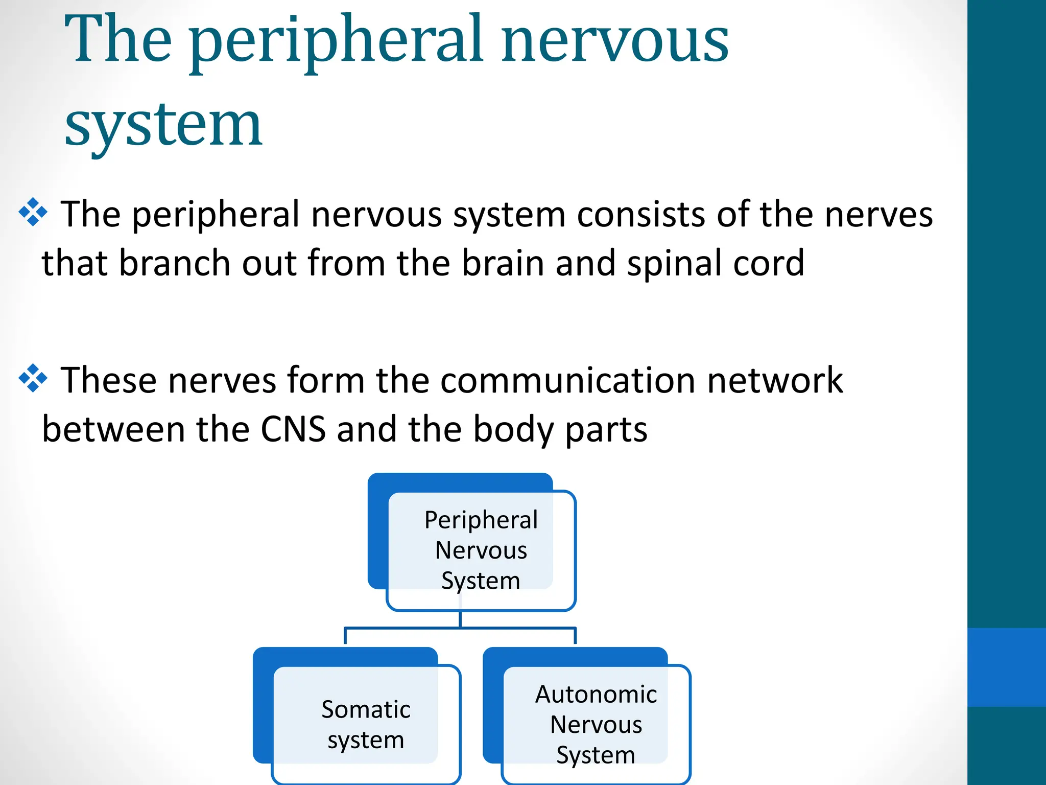
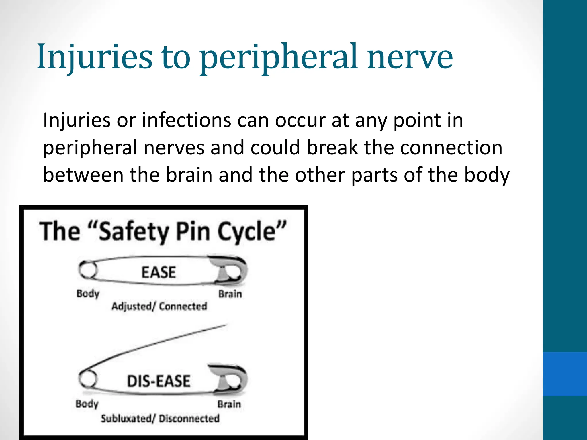
2. The peripheral nervous
system
The peripheral nervous system consists of the nerves
that branch out from the brain and spinal cord
These nerves form the communication network
between the CNS and the body parts
Peripheral
Nervous
System
Somatic
system
Autonomic
Nervous
System
3. Functions of PNS
• The primary role of the PNS is to connect the
CNS to the organs, limbs and skin
• The peripheral system allows the brain and
spinal cord to receive and send information to
other areas of the body, which allows us to
react to stimuli in our environment
4. Somatic and autonomous N.S
• The somatic nervous system consists of nerves
that go to the skin and muscles and is involved in
conscious activities.
• The autonomic nervous system consists of
nerves that connect the CNS to
the visceral organs such as the heart, stomach,
and intestines. It mediates unconscious
activities.
5. Spinal nerves
Thirty-one pairs of spinal nerves emerge
laterally from the spinal cord
Each pair of nerves corresponds to a
segment of the cord & they are named
accordingly
6. Spinal nerves
• This means there are 8 cervical nerves, 12
thoracic nerves, 5 lumbar nerves, 5 sacral
nerves, and 1 coccygeal nerve
7. Spinal nerves
Spinal nerves leave the vertebral column
through the intervertebral foramina located
between two successive vertebrae
Each spinal nerve then divides into two
branches called posterior/dorsal rami &
anterior/ventral rami
8. Spinal nerves
Both rami carry mixed fibers
The posterior rami travel backward and divide
into branches that supply muscles and sensation
to the back
The anterior rami supply the skin and muscles
of the limbs and anterior trunk
9. Plexus
Some groups of spinal nerves merge with each
other to form a large plexus, or network of
interlacing nerves.
Other spinal nerves divide into smaller branches
without forming a plexus
There are five main plexi formed by the spinal
nerves:
1. Cervical plexus
2. Brachial Plexus
3. Lumbar Plexus
4. Sacral Plexus
5. Coccegeal Plexus
10. Plexus
Cervical plexus: Composed of the merging of spinal
nerves C1 through C5, these divide into smaller nerves
that carry sensory messages and provide motor control
to the muscles of the neck and shoulders.
Brachial plexus: Formed by the merging of spinal
nerves C5 through T1, this plexus branches into nerves
that carry sensory messages and provide motor control
to the muscles of the arm and upper back.
11.
12. Brachial plexus
Divided into five parts:
Roots
Trunks
Divisions
Cords
Branches
Functionally there is no difference in all of the above
classifications
13. Brachial plexus
Roots: are formed by the anterior rami of spinal nerves
C5-T1
Trunks:
At the base of the neck, the roots of the brachial plexus
converge to form three trunks;
• Superior trunk – a combination of C5 and C6 roots.
• Middle trunk – continuation of C7.
• Inferior trunk – combination of C8 and T1 roots.
14. Brachial plexus
Divisons:
• Each trunk divides into two branches. One division
moves anteriorly (toward the front of the body) & the
other posteriorly (towards the back of the body).
Thus, they are known as the anterior and posterior
divisions
• There are three anterior and three posterior nerve
fibres. These divisions pass into the axilla.
They recombine into the cords of the brachial plexus.
15. Brachial plexus
Once the anterior and posterior divisions have
entered the axilla, they combine together to form
three cords, named by their position relative to
the axillary artery
The lateral cord is formed by:
• The anterior division of the superior trunk
• The anterior division of the middle trunk
16. Brachial plexus
The posterior cord is formed by:
• The posterior division of the superior trunk
• The posterior division of the middle trunk
• The posterior division of the inferior trunk
The medial cord is formed by:
• The anterior division of the inferior trunk.
• The cords give rise to the major branches of the
brachial plexus.
17. Brachial plexus
• In the axilla and the proximal aspect of the upper limb,
the three cords give rise to five major branches. These
nerves continue into the upper limb to provide
innervation to the muscles and skin present
18. Musculocutaneous Nerve
• Roots: C5, C6, C7
• Motor Functions: Innervates the brachialis, biceps
brachii and coracobrachialis muscles
19. Musculocutaneous Nerve
Sensory Functions: Gives off the lateral cutaneous
branch of the forearm, which innervates the lateral
half of the anterior forearm, and a small lateral
portion of the posterior forearm
20. Axillary Nerve
• Roots: C5 and C6
• Motor Functions: Innervates the teres minor and
deltoid muscles
• Sensory Functions: Gives off the superior lateral
cutaneous nerve of arm, which innervates the inferior
region of the deltoid
21. Median Nerve
• Roots: C6 – T1. (Also contains fibres from C5 in some
individuals)
• Motor Functions: Innervates most of the flexor
muscles in the forearm, the thenar muscles, and the
two lateral lumbricals associated with the index and
middle fingers
22. Median Nerve
Sensory Functions: Gives off the palmar cutaneous
branch, which innervates the lateral part of the palm,
and the digital cutaneous branch, which innervates
the lateral three and a half fingers on the anterior
(palmar) surface of the hand
23. Radial Nerve
• Roots: C5 – T1
• Motor Functions: Innervates the triceps brachii, and
the muscles in the posterior compartment of the
forearm (which are primarily, but not exclusively,
extensors of the wrist and fingers)
24. Radial Nerve
• Sensory Functions: Innervates the posterior aspect of
the arm and forearm, and the posterolateral aspect of
the hand
25. Ulnar Nerve
• Roots: C8 and T1
• Motor Functions: Innervates the muscles of the
hand (apart from the thenar muscles and two
lateral lumbricals), flexor carpi ulnaris and medial
half of flexor digitorum profundus
26. Ulnar Nerve
• Sensory Functions: Innervates the anterior and
posterior surfaces of the medial one and half fingers,
and associated palm area
27.
28.
29. • Lumbar plexus: Spinal nerves L1 through L4 converge to
form the lumbar plexus. This plexus splits into nerves
that carry sensory messages and provide motor control
to the muscles of the abdomen and leg
• Sacral plexus: Spinal nerves L4 through S4 join together.
They then branch out into nerves that carry sensory
messages and provide motor control to the muscles of
the legs
• Coccygeal plexus: The merging of nerves S4 through Co1,
this plexus supplies motor and sensory control of the
genitalia and the muscles that control defecation
Plexus
30. Motor functions of spinal
nerves
• C1–C6 Neck flexors
• C1–T1 Neck extensors
• C3, C4, C5 Supply diaphragm (mostly C4)
• C5, C6 Move shoulder, raise arm (deltoid); flex elbow (biceps)
• C6 Externally rotate (supinate) the arm
• C6, C7 Extend the elbow and wrist (triceps and wrist extensors);
pronate wrist
• C7, C8 Flex wrist; supply small muscles of the hand
• T1–T6 Intercostals and trunk above the waist
• T7–L1 Abdominal muscles
• L1–L4 Flex thigh
• L2, L3, L4 Adduct thigh; extend leg at the knee (quadriceps femoris)
• L4, L5, S1 Abduct thigh; flex leg at the knee (hamstrings); dorsiflex foot
(tibialis anterior); extend toes
• L5, S1, S2 Extend leg at the hip (gluteus maximus); plantar flex foot
and flex toes
• Branches of Spinal Nerves
31. The Somatic Nervous System
The somatic system is the part of the peripheral
nervous system responsible for carrying sensory
and motor information to and from the central
nervous system
The somatic system is responsible for
transmitting sensory information as well as for
voluntary movement
SENSORY
MOTOR
32. The Somatic Nervous System
Sensory neurons:
oAlso called afferent neurons
oCarry information from the nerves to the central
nervous system
o It is these sensory neurons that allow us to take
in sensory information and send it to the brain
and spinal cord
oE.g:
33. The Somatic Nervous System
oMotor neurons: Also called efferent neurons,
motor neurons carry information from the brain
and spinal cord to muscle fibers throughout the
body.
oThese motor neurons allow us to take physical
action in response to stimuli in the environment
E.g. when a phone rings you answer the call
34. The Autonomic Nervous
System
• It is the part of the peripheral nervous system
that's responsible for regulating involuntary body
functions such as blood flow, heartbeat, digestion,
and breathing
The autonomic system is further divided into two
branches:
Sympathetic Parasympathetic
35. Sympathetic system
By regulating the flight-or-fight response the
sympathetic system prepares the body to expend
energy to respond to environmental threats. When
action is needed, the sympathetic system triggers a
response by
accelerating heart rate
increasing breathing rate
boosting blood flow to muscles
activating sweat secretion
dilating the pupils
36. The Autonomic Nervous
System
Parasympathetic system: This helps maintain
normal body functions and conserve physical
resources. Once a threat has passed, this system
will slow the heart rate, slow breathing, reduce
blood flow to muscles, and constrict the pupils.
This allows us to return our bodies to a normal
resting state.
38. Injuries to peripheral nerve
Injuries or infections can occur at any point in
peripheral nerves and could break the connection
between the brain and the other parts of the body
39. Injuries to peripheral nerve
Symptoms: Can result in changes in sensation(numbness,
pain, tingling, burning, sensitivity to touch), muscle
weakness, or muscle paralysis
Some of the conditions that can affect the PNS include:
• Amyotrophic lateral sclerosis (ALS)
• Carpal tunnel syndrome
• Diabetes
• Guillain-Barre syndrome
• Peripheral neuropathy
• Peripheral nerve injuries
• Rheumatoid arthritis
• Tumors
• Vitamin deficiency
Editor's Notes
Visceral organs- internal organs
Inside the vertebral column anterior root , post. root outside the vertebral column, anterior rami and posterior rami
Rami means branch in latin
The somatic nervous system derives its name from the Greek word soma, which means "body."