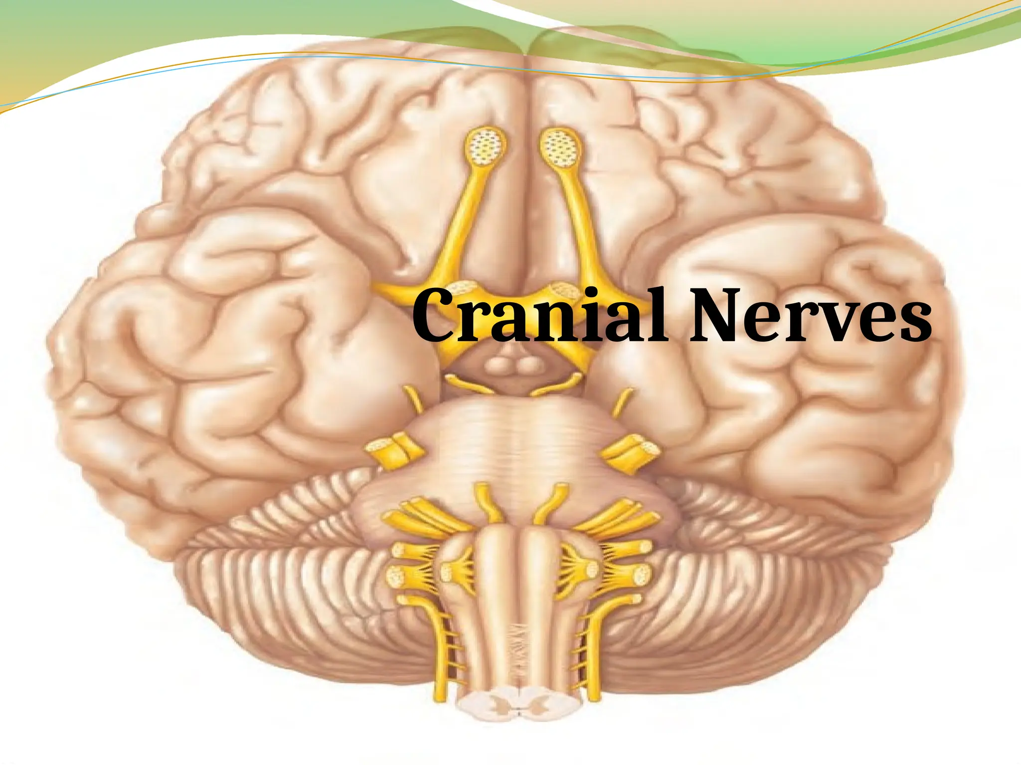
Cranial Nerves
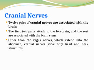
Twelvepairs of cranial nerves are associated with the
brain
The first two pairs attach to the forebrain, and the rest
are associated with the brain stem.
Other than the vagus nerves, which extend into the
abdomen, cranial nerves serve only head and neck
structures.
5.
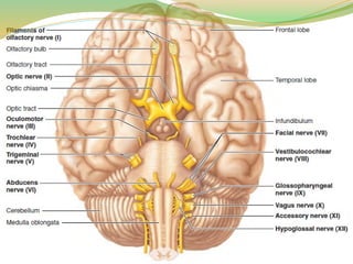
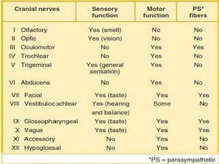
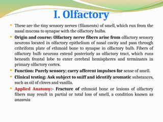
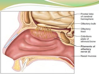
I. Olfactory
Theseare the tiny sensory nerves (filaments) of smell, which run from the
nasal mucosa to synapse with the olfactory bulbs.
Origin and course: Olfactory nerve fibers arise from olfactory sensory
neurons located in olfactory epithelium of nasal cavity and pass through
cribriform plate of ethmoid bone to synapse in olfactory bulb. Fibers of
olfactory bulb neurons extend posteriorly as olfactory tract, which runs
beneath frontal lobe to enter cerebral hemispheres and terminates in
primary olfactory cortex.
Function: Purely sensory; carry afferent impulses for sense of smell.
Clinical testing: Ask subject to sniff and identify aromatic substances,
such as oil of cloves and vanilla.
Applied Anatomy:- Fracture of ethmoid bone or lesions of olfactory
fibers may result in partial or total loss of smell, a condition known as
anosmia
7.
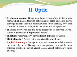
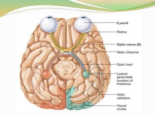
II. Optic.
Originand course: Fibers arise from retina of eye to form optic
nerve, which passes through optic canal of orbit. The optic nerves
converge to form the optic chiasma where fibers partially cross over,
continue on as optic tracts, enter thalamus, and synapse there.
Thalamic fibers run (as the optic radiation) to occipital (visual)
cortex, where visual interpretation occurs.
Function: Purely sensory; carry afferent impulses for vision.
Clinical testing: Assess vision and visual field with eye
Applied Anatomy:- Damage to optic nerve results in blindness in
eye served by nerve. Damage to visual pathway beyond the optic
chiasma results in partial visual losses. Visual defects are called
anopsias
9.
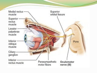
III Oculomotor Nerves
Origin and course: Fibers extend from ventral midbrain and pass
through bony orbit, via superior orbital fissure, to eye.
Function: Each nerve includes the following:
Somatic motor fibers to four of the six extrinsic eye muscles (inferior
oblique and superior, inferior, and medial rectus muscles) that help
direct eyeball, and to levator palpebrae superioris muscle, which raises
upper eyelid.
Parasympathetic (autonomic) motor fibers to sphincter pupillae
(circular muscles of iris), which cause pupil to constrict, and to ciliary
muscle, controlling lens shape for visual focusing. Some
parasympathetic cell bodies are in the ciliary ganglia.
Sensory (proprioceptor) afferents, which run from same four extrinsic
eye muscles to midbrain.
10.
Clinical testing:Examine pupils for size, shape, and
equality. Test pupillary reflex with penlight. Test
convergence for near vision and subject's ability to follow
objects with the eyes.
Applied anatomy:- In oculomotor nerve paralysis, eye
cannot be moved up, down, or inward. At rest, eye rotates
laterally [external strabismus] because the actions of the
two extrinsic eye muscles not served by cranial nerves III
are unopposed. Upper eyelid droops (ptosis), and the
person has double vision and trouble focusing on close
objects.
12.
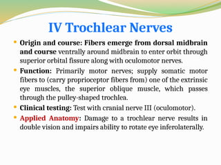
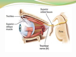
IV Trochlear Nerves
Origin and course: Fibers emerge from dorsal midbrain
and course ventrally around midbrain to enter orbit through
superior orbital fissure along with oculomotor nerves.
Function: Primarily motor nerves; supply somatic motor
fibers to (carry proprioceptor fibers from) one of the extrinsic
eye muscles, the superior oblique muscle, which passes
through the pulley-shaped trochlea.
Clinical testing: Test with cranial nerve III (oculomotor).
Applied Anatomy: Damage to a trochlear nerve results in
double vision and impairs ability to rotate eye inferolaterally.
14.
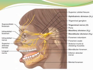
V Trigeminal Nerves
Largest cranial nerves; fibers extend from pons to face,
and form three divisions (trigemina 5 threefold):
Ophthalmic
maxillary,
mandibular
As main general sensory nerves of face, transmit afferent
impulses from touch, temperature, and pain receptors.
Cell bodies of sensory neurons of all three divisions are
located in large trigeminal ganglion.
The mandibular division also contains motor fibers that
innervate chewing muscles.
16.
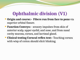
Ophthalmic division (V1)
Origin and course:- Fibers run from face to pons via
superior orbital fissure.
Function Conveys:- sensory impulses from skin of
anterior scalp, upper eyelid, and nose, and from nasal
cavity mucosa, cornea, and lacrimal gland.
Clinical testing Corneal reflex test:- Touching cornea
with wisp of cotton should elicit blinking.
17.
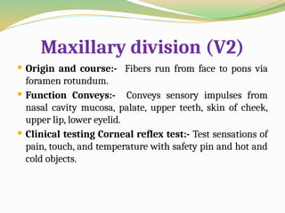
Maxillary division (V2)
Origin and course:- Fibers run from face to pons via
foramen rotundum.
Function Conveys:- Conveys sensory impulses from
nasal cavity mucosa, palate, upper teeth, skin of cheek,
upper lip, lower eyelid.
Clinical testing Corneal reflex test:- Test sensations of
pain, touch, and temperature with safety pin and hot and
cold objects.
18.
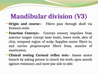
Mandibular division (V3)
Origin and course:- Fibers pass through skull via
foramen ovale.
Function Conveys:- Conveys sensory impulses from
anterior tongue (except taste buds), lower teeth, skin of
chin, temporal region of scalp. Supplies motor fibers to,
and carries proprioceptor fibers from, muscles of
mastication.
Clinical testing Corneal reflex test:- Assess motor
branch by asking person to clench his teeth, open mouth
against resistance, and move jaw side to side.
19.
Applied Anatomy
Trigeminalneuralgia:- caused by inflammation of trigeminal nerve,
is widely considered to produce most excruciating pain known.
The stabbing pain lasts for a few seconds to a minute, but it can be
relentless, occurring a hundred times a day. Usually provoked by
some sensory stimulus, such as brushing teeth or even a passing
breeze hitting the face. Thought to be caused by a loop of artery or
vein that compresses the trigeminal nerve near its exit from the
brain stem.
Analgesics and carbamazepine (an anticonvulsant) are only
partially effective. In severe cases, surgery relieves the agony either
by moving the compressing vessel or by destroying the nerve.
Nerve destruction results in loss of sensation on that side of face
20.
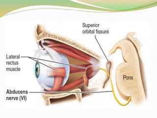
VI Abducens Nerves
Origin and course: Fibers leave inferior pons and enter
orbit via superior orbital fissure to run to eye.
Function: Primarily motor; supply somatic motor fibers to
lateral rectus muscle, an extrinsic muscle of the eye. Convey
proprioceptor impulses from same muscle to brain.
Clinical testing: Test in common with cranial nerve III
(oculomotor).
Applied Anatomy: In abducens nerve paralysis, eye
cannot be moved laterally. At rest, eyeball rotates medially
(internal strabismus).
22.
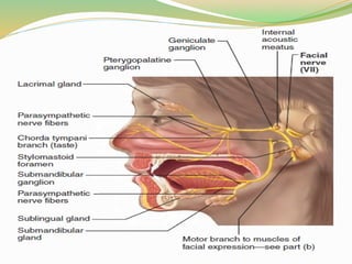
VII Facial Nerves
Origin and course: Fibers issue from pons, just lateral to abducens nerves enter
temporal bone via internal acoustic meatus, and run within bone (and through
inner ear cavity) before emerging through stylomastoid foramen. Nerve then
courses to lateral aspect of face.
Function: Mixed nerves that are the chief motor nerves of face. Five major
branches: temporal, zygomatic, buccal, mandibular, and cervical
Convey motor impulses to skeletal muscles of face (muscles of facial
expression), except for chewing muscles served by trigeminal nerves, and
transmit proprioceptor impulses from same muscles to pons.
Transmit parasympathetic (autonomic) motor impulses to lacrimal (tear)
glands, nasal and palatine glands, and submandibular and sublingual salivary
glands. Some of the cell bodies of these parasympathetic motor neurons are in
pterygopalatine and submandibular ganglia on the trigeminal nerve.
Convey sensory impulses from taste buds of anterior two-thirds of tongue; cell
bodies of these sensory neurons are in geniculate ganglion.
23.
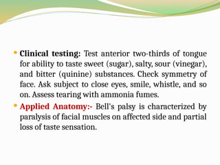
Clinical testing:Test anterior two-thirds of tongue
for ability to taste sweet (sugar), salty, sour (vinegar),
and bitter (quinine) substances. Check symmetry of
face. Ask subject to close eyes, smile, whistle, and so
on. Assess tearing with ammonia fumes.
Applied Anatomy:- Bell's palsy is characterized by
paralysis of facial muscles on affected side and partial
loss of taste sensation.
25.
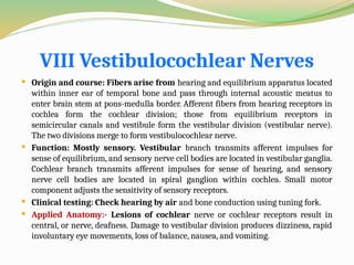
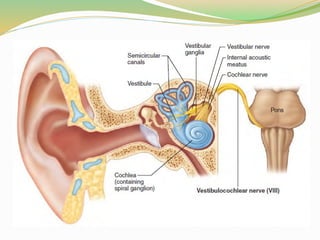
VIII Vestibulocochlear Nerves
Origin and course: Fibers arise from hearing and equilibrium apparatus located
within inner ear of temporal bone and pass through internal acoustic meatus to
enter brain stem at pons-medulla border. Afferent fibers from hearing receptors in
cochlea form the cochlear division; those from equilibrium receptors in
semicircular canals and vestibule form the vestibular division (vestibular nerve).
The two divisions merge to form vestibulocochlear nerve.
Function: Mostly sensory. Vestibular branch transmits afferent impulses for
sense of equilibrium, and sensory nerve cell bodies are located in vestibular ganglia.
Cochlear branch transmits afferent impulses for sense of hearing, and sensory
nerve cell bodies are located in spiral ganglion within cochlea. Small motor
component adjusts the sensitivity of sensory receptors.
Clinical testing: Check hearing by air and bone conduction using tuning fork.
Applied Anatomy:- Lesions of cochlear nerve or cochlear receptors result in
central, or nerve, deafness. Damage to vestibular division produces dizziness, rapid
involuntary eye movements, loss of balance, nausea, and vomiting.
27.
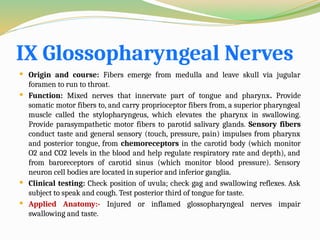
IX Glossopharyngeal Nerves
Origin and course: Fibers emerge from medulla and leave skull via jugular
foramen to run to throat.
Function: Mixed nerves that innervate part of tongue and pharynx. Provide
somatic motor fibers to, and carry proprioceptor fibers from, a superior pharyngeal
muscle called the stylopharyngeus, which elevates the pharynx in swallowing.
Provide parasympathetic motor fibers to parotid salivary glands. Sensory fibers
conduct taste and general sensory (touch, pressure, pain) impulses from pharynx
and posterior tongue, from chemoreceptors in the carotid body (which monitor
O2 and CO2 levels in the blood and help regulate respiratory rate and depth), and
from baroreceptors of carotid sinus (which monitor blood pressure). Sensory
neuron cell bodies are located in superior and inferior ganglia.
Clinical testing: Check position of uvula; check gag and swallowing reflexes. Ask
subject to speak and cough. Test posterior third of tongue for taste.
Applied Anatomy:- Injured or inflamed glossopharyngeal nerves impair
swallowing and taste.
29.
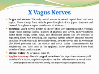
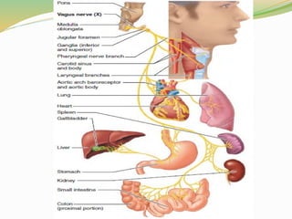
X Vagus Nerves
Origin and course: The only cranial nerves to extend beyond head and neck
region. Fibers emerge from medulla, pass through skull via jugular foramen, and
descend through neck region into thorax and abdomen.
Function: Mixed nerves. Nearly all motor fibers are parasympathetic efferents,
except those serving skeletal muscles of pharynx and larynx. Parasympathetic
motor fibers supply heart, lungs, and abdominal viscera and are involved in
regulating heart rate, breathing, and digestive system activity. Transmit sensory
impulses from thoracic and abdominal viscera, from the aortic arch baroreceptors
(for blood pressure) and the carotid and aortic bodies (chemoreceptors for
respiration), and taste buds on the epiglottis. Carry proprioceptor fibers from
muscles of larynx and pharynx.
Clinical testing: As for cranial nerve IX.
Applied Anatomy:- Since laryngeal branches of the vagus innervate nearly all
muscles of the larynx, vagal nerve paralysis can lead to hoarseness or loss of voice.
Other symptoms are difficulty swallowing and impaired digestive system motility.
31.
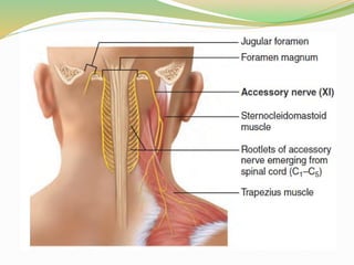
XI Accessory Nerves
Origin and course: Unique in that they form from rootlets that
emerge from the spinal cord, not the brain stem. These rootlets arise
laterally from superior region (C1–C5) of spinal cord, pass upward along
spinal cord, and enter the skull as the accessory nerves via foramen
magnum. The accessory nerves exit from skull through jugular foramen
together with the vagus nerves, and supply two large neck muscles.
Function: Mixed nerves, but primarily motor in function. Supply motor
fibers to trapezius and sternocleidomastoid muscles, which together move
head and neck, and convey proprioceptor impulses from same muscles.
Clinical testing: Check strength of sternocleidomastoid and trapezius
muscles by asking person to rotate head and shrug shoulders against
resistance.
Applied Anatomy:- Injury to one accessory nerve causes head to turn
toward injury side as result of sternocleidomastoid muscle paralysis.
Shrugging that shoulder (role of trapezius muscle) becomes difficult.
33.
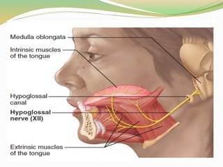
XII Hypoglossal Nerves
Origin and course: hypoglossal nerves mainly serve the tongue. Fibers
arise by a series of roots from medulla and exit from skull via
hypoglossal canal to travel to tongue.
Function: Mixed nerves, but primarily motor in function. Carry somatic
motor fibers to intrinsic and extrinsic muscles of tongue, and
proprioceptor fibers from same muscles to brain stem. Hypoglossal
nerve control allows tongue movements that mix and manipulate food
during chewing, and contribute to swallowing and speech.
Clinical testing: Ask subject to protrude and retract tongue. Note any
deviations in position.
Applied Anatomy:- Damage to hypoglossal nerves causes difficulties
in speech and swallowing. If both nerves are impaired, the person
cannot protrude tongue. If only one side is affected, tongue deviates
toward affected side.

![ Clinical testing: Examine pupils for size, shape, and
equality. Test pupillary reflex with penlight. Test
convergence for near vision and subject's ability to follow
objects with the eyes.
Applied anatomy:- In oculomotor nerve paralysis, eye
cannot be moved up, down, or inward. At rest, eye rotates
laterally [external strabismus] because the actions of the
two extrinsic eye muscles not served by cranial nerves III
are unopposed. Upper eyelid droops (ptosis), and the
person has double vision and trouble focusing on close
objects.](https://image.slidesharecdn.com/cranialnerves-181202085556-250328180011-5cbc04c3/85/In-this-ppt-we-have-made-carnial-nerves-10-320.jpg)