Downloaded 1,412 times


![Corneal Topometry
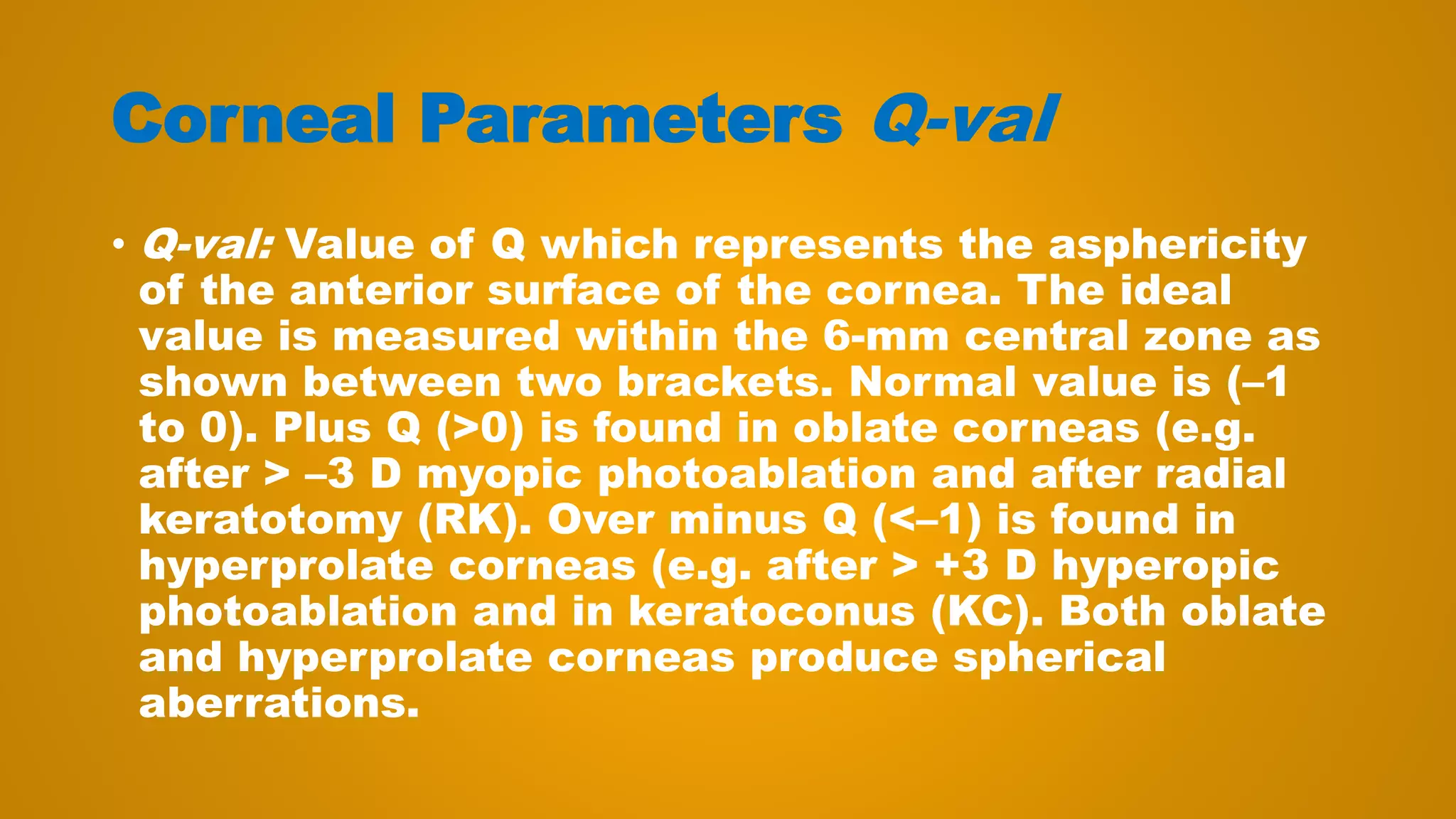
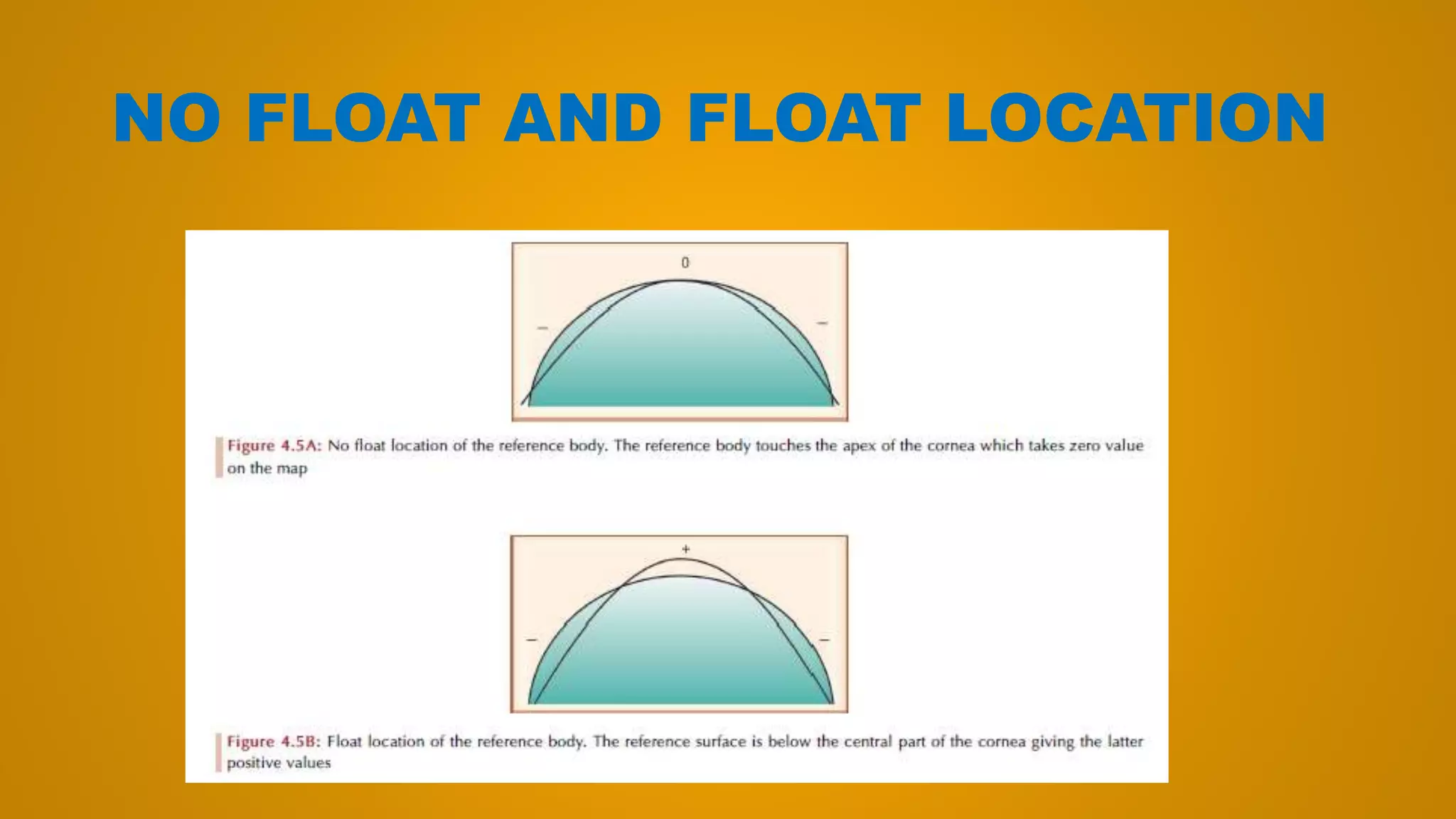
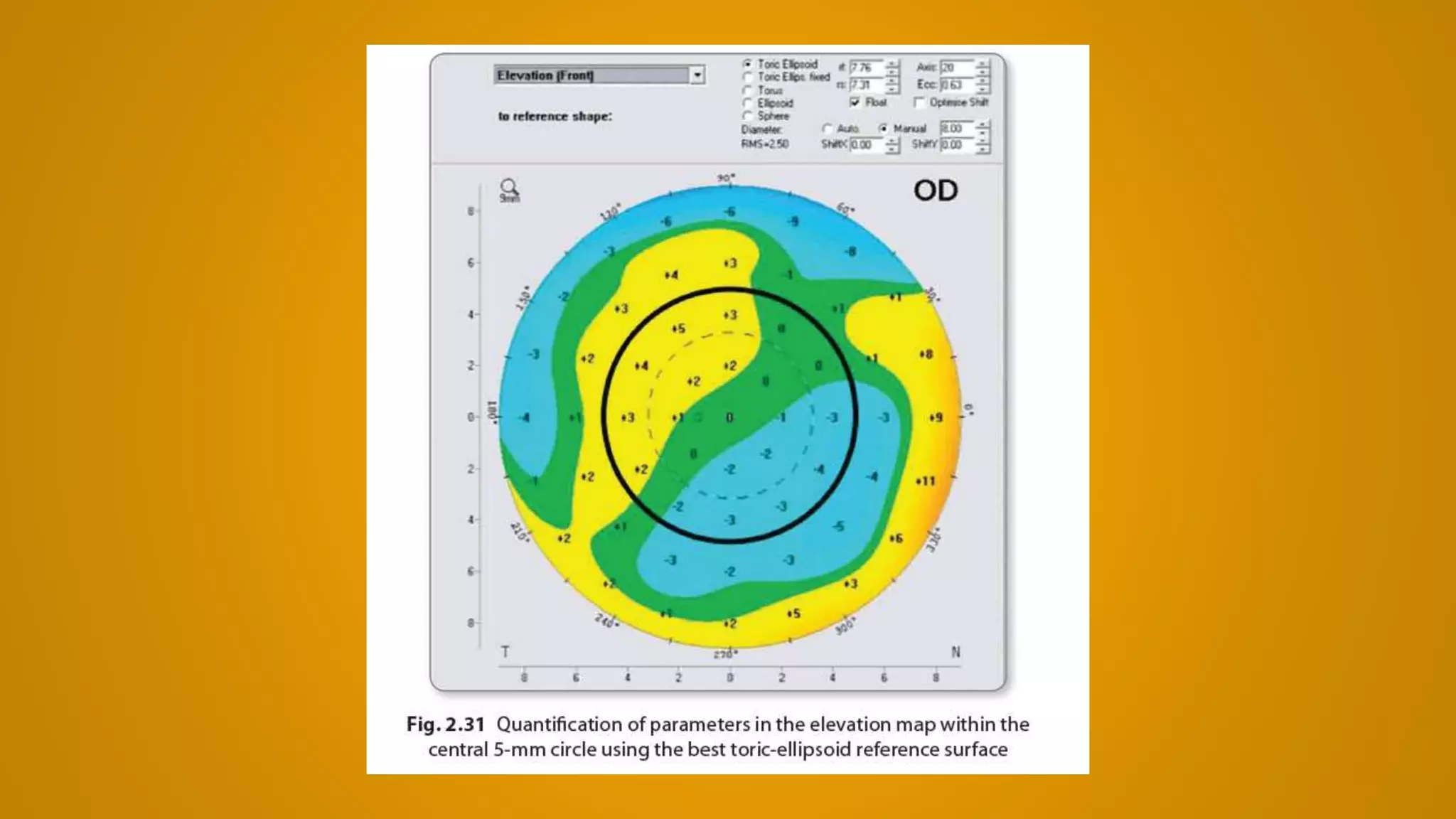
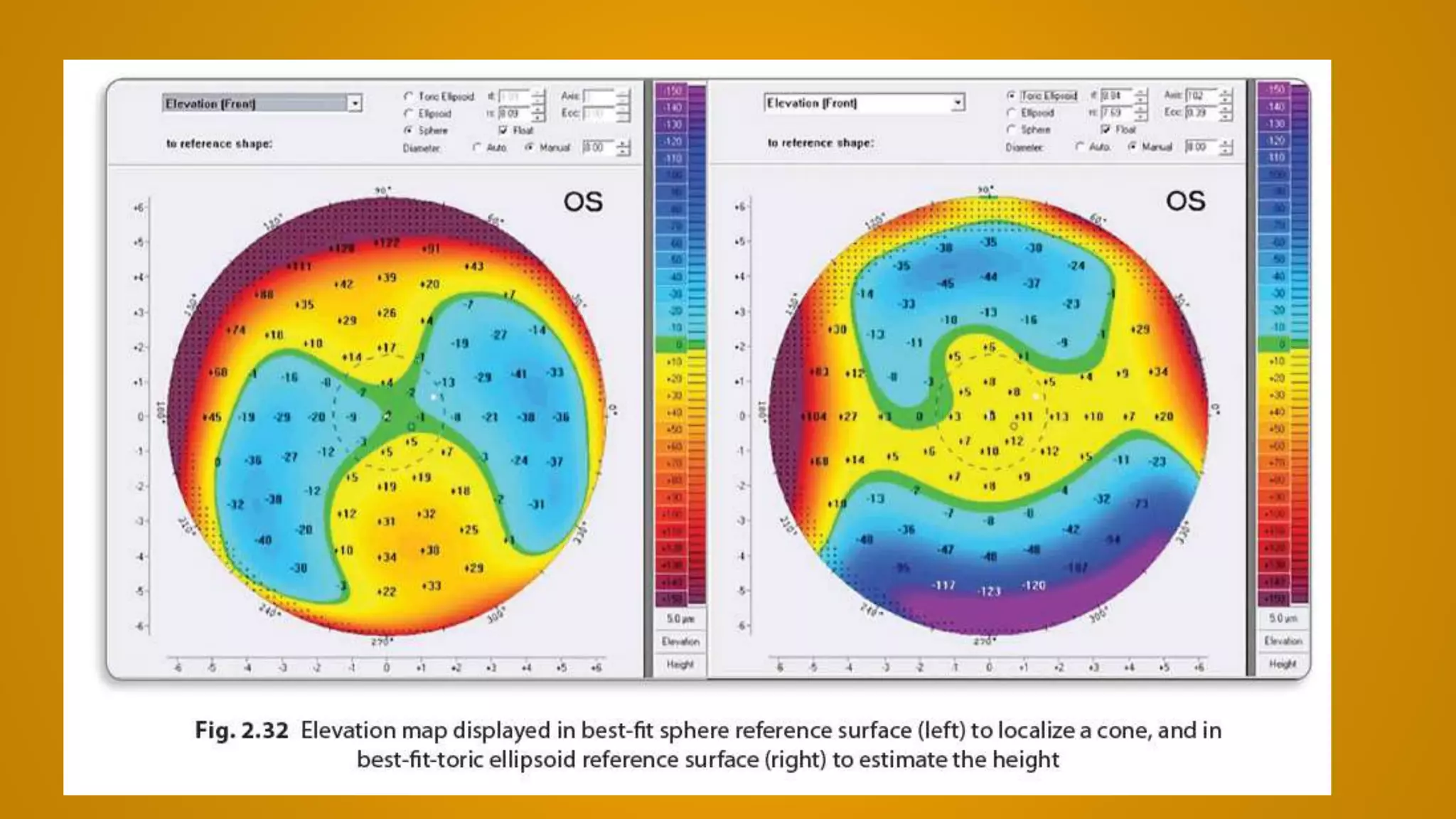
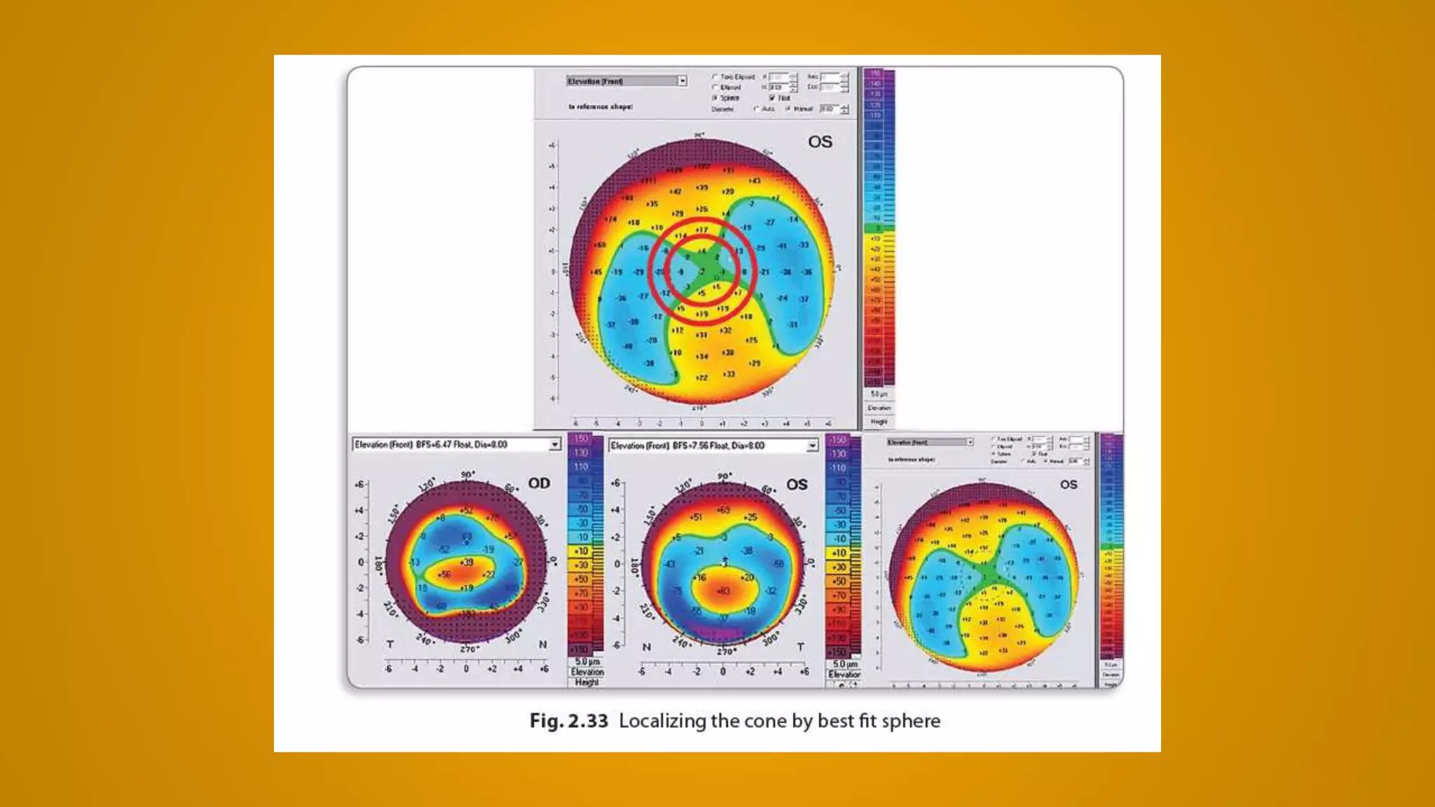
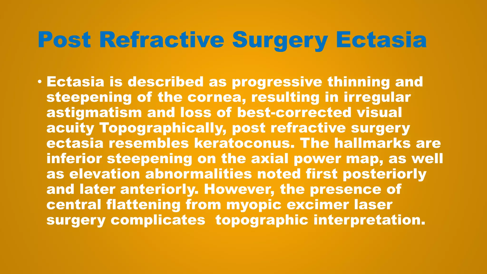
• Corneal topometry measures the slope of the
cornea. Corneal surface may take one of the four
main shapes: spheric, aspheric oblate, aspheric
prolate or aspheric hyperprolate . Q-value is
positive (> 0) when the cornea is oblate, negative
(< 0) when the cornea is prolate or hyperprolate,
and = 0 when the cornea is spheric. The normal
value is [–1 , 0]. In KC, Q-value is highly negative;
and after high myopic photoablation, Q-value is
positive. Abnormal Q-value causes spherical
aberrations. The least spherical aberrations are
found when Q-value = –0.27.](https://image.slidesharecdn.com/pentacamdemystified-160812173941/75/Pentacam-demystified-2016-198-2048.jpg)

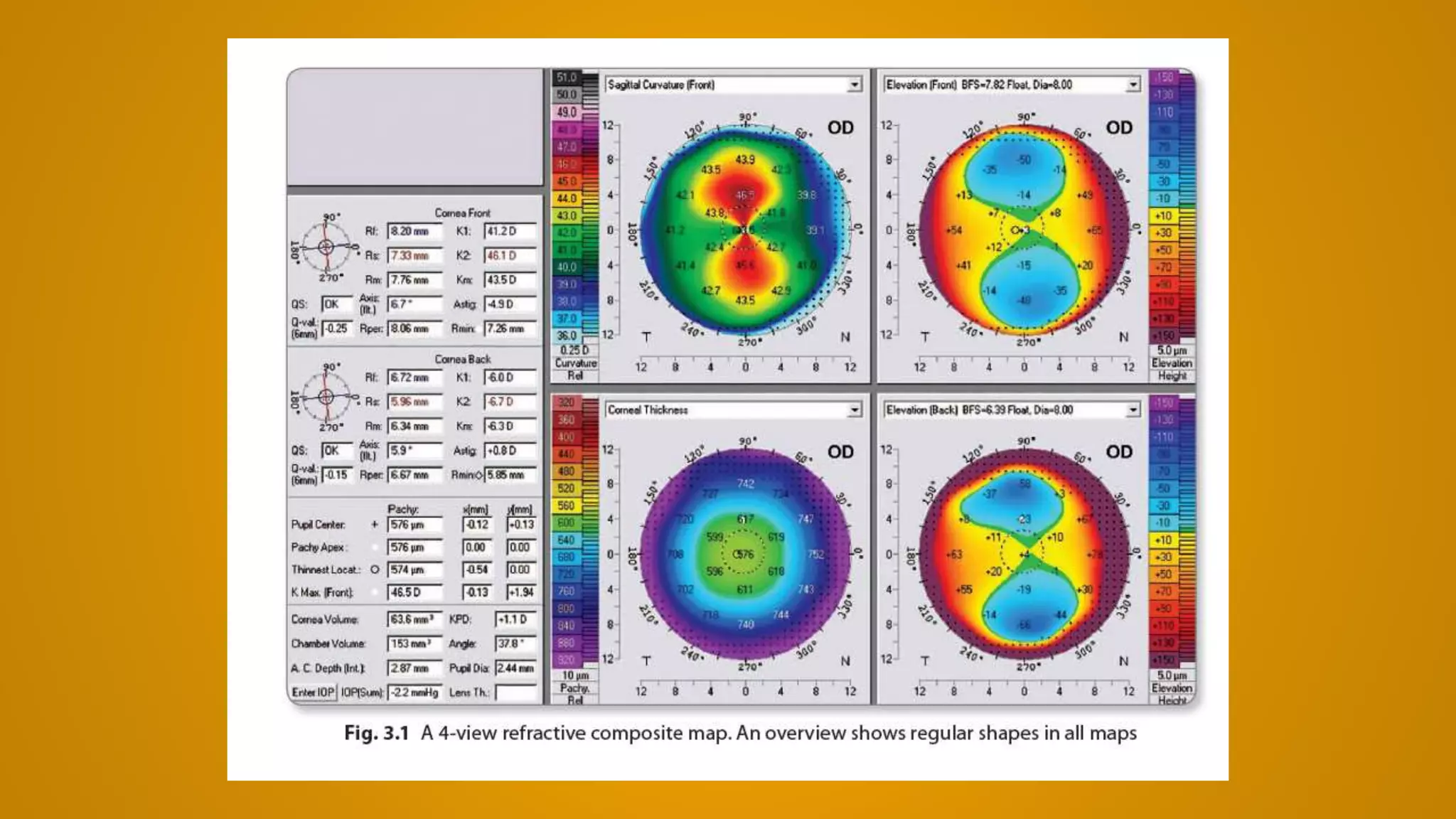


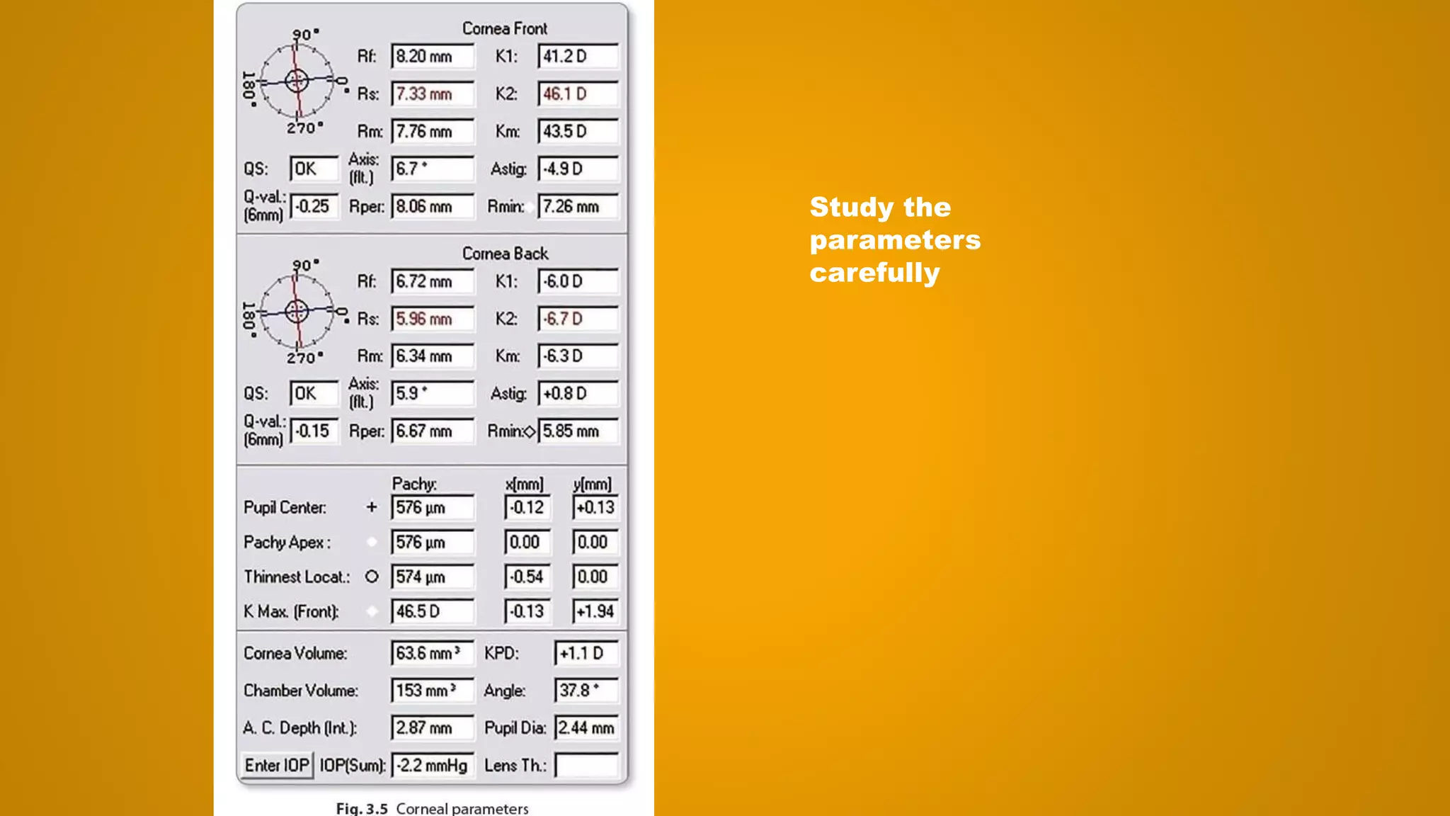
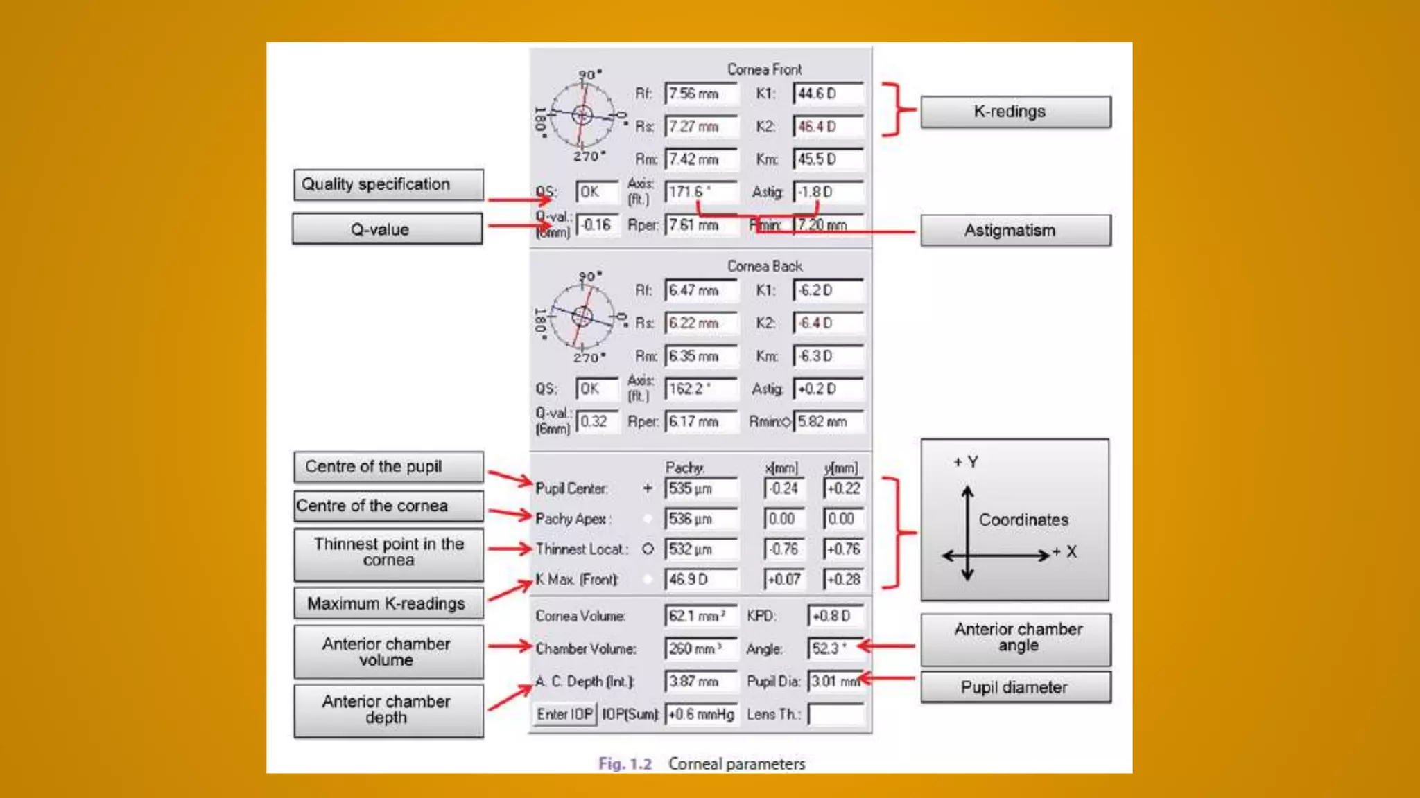
![Study the corneal parameters and focus on the
parameters of anterior corneal surface, corneal
thickness and anterior chamber
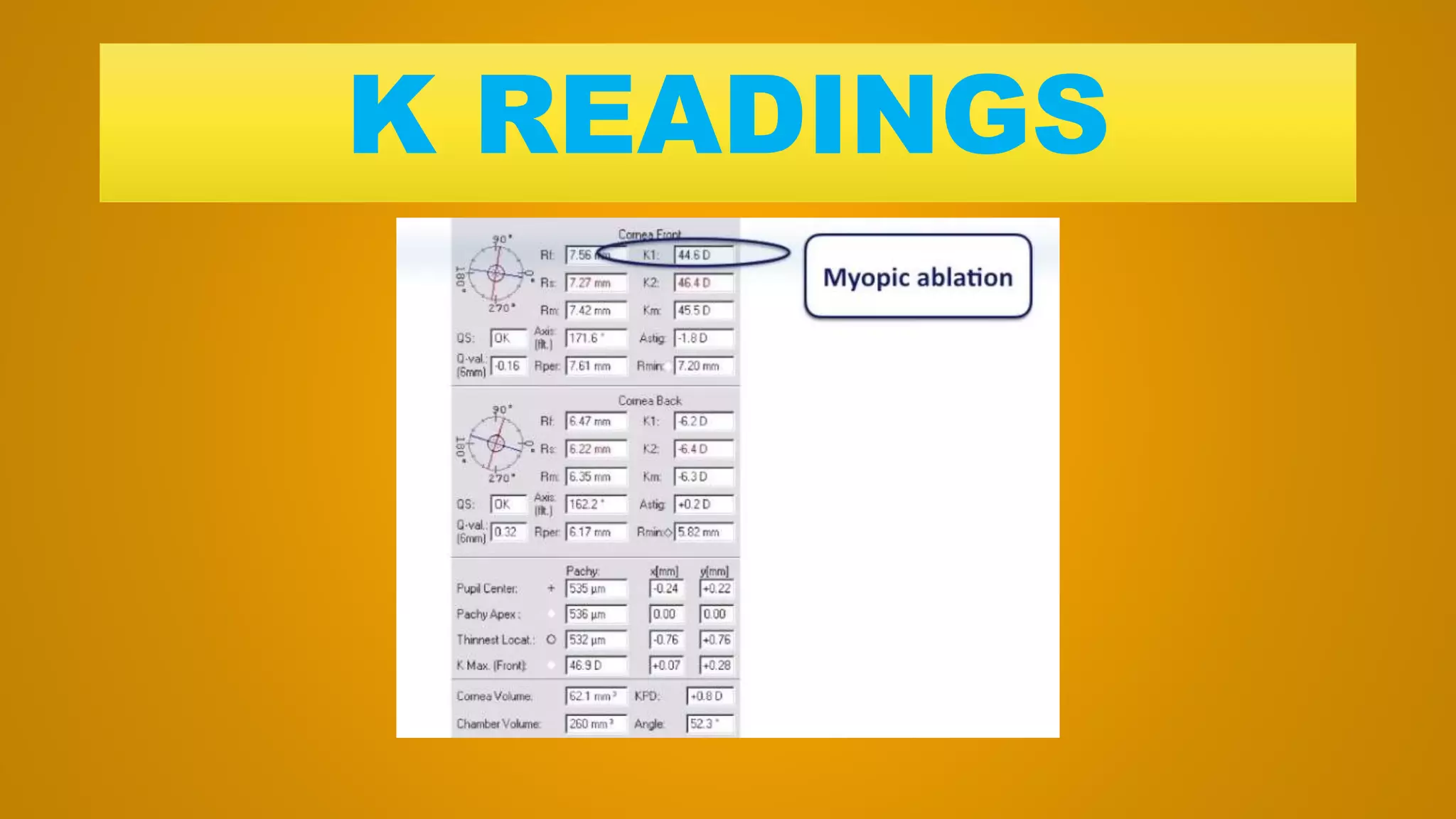
“Quality of the image (QS) is OK for both surfaces.
K-readings are within the normal range; both K2 and
K-max are < 49 D and (K-max—K2) is < 1 D. The
amount and axis of TA should be compared with MA.
Q-value of both surfaces is within the normal range
[–1 , 0]. TL thickness is > 500 μm. Difference in
thickness between the TL and pachy apex is < 10
μm. There is no vertical displacement of the TL .
Angle kappa is not significant ; x-coordinate is < 200
μm in absolute value. ACV is > 100 mm3, ACA is
normal and > 30°, ACD is normal (> 2.1 mm) but < 3.0
mm.”](https://image.slidesharecdn.com/pentacamdemystified-160812173941/75/Pentacam-demystified-2016-205-2048.jpg)

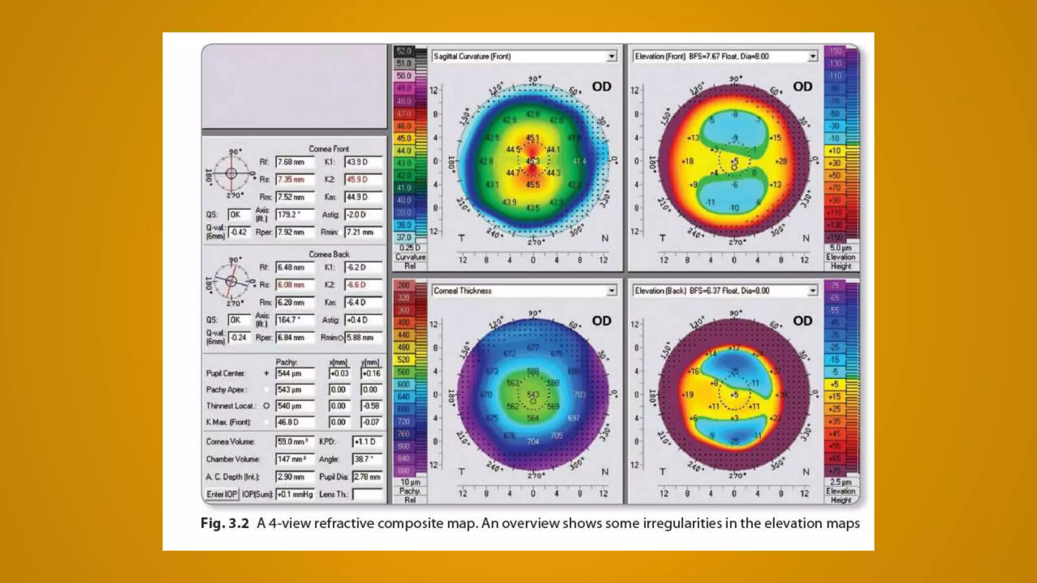
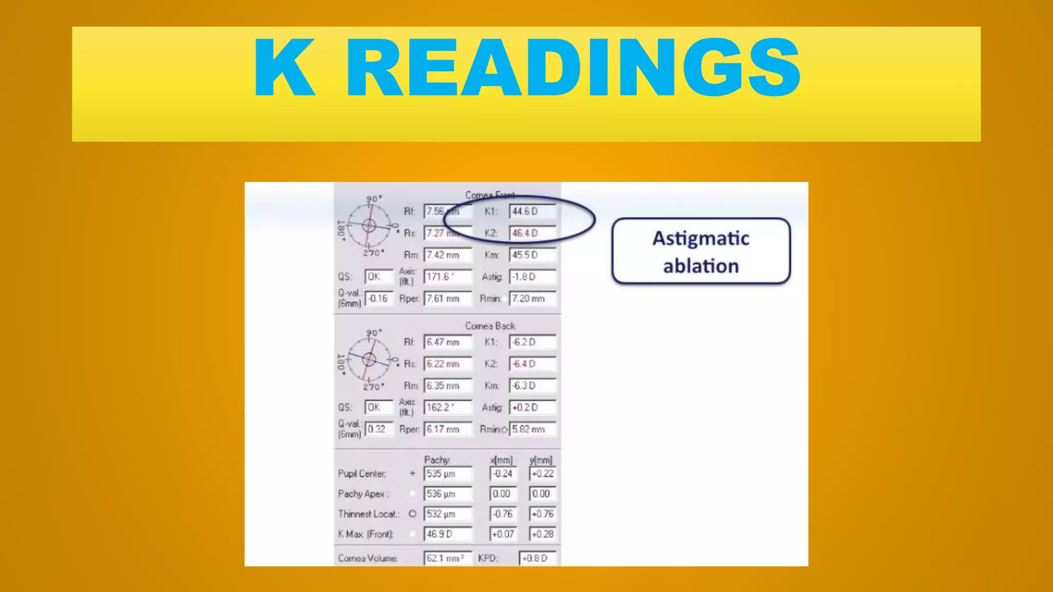
![• Quality of the image (QS) is OK for both surfaces.
K-max is > 49 D and (K-max—K2) is > 1 D. The
amount and axis of TA should be compared with
MA. Q-value of both surfaces is within the normal
range [–1 , 0], but on the upper limit indicating a
mild hyperprolate cornea. TL is < 500 μm.
Difference in thickness between the TL and pachy
apex is < 10 μm. The TL is < –500 μm in absolute
value. Angle kappa is not significant; x-coordinate
is < 200 μm in absolute value. ACV is 240 mm3,
which is very high; ACA is wide 39.2°; and ACD is
3.73 mm, which is quite deep.”](https://image.slidesharecdn.com/pentacamdemystified-160812173941/75/Pentacam-demystified-2016-207-2048.jpg)


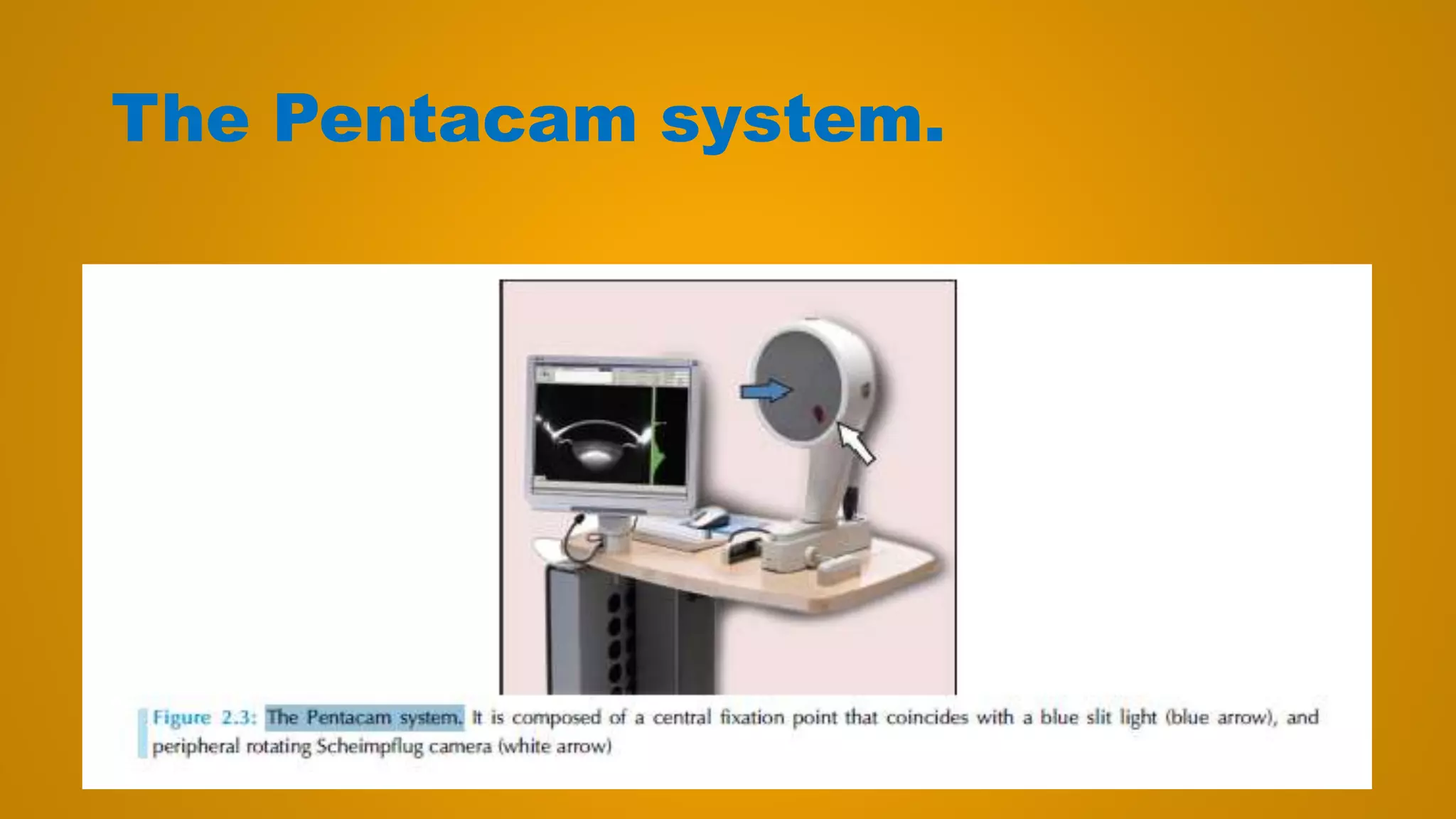
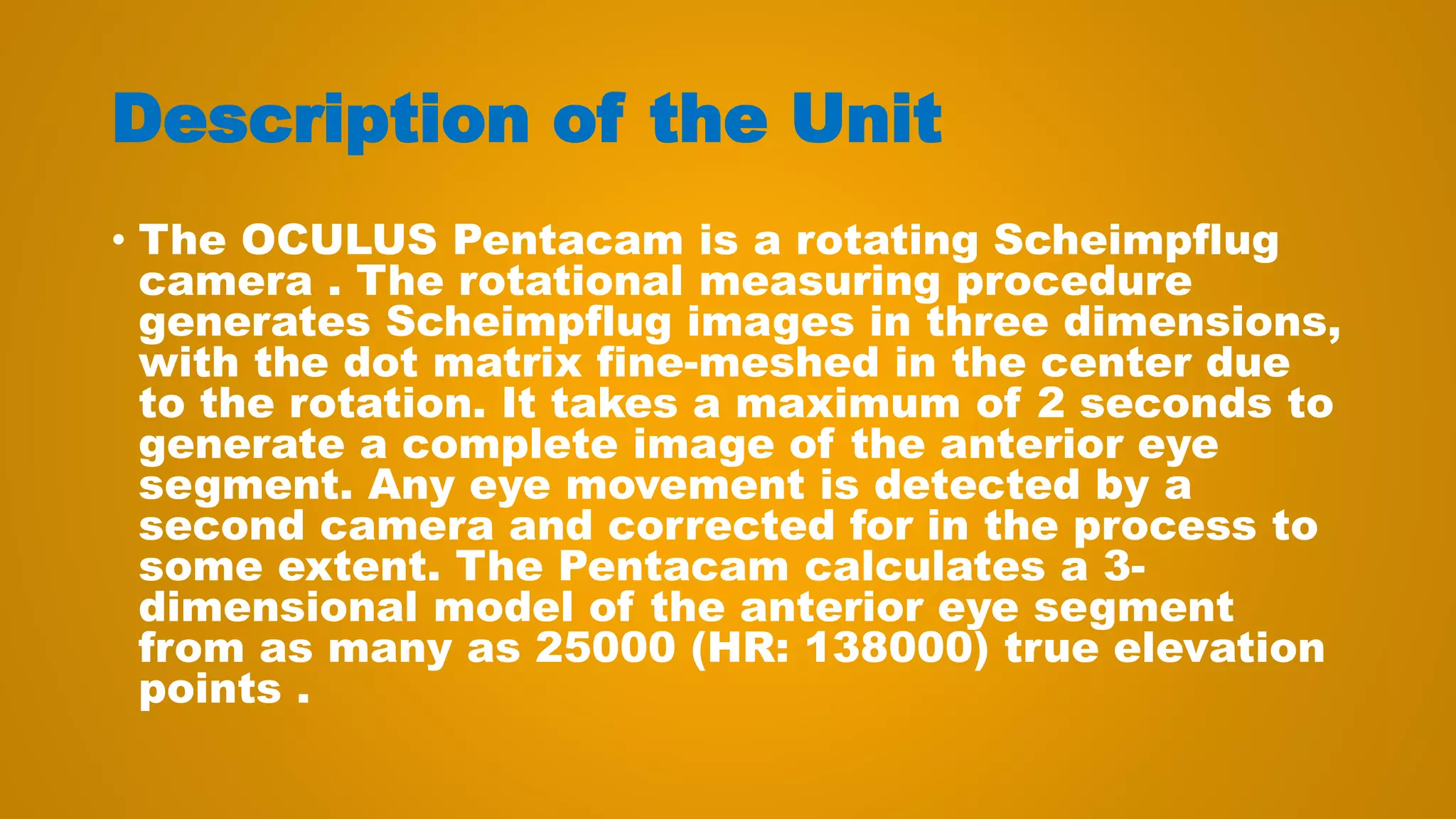
The document discusses corneal topography and tomography using the Pentacam device. It provides information on: 1) The Pentacam uses Scheimpflug imaging principles to capture 25,000 elevation points to create a 3D model of the anterior eye segment, allowing for analysis of the anterior and posterior corneal surfaces as well as pachymetry. 2) Compared to Placido disk-based systems, the Pentacam provides direct elevation data rather than deriving it from curvature, allowing for more accurate determination of corneal shape. It can also measure the entire corneal thickness through pachymetric maps. 3) The Pentacam examination involves capturing Scheimpflug images which are then used to generate


