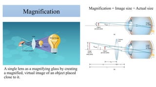
A single lensas a magnifying glass by creating
a magnified, virtual image of an object placed
close to it.
Magnification
Magnification = Image size ÷ Actual size
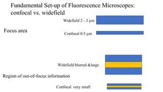
Region of out-of-focusinformation
Widefield blurred &large
Confocal very small
Fundamental Set-up of Fluorescence Microscopes:
confocal vs. widefield
Widefield 2 - 3 µm
Confocal 0.5 µm
Focus area
8.
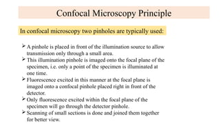
Confocal Microscopy Principle
Inconfocal microscopy two pinholes are typically used:
A pinhole is placed in front of the illumination source to allow
transmission only through a small area.
This illumination pinhole is imaged onto the focal plane of the
specimen, i.e. only a point of the specimen is illuminated at
one time.
Fluorescence excited in this manner at the focal plane is
imaged onto a confocal pinhole placed right in front of the
detector.
Only fluorescence excited within the focal plane of the
specimen will go through the detector pinhole.
Scanning of small sections is done and joined them together
for better view.
9.
Working
Confocal microscope incorporatestwo ideas:
1. Point by point illumination of the specimen.
2. Rejection of out of focus of light.
Light source of very high intensity is used-Zirconium arc lamp in
Minsky’s design and laser light source in modern design.
a)Laser provides intense blue excitation light.
b)The light reflects off a dichroic mirror, which directs it to an assembly
of vertically and horizontally scanning mirrors.
c)These motor driven mirrors scan the laser beam across the specimen.
d)The specimen is scanned by moving the stage back and forth in
vertical and horizontal directions and optics are kept stationary.
10.
Advantages
The specimen iseverywhere illuminated axially , rather than at
different angles, thereby avoiding optical aberrations.
Entire field of view is illuminated uniformly.
The field of view can be made larger than that of the static objective
by controlling the amplitude of the stage movements.
Image formed are of better resolution.
Cells can be live or fixed.
Serial optical sections can be collected.
Taking a series of optical slices from different focus levels in the
specimen generates a 3D data set.
11.
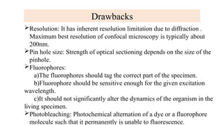
Drawbacks
Resolution: It hasinherent resolution limitation due to diffraction .
Maximum best resolution of confocal microscopy is typically about
200nm.
Pin hole size: Strength of optical sectioning depends on the size of the
pinhole.
Fluorophores:
a)The fluorophores should tag the correct part of the specimen.
b)Fluorophore should be sensitive enough for the given excitation
wavelength.
c)It should not significantly alter the dynamics of the organism in the
living specimen.
Photobleaching: Photochemical alternation of a dye or a fluorophore
molecule such that it permanently is unable to fluorescence.