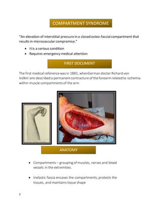
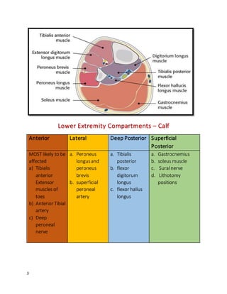
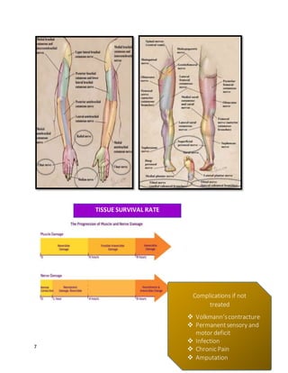
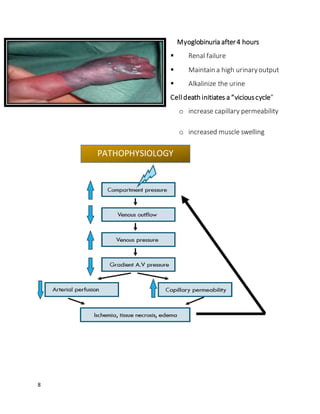
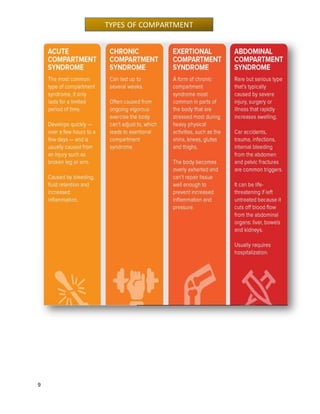
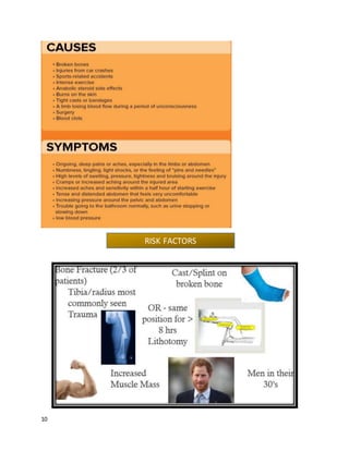
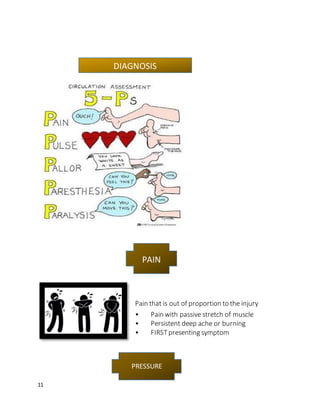
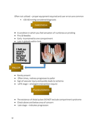
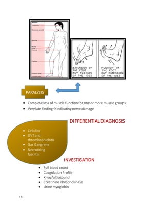
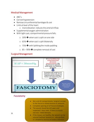
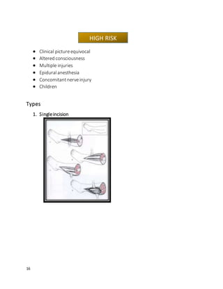
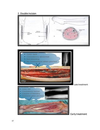
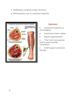
1
2
“An elevation of interstitial pressurein a closed osteo-fascialcompartment that
results in microvascular compromise.”
 Itis a serious condition
 Requires emergency medical attention
The first medical referencewasin 1881, whenGerman doctor Richard von
Volkm`ann described a permanent contractureof theforearm related to ischemia
within muscle compartmentsof the arm
 Compartments– grouping of muscles, nerves and blood
vessels in the extremities.
 Inelastic fascia encases the compartments, protects the
tissues, and maintains tissue shape
ANATOMY
COMPARTMENT SYNDROME
FIRST DOCUMENT
3
Lower Extremity Compartments – Calf
Anterior Lateral Deep Posterior Superficial
Posterior
MOST likely to be
affected
a) Tibialis
anterior
Extensor
muscles of
toes
b) Anterior Tibial
artery
c) Deep
peroneal
nerve
a. Peroneus
longusand
peroneus
brevis
b. superficial
peroneal
artery
a. Tibialis
posterior
b. flexor
digitorum
longus
c. flexor hallus
longus
a. Gastrocnemius
b. soleus muscle
c. Suralnerve
d. Lithotomy
positions
4
Lower Extremity Compartments – Thigh
5
Cross sectional anatomy of thigh
6
Lower Extremity - Calf Lower Extremity –
Thigh
Upper Extremity
1. Deep PeronealNerve
 (most commonlyaffected) -
Anterior compartment.
 Sensoryterritoryis confined to
webspace between 1st and 2nd
toes and activates dorsiflexion
2. SuperficialPeronealNerve
 runsalong lateralcompartment
and supplies dorsum of the foot
(except 1st webspace)
3. PosteriorTibialNerve
 is within deep posterior
compartmentand provides
sensation to plantar surfaceof
the foot – motor function is
flexion of the toes
 FemoralNerve
 Anterior
Compartment
 Most commonly
affected
 Obturator Nerve
 Medial
Compartmentof
thigh
 Sciatic Nerve
 Posterior
Compartmentof
thigh
1. Radial Nerve
Back of the arm
and wraps around
to skin of forearms
and hands
2. Median Nerve
 Main nerves
of arm that
runsfull
length
 Axilla injury
3. UlnarNerve
• Extends
from cervical
collar
• 4th and 5th
digits
PHYSICAL ASSESSMENT
7
Complications if not
treated
 Volkmann’scontracture
 Permanentsensory and
motor deficit
 Infection
 Chronic Pain
 Amputation
TISSUE SURVIVAL RATE
8
Myoglobinuria after4 hours
 Renal failure
 Maintain a high urinaryoutput
 Alkalinize the urine
Celldeath initiates a “viciouscycle”
o increase capillary permeability
o increased muscle swelling
PATHOPHYSIOLOGY
9
TYPES OF COMPARTMENT
10
RISK FACTORS
11
Pain that is out of proportion to the injury
• Pain with passive stretch of muscle
• Persistent deep ache or burning
• FIRSTpresenting symptom
DIAGNOSIS
PAIN
PRESSURE
12
Often not utilized – proper equipmentrequired and user errorsare common
• >30-40 mmHg considered diagnostic
 A condition in which you feel sensation of numbnessor prickling
 Pins & Needles
 Early →contained to one compartment
 Late→ globally within limb
 Rarely present
 Often times, redness progressesto pallor
 Sign of vascular injury and quickly leads to ischemia
 LATE stage – emergent intervention require
 The existence of distal pulses DO NOT exclude compartmentsyndrome
 Check above and below area of concern
 Late stage – indicates progression
PARESTHESIA
PALLOR
PULSELESSNESS
13
 Complete loss of muscle function for one or moremuscle groups
 Verylate finding→indicating nerve damage
DIFFERENTIALDIAGNOSIS
INVESTIGATION
 Full blood count
 Coagulation Profile
 X-ray/ultrasound
 Creatinine Phosphokinase
 Urine myoglobin
PARALYSIS
 Cellulitis
 DVTand
thrombophlebitis
 Gas Gangrene
 Necrotizing
fasciitis
 Peripheral
vascular injuries
14
EVALUATION
1. Stryker Manometer
2. Mercurymanometer
3. Wick hand instrument
 Surgical(Should notbedelayed)
 Medical Management
 Physical therapyManagement
TREATMENT
15
Medical Management
 ABC’s.
 Correcthypotension
 Remove circumferentialbandages& cast
 Limb at level of the heart
o moreelevation reduces the arterialinflow
 Supplementaloxygen administration
 With tight cast, compartmentalpressurefalls:
o 30% when cast is split on one side
o 65% when cast is split Bilaterally
o 75% with Splitting the inside padding
o 85 – 90% complete removal of cast
Surgical Management
Fasciotomy
 Skin and All compartments. Surgical incision
to the fascia to relieve tension or pressure.
 Complete opening of all fascial envelopes.
 The wound should be left open and
inspected 2 days later. If there is muscle
necrosis →debridement.
 If the tissues are healthy, the wound can be -
sutured (without tension) or - skin-grafted
or- allowed to heal by secondary intention
16
 Clinical pictureequivocal
 Altered consciousness
 Multiple injuries
 Epiduralanesthesia
 Concomitantnerveinjury
 Children
Types
1. Singleincision
HIGH RISK
17
2. Double incision
Late treatment
Early treatment
18
Contra-indications to fasciotomy
 Confirmed acute compartmentsyndromediagnosisfor > 48 hours
o damagecannotbe reversed and
o significantinfection rate when dead tissue exposed
o Alreadydead muscles, as in crush injuries
EDUCATION:
 Wearing moreappropriatefootwear
 Choosing more appropriatesurfacesand terrain for exercise
 Pacing your activities
 Avoiding certain activities altogether
PT
MANAGEMENT
Stretching.
ROM
Muscle
Stengthenin
g
Manual
therapy
Education
Modalitie
s
19
 Modifying your workplace to lower risk of injury
 PRICE(protection, rest, ice, compression, elevation)
Summary
 Compartmentsyndromeis a
clinical diagnosis
 should not be missed - disaster
 Requires urgenttreatment
 “Time” is the most important
factor to avoid irreversible
complications
 Do NOT applycircumferential
dressings

Compartment syndrome

  • 1.
  • 2.
    2 “An elevation ofinterstitial pressurein a closed osteo-fascialcompartment that results in microvascular compromise.”  Itis a serious condition  Requires emergency medical attention The first medical referencewasin 1881, whenGerman doctor Richard von Volkm`ann described a permanent contractureof theforearm related to ischemia within muscle compartmentsof the arm  Compartments– grouping of muscles, nerves and blood vessels in the extremities.  Inelastic fascia encases the compartments, protects the tissues, and maintains tissue shape ANATOMY COMPARTMENT SYNDROME FIRST DOCUMENT
  • 3.
    3 Lower Extremity Compartments– Calf Anterior Lateral Deep Posterior Superficial Posterior MOST likely to be affected a) Tibialis anterior Extensor muscles of toes b) Anterior Tibial artery c) Deep peroneal nerve a. Peroneus longusand peroneus brevis b. superficial peroneal artery a. Tibialis posterior b. flexor digitorum longus c. flexor hallus longus a. Gastrocnemius b. soleus muscle c. Suralnerve d. Lithotomy positions
  • 4.
  • 5.
  • 6.
    6 Lower Extremity -Calf Lower Extremity – Thigh Upper Extremity 1. Deep PeronealNerve  (most commonlyaffected) - Anterior compartment.  Sensoryterritoryis confined to webspace between 1st and 2nd toes and activates dorsiflexion 2. SuperficialPeronealNerve  runsalong lateralcompartment and supplies dorsum of the foot (except 1st webspace) 3. PosteriorTibialNerve  is within deep posterior compartmentand provides sensation to plantar surfaceof the foot – motor function is flexion of the toes  FemoralNerve  Anterior Compartment  Most commonly affected  Obturator Nerve  Medial Compartmentof thigh  Sciatic Nerve  Posterior Compartmentof thigh 1. Radial Nerve Back of the arm and wraps around to skin of forearms and hands 2. Median Nerve  Main nerves of arm that runsfull length  Axilla injury 3. UlnarNerve • Extends from cervical collar • 4th and 5th digits PHYSICAL ASSESSMENT
  • 7.
    7 Complications if not treated Volkmann’scontracture  Permanentsensory and motor deficit  Infection  Chronic Pain  Amputation TISSUE SURVIVAL RATE
  • 8.
    8 Myoglobinuria after4 hours Renal failure  Maintain a high urinaryoutput  Alkalinize the urine Celldeath initiates a “viciouscycle” o increase capillary permeability o increased muscle swelling PATHOPHYSIOLOGY
  • 9.
  • 10.
  • 11.
    11 Pain that isout of proportion to the injury • Pain with passive stretch of muscle • Persistent deep ache or burning • FIRSTpresenting symptom DIAGNOSIS PAIN PRESSURE
  • 12.
    12 Often not utilized– proper equipmentrequired and user errorsare common • >30-40 mmHg considered diagnostic  A condition in which you feel sensation of numbnessor prickling  Pins & Needles  Early →contained to one compartment  Late→ globally within limb  Rarely present  Often times, redness progressesto pallor  Sign of vascular injury and quickly leads to ischemia  LATE stage – emergent intervention require  The existence of distal pulses DO NOT exclude compartmentsyndrome  Check above and below area of concern  Late stage – indicates progression PARESTHESIA PALLOR PULSELESSNESS
  • 13.
    13  Complete lossof muscle function for one or moremuscle groups  Verylate finding→indicating nerve damage DIFFERENTIALDIAGNOSIS INVESTIGATION  Full blood count  Coagulation Profile  X-ray/ultrasound  Creatinine Phosphokinase  Urine myoglobin PARALYSIS  Cellulitis  DVTand thrombophlebitis  Gas Gangrene  Necrotizing fasciitis  Peripheral vascular injuries
  • 14.
    14 EVALUATION 1. Stryker Manometer 2.Mercurymanometer 3. Wick hand instrument  Surgical(Should notbedelayed)  Medical Management  Physical therapyManagement TREATMENT
  • 15.
    15 Medical Management  ABC’s. Correcthypotension  Remove circumferentialbandages& cast  Limb at level of the heart o moreelevation reduces the arterialinflow  Supplementaloxygen administration  With tight cast, compartmentalpressurefalls: o 30% when cast is split on one side o 65% when cast is split Bilaterally o 75% with Splitting the inside padding o 85 – 90% complete removal of cast Surgical Management Fasciotomy  Skin and All compartments. Surgical incision to the fascia to relieve tension or pressure.  Complete opening of all fascial envelopes.  The wound should be left open and inspected 2 days later. If there is muscle necrosis →debridement.  If the tissues are healthy, the wound can be - sutured (without tension) or - skin-grafted or- allowed to heal by secondary intention
  • 16.
    16  Clinical pictureequivocal Altered consciousness  Multiple injuries  Epiduralanesthesia  Concomitantnerveinjury  Children Types 1. Singleincision HIGH RISK
  • 17.
    17 2. Double incision Latetreatment Early treatment
  • 18.
    18 Contra-indications to fasciotomy Confirmed acute compartmentsyndromediagnosisfor > 48 hours o damagecannotbe reversed and o significantinfection rate when dead tissue exposed o Alreadydead muscles, as in crush injuries EDUCATION:  Wearing moreappropriatefootwear  Choosing more appropriatesurfacesand terrain for exercise  Pacing your activities  Avoiding certain activities altogether PT MANAGEMENT Stretching. ROM Muscle Stengthenin g Manual therapy Education Modalitie s
  • 19.
    19  Modifying yourworkplace to lower risk of injury  PRICE(protection, rest, ice, compression, elevation) Summary  Compartmentsyndromeis a clinical diagnosis  should not be missed - disaster  Requires urgenttreatment  “Time” is the most important factor to avoid irreversible complications  Do NOT applycircumferential dressings