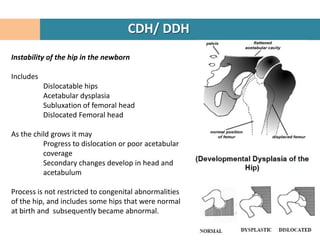
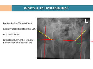
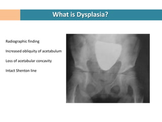
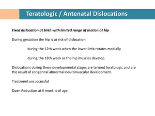
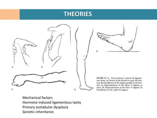
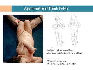
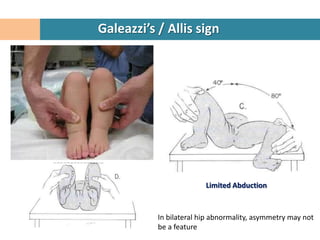
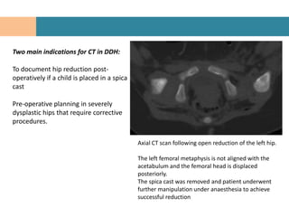
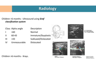
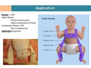
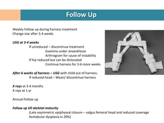
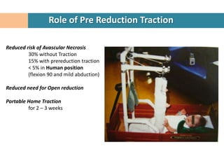
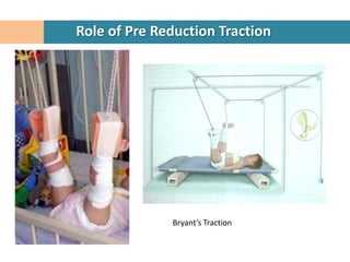
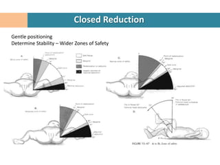
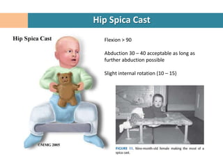
The document discusses developmental dysplasia of the hip (DDH), including: definitions; clinical detection from birth to 6 months using tests like Ortolani's and Barlow's; treatment from birth to 6 months using a Pavlik harness or closed reduction and hip spica casting; and treatment from 6 to 18 months also using closed reduction and hip spica casting, with the goal of obtaining and maintaining reduction without damaging the femoral head. Obstacles to reduction like hypertrophic soft tissues are also mentioned.
Presentation contain etiology, blood supply of femoral head & neck,pathogenesis ,classification system ,clinical features,diagnosis,managment, pelvic & femoral osteotomies in detail
Avascular necrosis (AVN) of the femoral head is a pathologic process that results from interruption of blood supply to the bone. AVN of the hip is poorly understood, but this process is the final common pathway of traumatic or nontraumatic factors that compromise the already precarious circulation of the femoral head. Femoral head ischemia results in the death of marrow and osteocytes and usually results in the collapse of the necrotic segment
A course Review from James Moore's Sporting Hip and Groin Course - February 2016 (Highly Recommend!). Following my attendance of the course, i performed my own research on 'The Sporting Hip and Groin' and incorporated this into the course review which I presented to the Sports Science and Medicine staff at Wigan Athletic FC. Further references available upon request.
Planning and performance of a total hip replacement for a case of neglected acetabular fracture. Surgery performed by Dr.A.K.Venkatachalam of www.hipsurgery.in.
developmental dyspepsia of the hip is the most common pediatric hip problem. often occurs in first born female baby, in left side more than right side in cases of breech presentation. it may be bilateral in 20% of cases.
- Video recording of this lecture in English language: https://youtu.be/lK81BzxMqdo
- Video recording of this lecture in Arabic language: https://youtu.be/Ve4P0COk9OI
- Link to download the book free: https://nephrotube.blogspot.com/p/nephrotube-nephrology-books.html
- Link to NephroTube website: www.NephroTube.com
- Link to NephroTube social media accounts: https://nephrotube.blogspot.com/p/join-nephrotube-on-social-media.html
New Directions in Targeted Therapeutic Approaches for Older Adults With Mantl...i3 Health
i3 Health is pleased to make the speaker slides from this activity available for use as a non-accredited self-study or teaching resource.
This slide deck presented by Dr. Kami Maddocks, Professor-Clinical in the Division of Hematology and
Associate Division Director for Ambulatory Operations
The Ohio State University Comprehensive Cancer Center, will provide insight into new directions in targeted therapeutic approaches for older adults with mantle cell lymphoma.
STATEMENT OF NEED
Mantle cell lymphoma (MCL) is a rare, aggressive B-cell non-Hodgkin lymphoma (NHL) accounting for 5% to 7% of all lymphomas. Its prognosis ranges from indolent disease that does not require treatment for years to very aggressive disease, which is associated with poor survival (Silkenstedt et al, 2021). Typically, MCL is diagnosed at advanced stage and in older patients who cannot tolerate intensive therapy (NCCN, 2022). Although recent advances have slightly increased remission rates, recurrence and relapse remain very common, leading to a median overall survival between 3 and 6 years (LLS, 2021). Though there are several effective options, progress is still needed towards establishing an accepted frontline approach for MCL (Castellino et al, 2022). Treatment selection and management of MCL are complicated by the heterogeneity of prognosis, advanced age and comorbidities of patients, and lack of an established standard approach for treatment, making it vital that clinicians be familiar with the latest research and advances in this area. In this activity chaired by Michael Wang, MD, Professor in the Department of Lymphoma & Myeloma at MD Anderson Cancer Center, expert faculty will discuss prognostic factors informing treatment, the promising results of recent trials in new therapeutic approaches, and the implications of treatment resistance in therapeutic selection for MCL.
Target Audience
Hematology/oncology fellows, attending faculty, and other health care professionals involved in the treatment of patients with mantle cell lymphoma (MCL).
Learning Objectives
1.) Identify clinical and biological prognostic factors that can guide treatment decision making for older adults with MCL
2.) Evaluate emerging data on targeted therapeutic approaches for treatment-naive and relapsed/refractory MCL and their applicability to older adults
3.) Assess mechanisms of resistance to targeted therapies for MCL and their implications for treatment selection
HOT NEW PRODUCT! BIG SALES FAST SHIPPING NOW FROM CHINA!! EU KU DB BK substit...GL Anaacs
Contact us if you are interested:
Email / Skype : kefaya1771@gmail.com
Threema: PXHY5PDH
New BATCH Ku !!! MUCH IN DEMAND FAST SALE EVERY BATCH HAPPY GOOD EFFECT BIG BATCH !
Contact me on Threema or skype to start big business!!
Hot-sale products:
NEW HOT EUTYLONE WHITE CRYSTAL!!
5cl-adba precursor (semi finished )
5cl-adba raw materials
ADBB precursor (semi finished )
ADBB raw materials
APVP powder
5fadb/4f-adb
Jwh018 / Jwh210
Eutylone crystal
Protonitazene (hydrochloride) CAS: 119276-01-6
Flubrotizolam CAS: 57801-95-3
Metonitazene CAS: 14680-51-4
Payment terms: Western Union,MoneyGram,Bitcoin or USDT.
Deliver Time: Usually 7-15days
Shipping method: FedEx, TNT, DHL,UPS etc.Our deliveries are 100% safe, fast, reliable and discreet.
Samples will be sent for your evaluation!If you are interested in, please contact me, let's talk details.
We specializes in exporting high quality Research chemical, medical intermediate, Pharmaceutical chemicals and so on. Products are exported to USA, Canada, France, Korea, Japan,Russia, Southeast Asia and other countries.
Ozempic: Preoperative Management of Patients on GLP-1 Receptor Agonists Saeid Safari
Preoperative Management of Patients on GLP-1 Receptor Agonists like Ozempic and Semiglutide
ASA GUIDELINE
NYSORA Guideline
2 Case Reports of Gastric Ultrasound
Title: Sense of Taste
Presenter: Dr. Faiza, Assistant Professor of Physiology
Qualifications:
MBBS (Best Graduate, AIMC Lahore)
FCPS Physiology
ICMT, CHPE, DHPE (STMU)
MPH (GC University, Faisalabad)
MBA (Virtual University of Pakistan)
Learning Objectives:
Describe the structure and function of taste buds.
Describe the relationship between the taste threshold and taste index of common substances.
Explain the chemical basis and signal transduction of taste perception for each type of primary taste sensation.
Recognize different abnormalities of taste perception and their causes.
Key Topics:
Significance of Taste Sensation:
Differentiation between pleasant and harmful food
Influence on behavior
Selection of food based on metabolic needs
Receptors of Taste:
Taste buds on the tongue
Influence of sense of smell, texture of food, and pain stimulation (e.g., by pepper)
Primary and Secondary Taste Sensations:
Primary taste sensations: Sweet, Sour, Salty, Bitter, Umami
Chemical basis and signal transduction mechanisms for each taste
Taste Threshold and Index:
Taste threshold values for Sweet (sucrose), Salty (NaCl), Sour (HCl), and Bitter (Quinine)
Taste index relationship: Inversely proportional to taste threshold
Taste Blindness:
Inability to taste certain substances, particularly thiourea compounds
Example: Phenylthiocarbamide
Structure and Function of Taste Buds:
Composition: Epithelial cells, Sustentacular/Supporting cells, Taste cells, Basal cells
Features: Taste pores, Taste hairs/microvilli, and Taste nerve fibers
Location of Taste Buds:
Found in papillae of the tongue (Fungiform, Circumvallate, Foliate)
Also present on the palate, tonsillar pillars, epiglottis, and proximal esophagus
Mechanism of Taste Stimulation:
Interaction of taste substances with receptors on microvilli
Signal transduction pathways for Umami, Sweet, Bitter, Sour, and Salty tastes
Taste Sensitivity and Adaptation:
Decrease in sensitivity with age
Rapid adaptation of taste sensation
Role of Saliva in Taste:
Dissolution of tastants to reach receptors
Washing away the stimulus
Taste Preferences and Aversions:
Mechanisms behind taste preference and aversion
Influence of receptors and neural pathways
Impact of Sensory Nerve Damage:
Degeneration of taste buds if the sensory nerve fiber is cut
Abnormalities of Taste Detection:
Conditions: Ageusia, Hypogeusia, Dysgeusia (parageusia)
Causes: Nerve damage, neurological disorders, infections, poor oral hygiene, adverse drug effects, deficiencies, aging, tobacco use, altered neurotransmitter levels
Neurotransmitters and Taste Threshold:
Effects of serotonin (5-HT) and norepinephrine (NE) on taste sensitivity
Supertasters:
25% of the population with heightened sensitivity to taste, especially bitterness
Increased number of fungiform papillae
The prostate is an exocrine gland of the male mammalian reproductive system
It is a walnut-sized gland that forms part of the male reproductive system and is located in front of the rectum and just below the urinary bladder
Function is to store and secrete a clear, slightly alkaline fluid that constitutes 10-30% of the volume of the seminal fluid that along with the spermatozoa, constitutes semen
A healthy human prostate measures (4cm-vertical, by 3cm-horizontal, 2cm ant-post ).
It surrounds the urethra just below the urinary bladder. It has anterior, median, posterior and two lateral lobes
It’s work is regulated by androgens which are responsible for male sex characteristics
Generalised disease of the prostate due to hormonal derangement which leads to non malignant enlargement of the gland (increase in the number of epithelial cells and stromal tissue)to cause compression of the urethra leading to symptoms (LUTS
Tom Selleck Health: A Comprehensive Look at the Iconic Actor’s Wellness Journeygreendigital
Tom Selleck, an enduring figure in Hollywood. has captivated audiences for decades with his rugged charm, iconic moustache. and memorable roles in television and film. From his breakout role as Thomas Magnum in Magnum P.I. to his current portrayal of Frank Reagan in Blue Bloods. Selleck's career has spanned over 50 years. But beyond his professional achievements. fans have often been curious about Tom Selleck Health. especially as he has aged in the public eye.
Follow us on: Pinterest
Introduction
Many have been interested in Tom Selleck health. not only because of his enduring presence on screen but also because of the challenges. and lifestyle choices he has faced and made over the years. This article delves into the various aspects of Tom Selleck health. exploring his fitness regimen, diet, mental health. and the challenges he has encountered as he ages. We'll look at how he maintains his well-being. the health issues he has faced, and his approach to ageing .
Early Life and Career
Childhood and Athletic Beginnings
Tom Selleck was born on January 29, 1945, in Detroit, Michigan, and grew up in Sherman Oaks, California. From an early age, he was involved in sports, particularly basketball. which played a significant role in his physical development. His athletic pursuits continued into college. where he attended the University of Southern California (USC) on a basketball scholarship. This early involvement in sports laid a strong foundation for his physical health and disciplined lifestyle.
Transition to Acting
Selleck's transition from an athlete to an actor came with its physical demands. His first significant role in "Magnum P.I." required him to perform various stunts and maintain a fit appearance. This role, which he played from 1980 to 1988. necessitated a rigorous fitness routine to meet the show's demands. setting the stage for his long-term commitment to health and wellness.
Fitness Regimen
Workout Routine
Tom Selleck health and fitness regimen has evolved. adapting to his changing roles and age. During his "Magnum, P.I." days. Selleck's workouts were intense and focused on building and maintaining muscle mass. His routine included weightlifting, cardiovascular exercises. and specific training for the stunts he performed on the show.
Selleck adjusted his fitness routine as he aged to suit his body's needs. Today, his workouts focus on maintaining flexibility, strength, and cardiovascular health. He incorporates low-impact exercises such as swimming, walking, and light weightlifting. This balanced approach helps him stay fit without putting undue strain on his joints and muscles.
Importance of Flexibility and Mobility
In recent years, Selleck has emphasized the importance of flexibility and mobility in his fitness regimen. Understanding the natural decline in muscle mass and joint flexibility with age. he includes stretching and yoga in his routine. These practices help prevent injuries, improve posture, and maintain mobilit
Anti ulcer drugs and their Advance pharmacology ||
Anti-ulcer drugs are medications used to prevent and treat ulcers in the stomach and upper part of the small intestine (duodenal ulcers). These ulcers are often caused by an imbalance between stomach acid and the mucosal lining, which protects the stomach lining.
||Scope: Overview of various classes of anti-ulcer drugs, their mechanisms of action, indications, side effects, and clinical considerations.
MANAGEMENT OF ATRIOVENTRICULAR CONDUCTION BLOCK.pdfJim Jacob Roy
Cardiac conduction defects can occur due to various causes.
Atrioventricular conduction blocks ( AV blocks ) are classified into 3 types.
This document describes the acute management of AV block.
Ethanol (CH3CH2OH), or beverage alcohol, is a two-carbon alcohol
that is rapidly distributed in the body and brain. Ethanol alters many
neurochemical systems and has rewarding and addictive properties. It
is the oldest recreational drug and likely contributes to more morbidity,
mortality, and public health costs than all illicit drugs combined. The
5th edition of the Diagnostic and Statistical Manual of Mental Disorders
(DSM-5) integrates alcohol abuse and alcohol dependence into a single
disorder called alcohol use disorder (AUD), with mild, moderate,
and severe subclassifications (American Psychiatric Association, 2013).
In the DSM-5, all types of substance abuse and dependence have been
combined into a single substance use disorder (SUD) on a continuum
from mild to severe. A diagnosis of AUD requires that at least two of
the 11 DSM-5 behaviors be present within a 12-month period (mild
AUD: 2–3 criteria; moderate AUD: 4–5 criteria; severe AUD: 6–11 criteria).
The four main behavioral effects of AUD are impaired control over
drinking, negative social consequences, risky use, and altered physiological
effects (tolerance, withdrawal). This chapter presents an overview
of the prevalence and harmful consequences of AUD in the U.S.,
the systemic nature of the disease, neurocircuitry and stages of AUD,
comorbidities, fetal alcohol spectrum disorders, genetic risk factors, and
pharmacotherapies for AUD.
micro teaching on communication m.sc nursing.pdfAnurag Sharma
Microteaching is a unique model of practice teaching. It is a viable instrument for the. desired change in the teaching behavior or the behavior potential which, in specified types of real. classroom situations, tends to facilitate the achievement of specified types of objectives.
2. CDH/ DDH
Instability of the hip in the newborn
Includes
Dislocatable hips
Acetabular dysplasia
Subluxation of femoral head
Dislocated Femoral head
As the child grows it may
Progress to dislocation or poor acetabular
coverage
Secondary changes develop in head and
acetabulum
Process is not restricted to congenital abnormalities
of the hip, and includes some hips that were normal
at birth and subsequently became abnormal.
4. Which is an Unstable Hip?
Positive Barlow/ Ortolani Tests
Clinically stable but abnormal USG
Acetabular Index
Lateral displacement of femoral
beak in relation to Perkin’s line
6. What is Subluxation?
Femoral head not fully in contact
with acetabulum
Widened Teardrop – femoral head
distance
Reduced CE angle
Break in Shenton line
Dysplastic changes in acetabulum
8. Teratologic / Antenatal Dislocations
Fixed dislocation at birth with limited range of motion at hip
During gestation the hip is at risk of dislocation
during the 12th week when the lower limb rotates medially,
during the 18th week as the hip muscles develop.
Dislocations during these developmental stages are termed teratologic and are
the result of congenital abnormal neuromuscular development.
Treatment unsuccessful
Open Reduction at 6 months of age
9. Secondary Hip Dysplasia
Strictly, DDH applies to idiopathic hip dysplasia
Secondary Hip dysplasia
Neurological conditions
Myelomeningocele
Cerebral palsy
Connective tissue diseases
Ehlers-Danlos syndrome
Myopathic disorders
Arthrogryposis multiplex congenita.
11. INCIDENCE
Breech delivery
Frank Breech
20% incidence
of DDH
Left hip > Bilateral > Right hip
Complete or
Lowry, et al, showed that breech infants footling breech
delivered by elective caesarean section (pre- lower incidence
labor) had a lower incidence of DDH than of DDH (2%)
those breech babies delivered vaginally
12. ASSOCIATED CONDITIONS
Other positional abnormalities including:
Torticollis (20%)
Metatarsus adductus (10%)
Positional club foot
Congenital Knee dislocation
15. Asymmetrical Thigh Folds
Indicative of Abnormal hips
Also seen in infants with normal hips
Widened perineum
Prominent Greater trochanter
16. Galeazzi’s / Allis sign
Limited Abduction
In bilateral hip abnormality, asymmetry may not
be a feature
17. Ortolani’s sign
(Ortolani 1937, 1976).
Hold the knees and abduct the hip while lifting
up on the greater trochanter
A positive test is feeling the dislocated hip
clunk into the acetabulum
18. Barlow’s Provocative Test
(Barlow,1962)
Adduct and push posteriorly on the hip
A positive test is feeling the hip push out of the
acetabulum
19. Klisic sign in bilateral DDH
The Ortolani and Barlow manoeuvres are the mainstay of clinical diagnosis in
the first months of life
Even in the best hands physical examination can fail to detect DDH, and after 3 months
of age the Ortolani and Barlow tests become negative due to progressive soft tissue
contractures.
21. Abduction becomes more limited which is particularly noticeable when there is
unilateral disease.
Femoral shortening (positive Galeazzi test) may be apparent but this can be difficult
to appreciate with bilateral disease.
Ambulatory children will have a Trendelenburg gait in addition to limited hip
abduction
Trendelenburg sign
Telescopy positive
Exaggerated lumbar lordosis
23. What happens to a Dysplastic hip without subluxation ?
Usually become painful
Degenerative Arthritis over time
Often subluxate as degeneration progresses
Hips with Acetabular index > 35 will likely require hip replacement later
If these hips are well reduced after primary treatment of DDH, with a greater
than normal CE angle, risk of Degenerative arthritis decreases
24. What happens to a Subluxated hip?
Always lead to symptomatic degenerative arthritis
Rapid progression
Severe subluxation – symptoms in 2nd decade
Moderate subluxation – in 3rd to 4th decade
Mild subluxation – in 5th to 6th decade
25. What happens to a completely dislocated hip?
Symptoms much later than a subluxated hip
May never become painful
False acetabulum
False acetabulum
26. Consequences of Persistent Dysplasia
Abnormal gait
Restricted abduction
Reduced strength
Increased rate of degenerative joint disease.
Outcome of untreated unilateral DDH is less favorable compared with bilateral
disease
Limb length discrepancy,
Asymmetrical movement, strength
Ipsilateral valgus knee.
Degenerative joint disease tends to present earlier in subluxated hips than in
dysplastic hips without subluxation
27. Natural History
Over the age of 6 months spontaneous resolution of dysplasia is unlikely
Usually more aggressive treatment is required compared with younger children.
Older children tend to have more advanced changes in the soft tissues and bony
structures.
Changes in acetabulum
Ossification is delayed
Shallow,
Anteverted
Deficient anterolaterally.
Changes in Femoral Head
Delayed ossification
Exaggerated femoral anteversion.
28. Natural History
A 16y /F with longstanding “missed” left DDH.
The left femoral head articulates with a neo-acetabulum in the iliac bone.
The left femoral shaft is reduced in diameter and there is wasting of the thigh muscles
32. Normal Hip
Perkins’ line
acetabular
index
Hilgenreiner’s line
Shenton’s line
33. Normal Hip
Acetabular index
Angle between the acetabulum
and hilgenreiner’s line
It should be < 30 degrees in a
newborn
Center-edge angle of Wiberg
Cannot be measured until the ossific
nucleus appears
Normal is > 10 degrees in children 6-
13 yrs
34. DDH
Ossification centre
Head of femur
2 and 7 months
35. DDH
8 month /F with left DDH.
Left acetabulum is shallow and the
hip is dislocated.
Left proximal femoral ossification
centre is small compared with the
normal right side.
The normal right acetabulum has a
central depression and well-defined
lateral edge.
The acetabular teardrop is
developing on the right but not on
the dysplastic left side
37. L
β
Iliac Wing Femoral Head
α
T
Bony
Diagram showing hip anatomy on standard coronal US plane and lines Acetabular
used to evaluate hip dysplasia using the Graf method. roof
Femoral head (FH) is centred over the hypoechoic triradiate cartilage
(T). The promontory is at the junction of the iliac wing (IL) and the
bony acetabular roof (A). L, labrum.
The alpha angle reflects the depth of the bony acetabular roof and is Standard coronal ultrasound of the normal
formed by the intersection of the baseline (BL) and acetabular roof infant hip joint. The promontory is sharply
line (AL). In a normal mature hip the alpha angle is greater than 60°.
defined and the bony acetabular roof is
The beta angle reflects cartilaginous coverage and is formed by the steep. The ossification centre for the femoral
intersection of the baseline with the labral line (LL). A normal beta head has not yet developed
angle is less than 55°
40. Not routinely performed
Either performed before surgery or intraoperatively.
Goal of arthrography in DDH is to demonstrate the position of the femoral head with
respect to other joint structures both at rest and during reduction or stress manoeuvres
It also outlines any deformities or obstructions to concentric reduction
Inverted limbus,
Capsular constriction,
Pulvinar Hypertrophy
Hypertrophied ligamentum teres
41. Arthrogram, made after
fifteen days of overhead
traction, showing the hip
to be reduced. A square-
shaped limbus (arrow) at
the superior pole of the
epiphysis and marked
capsular distension are
evident.
43. Two main indications for CT in DDH:
To document hip reduction post-
operatively if a child is placed in a spica
cast
Pre-operative planning in severely
dysplastic hips that require corrective
procedures.
Axial CT scan following open reduction of the left hip.
The left femoral metaphysis is not aligned with the
acetabulum and the femoral head is displaced
posteriorly.
The spica cast was removed and patient underwent
further manipulation under anaesthesia to achieve
successful reduction
45. Distinct advantages over both CT and arthrography in pre-operative imaging in
patients with DDH.
Ability to differentiate between different types of soft tissue which enables
visualisation of unossified cartilage, ligaments, fat, muscle and fluid including those soft
tissues that present an obstruction to concentric reduction such as pulvinar, the labrum
and joint capsule.
Lack of ionising radiation
MR imaging is more time-consuming than CT or arthrography.
In newborns and infants sedation may be required, except if patents are in a spica cast
since this limits movement.
The main disadvantages of MR imaging is increased cost and limited availability
48. Hip Examination
Barlow’s test (<3-4 months of age)
Ortolani’s test (<3-4 months of age)
Limitation of Abduction
Leg length discrepancy (Galleazi test)
Asymmetry of thigh folds
Limp in a walking child
49. Radiology
Children <6 months - Ultrasound using Graf
classification system
Class Alpha angle Description
I >60 Normal
II 60-43 Immature/Dysplastic
III <43 Subluxed/Dislocated
IV Unmeasurable Dislocated
Children >6 months - Xrays.
50. Criteria for Screening
Clinical Screening for all neonates for Hip Instability.
Careful screening for infants with risk factors:
Family history of DDH
Breech
Torticollis
Metatarsus adductus
Oligohydomnios
Screening with USG
Controversial
As per AAP guidelines
Female infants delivered breech
Family history of DDH +
53. Indications
All Dislocated hip that can be reduced (Ortolani’s sign)
Start harness at the time of diagnosis
Hips that are located but can be subluxated (Barlow’s sign)
Some may spontaneously stabilize, some may dislocate.
Re-examine after few weeks before starting treatment
Hips that are normal on clinical examination but abnormal on USG
Close observation
Repeat USG at 6 weeks of age
Abnormal hips treated
Graf II hips more likely to improve without treatment than Graf III/ IV hips
Irreducible hips
Likely to fail treatment if initial coverage was < 20%
54. Application
Flexion – 120°
Hyper flexion
Femoral nerve palsy
Inferior dislocation of head
Inadequate flexion < 90°
Fails to reduce hip
Abduction by gravity
1
4
3
2
55. Follow Up
Weekly Follow up during harness treatment
Change size after 3-4 weeks
USG at 3-4 weeks
If unreduced – discontinue treatment
Examine under anaesthesia
Arthrogram for cause of instability
If hip reduced but can be dislocated
Continue harness for 3-6 more weeks
After 6 weeks of harness – USG with child out of harness.
If reduced head – Wean/ discontinue harness
X rays at 3-4 months
X rays at 1 yr
Annual Follow up
Follow up till skeletal maturity
(Late asymmetric epiphyseal closure – valgus femoral head and reduced coverage
Acetabular dysplasia in 20%)
56. Problems in Harness
If reduction not achieved with harness in 3 – 4 weeks then discontinue harness and
other treatment started.
PAVLIK DISEASE
Prolonged positioning of dislocated hip in flexion and abduction
Dysplasia increased - Flattening of posterior acetabulum
Open reduction needed
Femoral nerve palsy
58. Goal of Treatment
Obtain and maintain reduction without damaging femoral head
Closed Reduction
Open Reduction
Problems
Risk of Avascular Necrosis
Growth potential of acetabulum declines with age (and hence remodelling
potential)
59. Role of Pre Reduction Traction
Reduced risk of Avascular Necrosis
30% without Traction
15% with prereduction traction
< 5% in Human position
(flexion 90 and mild abduction)
Reduced need for Open reduction
Portable Home Traction
for 2 – 3 weeks
60. Role of Pre Reduction Traction
Bryant’s Traction
63. Hip Spica Cast
Flexion > 90
Abduction 30 – 40 acceptable as long as
further abduction possible
Slight internal rotation (10 – 15)
64. Hip Spica Cast
Intraop radiograph
Confirm reduction – USG/ CT Scan
MRI – reduction and vascular status of head can both be assessed
Remove cast after 6 weeks under anesthesia
Assess stability through moderate range of motion/ do not dislocate.
Xray AP pelvis to confirm reduction
Reapply second cast for 6 weeks.
Third cast/ Abduction splinting
Usually applied for 6 more weeks and discontinue immobilization
65. Obstacles to Reduction
• Extra- articular
– Iliopsoas tendon
– adductors
• Intra-articular
– inverted hypertrophic labrum
– tranverse acetabular ligament
– pulvinar, ligamentum teres
– constricted anteromedial capsule espec in late cases
• neolimbus is not an obstacle to reduction and represents epiphyseal cartilage
that must not be removed as this impairs acetabular development
66. Open Reduction +/- Capsulorrhaphy
For unstable hips
Widened joint space
Approaches
Medial Approch v/s Anterior approach
Minimum dissection Better exposure
Obstructions approached directly Capsulorrhaphy can be performed
Limited exposure
Medial femoral Cx Artery
< 1 yr of age in older children
67. Open Reduction + Femoral Shortening
If there is excessive pressure on femoral head when it is reduced
Consider in a dislocated hip in > 2 yrs age
To correct excessive anteversion
To place femoral head into Varus position
Intertrochanteric/ Subtrochanteric
osteotomy
68. Open Reduction + Innominate Osteotomy
If there is need for added coverage
Assess need:
Hip in extension – neutral rotation –
abduction
If > 30% head visible then innominate
osteotomy required
Innominate osteotomy in > 18 months.
Salter / Pemberton
Consider femoral shortening for
excessive pressure on head
70. Open Reduction
Evaluate stability of hip during open reduction
Assess need for added coverage
Salter/ Pemberton pelvic osteotomy
For Children > 3 yrs, usually an acetabular procedure is needed to cover femoral
head adequately
73. Problems
Proximal head location
Contracted muscles
Femoral shortening required, greater shortening required for higher dislocations
Primary acetabular repositioning osteotomy often needed
Salter/ Pemberton
In > 3 yr, an acetabular procedure needed to adequately cover the head
74. Open Reduction
Upper age for Successful Reduction
Unilateral dislocations
Open reduction should be attempted till 9 – 10 yrs of age
Gait asymmetry
Bilateral dislocations
Upto 8 yrs
Increased complication if both hips reduced
Natural outcome of untreated B/L Dislocation better than results of treatment
75. PELVIC OSTEOTOMIES
Simple osteotomies that Reposition the Acetabulum
Salter Innominate Ostetomy
Pemberton’s Acetabuloplasty
Dega Osteotomy
Complex Osteotomies that Reposition the Acetabulum
Steel osteotomy
Tonnis Osteotomy
Ganz Osteotomy
Spherical Acetabular osteotomy
Osteotomies that Augment the Acetabulum
Chiari Osteotomy
Shelf Procedures
Staheli Slotted Acetabular Augmentation
77. Salter Innominate Osteotomy
Prerequisite – concentrically reduced hip
Acetabular dysplasia
Failure of acetabular index to improve within 2 yrs after
reduction
Persistent dysplasia after 5 yrs of age
Likely hood of Degenerative osteoarthritis high
For 2 – 9 yrs of age
(< 2 yrs – not enough iliac wings that are thick enough to
support bone graft,
> 9 yrs – acetabular fragment not adequately mobilized.
Improves Acetabular index by avg 10°.
80. Pemberton’s Acetabuloplasty
Improves anterior and lateral coverage of femoral head
Stable osteotomy, does not require Internal Fixation
Hinges through triradiate cartilage, reduces the volume
of acetabulum
Contraindicated if acetabulum is small relative to femoral
head.
Complication
Closure of triradiate cartilage (very rare)
Damage to acetabular growth areas if
osteotomy too close to acetabulum
82. Pemberton’s Acetabuloplasty
6-year-old child underwent
open reduction with
capsular placation, femoral
shortening, and a pelvic
(Pemberton) osteotomy.
83. Dega Osteotomy
Increases acetabular coverage anteriorly, centrally and posteriorly.
Placement of wedges determine the areas of coverage to be improved
86. Chiari Osteotomy
Used when other procedures cannot
achieve a concentric reduction of hip.
Controlled fracture through ilium with
medial displacement of acetabular
fragment and the intact hip capsule
under the ilium.
Over time the hip capsule converts into
fibrocartilage and becomes the new
acetabular coverage.
A Salvage surgery
91. If Femoral head cannot be repositioned distally to the level of Acetabulum
Palliative Salvage Surgeries
Rarely, Femoral shortening + Pelvic Osteotomy
Degenerative Arthritic Painful Hip
Total Hip Replacement
Role of Arthrodesis
Rarely used now for old unreduced dislocations
Contraindicated in bilateral dislocations
Bilateral Dislocations
Leave hip unreduced
Later do THR
Reduced hip but painful acetabular dysplasia
Pelvic osteotomy
93. Complications
Avascular Necrosis of Femoral Head
Trochanteric overgrowth
Inadequate Reduction and Redislocation
Residual Acetabular Dysplasia
Acetabular dysplasia Presenting late