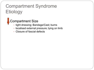
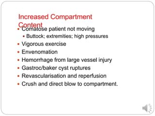
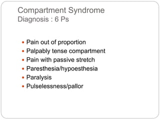
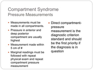
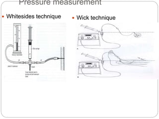
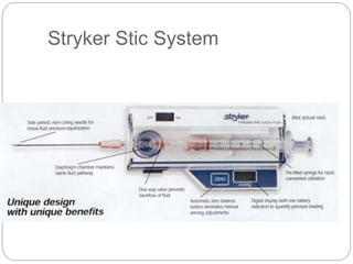
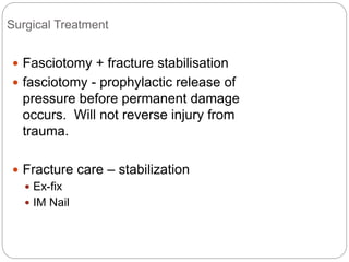
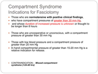
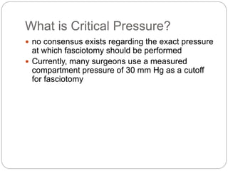
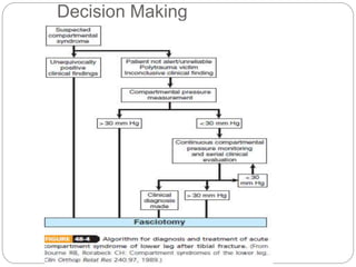
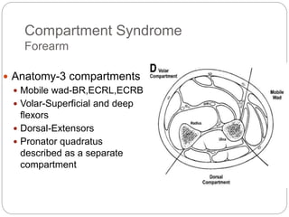
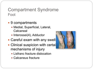
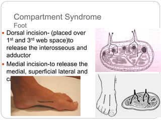
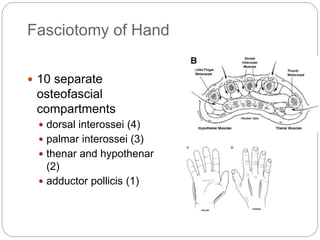
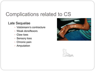
Compartment Syndrome In
Orthopaedics
Dr.Pradeep Pathak
PGIMS Rohtak
dr.pathak09@gmail.com
Definition
2
 An elevation of the interstitial pressure in a closed
osteo-fascial compartment that results in micro-
vascular compromise. If left untreated will cause
tissue damage.
 Compartments with relatively noncompliant fascial
or osseous structures are most commonly
involved ,especially the anterior compartment of
the thigh, leg and the volar compartment of the
forearm.
Demographics
 Incidence: 3.1 per 100000 population
 10 times more commonly seen in men as
compared to women
 69% due to trauma
 36% # tibia
 9.8% distal radius
 23% soft tissue injury without #
 10% on anticoagulants
Compartment Syndrome
Etiology
Compartment Size
 tight dressing; Bandage/Cast; burns
 localised external pressure; lying on limb
 Closure of fascial defects
 Comatose patient not moving
 Buttock; extremities; high pressures
 Vigorous exercise
 Envenomation
 Hemorrhage from large vessel injury
 Gastroc/baker cyst ruptures
 Revascularisation and reperfusion
 Crush and direct blow to compartment.
Increased Compartment
Content
Fracture
 The most common cause
 incidence of accompanying compartment syndrome of 9.1%
 The incidence is directly proportional to the degree of injury to
soft tissue and bone
 Blunt trauma is 2nd most common cause
A study by Shadgan et al indicated that in adult
patients with tibial diaphyseal fractures,
younger age is a risk factor for acute
compartment syndrome. The study, of 1125
patients, found that the mean age of those who
developed the syndrome was significantly
below that of the rest of the cohort (33.08 years
vs 42.01 years). Patient sex, whether the
fracture was open or closed, were not found to
be risk factors
 Shadgan B, Pereira G, Menon M, et al. Risk factors for acute
compartment syndrome of the leg associated with tibial diaphyseal
fractures in adults. J Orthop Traumatol. 2014 Dec 28
radiographic predictors of
compartment syndrome
 each 10% increase in
the ratio of fracture
length to tibial length
increased the odds of
compartment
syndrome by 1.67
 likelihood of
compartment
syndrome after
plateau fracture was
12%, compared with
3% and 2% for shaft
and pilon fractures,
respectively
 patients with Schatzker
VI fractures were at
greater risk for
compartment syndrome
than were those with
other types of Schatzker
fractures
 in patients with plateau
fractures, accompanying
fibular fractures also
raised the likelihood of
compartment syndrome
 Allmon C, Greenwell P, Paryavi E, Dubina A, O'Toole RV. Radiographic
Predictors of Compartment Syndrome Occurring after Tibial Fracture. J
Orthop Trauma. 2016 Feb 24
 Compartment
syndrome may follow
operations for
orthopedic fixation
 risks can usually be
minimized by
releasing the
tourniquet before
wound closure to
ensure that
hemostasis is
adequate and by
closing only the
subcutaneous tissue
and skin.
 According to one
prospective observational
study involving distal radius
fractures, pressures peaks
immediately after
reduction. A second
pressure peak is seen
approximately four hours
after reduction and
 Compartment Pressures
during intramedullary
nailing of the tibia appear
to peak during the
procedure and decrease
over the following 36 hours
.
 Compartment pressures
following volar plating of
distal radius fractures
appear to diminish
substantially during the 24
hour period following
surgery
Pathophysiology
 This follows the path of ischemic injury. When
fluid is introduced into a fixed volume or when
volume decreases, pressure rises.
 In the case of CS, compartments have a
relatively fixed volume. An introduction of
excess fluid or extraneous constriction
increases pressure and decreases tissue
perfusion until no O2 is available for cellular
metabolism.
Pathophysiology cont.
 Elevated perfusion pressure is the physiological
response to rising intracompartmental pressure
(IP). When IP rises, autoregulatory mechanisms
are overwhelmed and a cascade of injury
develops.
 Tissue perfusion pressure is measured by
subtracting the interstitial fluid pressure from the
capillary perfusion pressure. When this pressure
falls below a critical level, injury results.
Pathophysiology cont.
 When intracompartmentalpresssure rises,
venous pressure rises. When venous pressure
exceeds CPP, capillaries collapse. Generally,
an intracompartmental pressure greater than
30mmHg requires intervention.
 At this point, blood flow stops, resulting in
decreased O2 delivery. Hypoxic injury causes
cells to release vasoactive substances which
increases endothelial permeability.
Pathophysiology cont.
 Capillaries allow continued fluid loss which
increases tissue pressures and advances
injury.
 Nerve conduction slows,tissue ph falls due to
anaerobic metabolism,surrounding tissue
suffers further damage, and muscle tissue
suffers necrosis releasing myoglobin.
 The end is loss of the extremity and possibly,
the loss of life.
Tissue Survival
 Muscle
 3-4 hours - reversible changes
 6 hours - variable damage
 8 hours - irreversible changes
 Nerve
 2 hours - looses nerve conduction
 4 hours - neuropraxia
 8 hours - irreversible changes
Compartment Syndrome
Diagnosis : 6 Ps
 Pain out of proportion
 Palpably tense compartment
 Pain with passive stretch
 Paresthesia/hypoesthesia
 Paralysis
 Pulselessness/pallor
Pain
 Earliest symptom
 Classically out of proportion to injury
 Burning or deep and aching in nature
 Exaggerated with passive stretch of the involved
muscles in compartment
Paresthesia
 Paresthesia or numbness is an unreliable early
complaint
 in acute anterior lower leg compartment
syndrome, the first sign to develop may be
numbness between the first 2 toes (superficial
peroneal nerve).
 decreased 2-point discrimination is a more
reliable early test and can be helpful to make the
diagnosis
 Botte and Gelberman reported that 4 of 9 awake
patients with compartment pressures higher than
30 mm Hg had median nerve 2-point
discrimination of more than 1 cm
Paralysis
 Very late finding
 Irreversible nerve and muscle damage present
 Paresis may be present early but Difficult to
evaluate because of pain
 If objective evidence of a major sensory deficit, a
motor deficit, or loss of peripheral pulse is found,
the syndrome is far advanced
Pallor & Pulselessness
 Rarely present
 Indicates direct damage to vessels rather than
compartment syndrome
 Vascular injury may be more of contributing factor
to syndrome rather than result
“Pain out of proportion to the injury and the
aggravation of pain by passive stretching of the
muscles in the compartment in question are the
most sensitive (and generally the only) clinical
finding before the onset of ischemic dysfunction
in the nerves and muscles.”
DDx of ACS
22
 Cellulitis
 DVT
 Gas gangrene
 Necrotizing fasciitis
 Peripheral vascular injury
 Rhabdomyolysis
Compartment Syndrome
Pressure Measurements
 Measurements must be
made in all compartments.
 Pressure in anterior and
deep posterior
compartment are usually
highest
 Measurement made within
5 cm of #
 Marginal readings must be
followed with repeat
physical exam and repeat
compartment pressure
measurement
 Direct compartment-
pressure
measurement is the
diagnostic criterion
standard and should
be the first priority if
the diagnosis is in
question
Pressure measurement
 Whitesides technique  Wick technique
Stryker Stic System
Non invasive methods
 Ultrasonography: displacement of fascial wall
caused by increased volume.
 Sn: 77%, Sp: 93%
 Infrared imaging: determine temperature
difference between proximal and distal skin
surface.
Medical Management
 Remove cicumferential
bandages and cast
 Maintain the limb at level
of the heart as elevation
reduces the arterial inflow
and the arterio-venous
pressure gradient on
which perfusion depends.
 Ensure patient is
normotensive ,as
hypotension reduces
prefusion pressure
and facilitates further
tissue injury.
 Correct hypoperfusion
with i.v fluids and
blood products.
 Relative hypertension
and correction of
acute anemia

 The role of mannitol in
decreasing tissue edema is
still under investigation; it
may reduce compartment
pressures and lessen
reperfusion injury.
? Traction
 Pressure increases linear with increasing weight
 Posterior compartment of leg most effected
 1 kg added weight
 5% increase in posterior compartment
 <2% increase in anterior compartment
 Calcaneal traction increases dorsiflexion and it
will increase pressure in post compartment
Surgical Treatment
 Fasciotomy + fracture stabilisation
 fasciotomy - prophylactic release of
pressure before permanent damage
occurs. Will not reverse injury from
trauma.
 Fracture care – stabilization
 Ex-fix
 IM Nail
Compartment Syndrome
Indications for Fasciotomy
 Those who are normotensive with positive clinical findings,
 who have compartment pressures of greater than 30 mm Hg,
 and whose duration of increased pressure is unknown or thought to
be longer than 8 hours
 Those who are uncooperative or unconscious, with a compartment
pressure of greater than 30 mm Hg
 Those with low blood pressure and a compartment pressure of
greater than 20 mm Hg
 In hand compartmental pressure of greater than 15-20 mm Hg is a
relative indication for release.
 CONTRAINDICATION - Missed compartment
syndrome (>24-48 hrs)
What is Critical Pressure?
 no consensus exists regarding the exact pressure
at which fasciotomy should be performed
 Currently, many surgeons use a measured
compartment pressure of 30 mm Hg as a cutoff
for fasciotomy
Decision Making
Fasciotomy Principles
 Make early diagnosis
 Long extensile incisions
 Release all fascial compartments
 Preserve neurovascular structures
 Debride necrotic tissues
 Coverage within 7-10 days
Prophylactic Fasciotomy
34
Prophylactic fasciotomy of the forearm or
leg should be performed if arterial
ischemia has been present for >4 –6 h
Compartment Syndrome
Lower Leg
 4 compartments
 Lateral: Peroneus longus and
brevis
 Anterior: EHL, ED, Tibialis
anterior, Peroneus tertius
 Supeficial posterior-
Gastrocnemius, Soleus
 Deep posterior-Tibialis
posterior, FHL, FDL
Single Incision fasciotomy
Double Incision
Fasciotomy:
Compartment Syndrome
Forearm
 Anatomy-3 compartments
 Mobile wad-BR,ECRL,ECRB
 Volar-Superficial and deep
flexors
 Dorsal-Extensors
 Pronator quadratus
described as a separate
compartment
Forearm Fasciotomy
 Volar-Henry
approach
 Include a carpal tunnel
release
 Protect median
nerve, brachial artery
and tendons after
release
Compartment Syndrome
Foot
 9 compartments
 Medial, Superficial, Lateral,
Calcaneal
 Interossei(4), Adductor
 Careful exam with any swelling
 Clinical suspicion with certain
mechanisms of injury
 Lisfranc fracture dislocation
 Calcaneus fracture
Compartment Syndrome
Foot
 Dorsal incision- (placed over
1st and 3rd web space)to
release the interosseous and
adductor
 Medial incision-to release the
medial, superficial lateral and
calcaneal compartments
Compartment Syndrome
Hand
 non specific aching
of the hand
 disproportionate pain
 loss of digital motion
& continued swelling
 MP extension and PIP
flexion
 difficult to measure
tissue pressure
Fasciotomy of Hand
 10 separate
osteofascial
compartments
 dorsal interossei (4)
 palmar interossei (3)
 thenar and hypothenar
(2)
 adductor pollicis (1)
Compartment Syndrome
Thigh
 Mc cause: blunt
trauma
 Mc in ant
compartment
 Lateral to release
anterior and posterior
compartments
 May require medial
incision for adductor
compartment
Lateral septum
Vastus lateralis
Delayed Fasciotomy
is contraindicated
 Sheridan, Matsen.JBJS 1976
 infection rate of 46% and amputation rate of 21%
after a delay of 12 hours
 4.5 % complications for early fasciotomies and 54%
for delayed ones
 Recommendations
 If the CS has existed for more than 8-10 hrs,
supportive treatment of acute renal failure should be
considered.
 Skin is left intact and late reconstructions maybe
planned.
Wound Management
 After the fasciotomy, a bulky compression dressing
and a splint are applied.
 “VAC” (Vacuum Assisted Closure) can be used
 Foot should be placed in neutral to prevent equinus
contracture.
 Incision for the fasciotomy usually can be closed after
three to five days
Interim Coverage Techniques
 Simple absorbent
dressing
 Semipermeable skin-
like membrane
 Vessel loop
“bootlace”
 “VAC” (Vacuum
Assisted Closure)
Wound Management
 Second look debridement with consideration for
coverage after 48-72 hrs
 Limb should not be at risk for further swelling
 Pt should be adequately stabilized
 Usually requires skin graft
 Delayed Primary Closureis possible if residual
swelling is minimal
 Flap coverage needed if nerves, vessels, or bone
exposed
 Goal is to obtain definitive coverage within 7-10
days
Wound Closure
 STSG
 Delayed primary
closure with relaxing
incisions
Complications related to CS
Late Sequelae
 Volckmann’s contracture
 Weak dorsiflexors
 Claw toes
 Sensory loss
 Chronic pain
 Amputation
Myoglobinemia
 Released in high levels at reperfusion
 Toxic to glomeruli
 Metabolic acidosis & hperkalemia
 Together lead to:
 Renal failure
 Cardiac arrhythmia & failure
 Hypothermia
 Shock
Summary
 Keep a high index of suspicion
 Treat as soon as you suspect CS
 If clinically evident, do not measure
 Fasciotomy
Reliable, safe, and effective
The only treatment for compartment
syndrome,
when performed in time
THANK YOU

Compartment syndrome in orthopaedics

  • 1.
    Compartment Syndrome In Orthopaedics Dr.PradeepPathak PGIMS Rohtak dr.pathak09@gmail.com
  • 2.
    Definition 2  An elevationof the interstitial pressure in a closed osteo-fascial compartment that results in micro- vascular compromise. If left untreated will cause tissue damage.  Compartments with relatively noncompliant fascial or osseous structures are most commonly involved ,especially the anterior compartment of the thigh, leg and the volar compartment of the forearm.
  • 3.
    Demographics  Incidence: 3.1per 100000 population  10 times more commonly seen in men as compared to women  69% due to trauma  36% # tibia  9.8% distal radius  23% soft tissue injury without #  10% on anticoagulants
  • 4.
    Compartment Syndrome Etiology Compartment Size tight dressing; Bandage/Cast; burns  localised external pressure; lying on limb  Closure of fascial defects
  • 5.
     Comatose patientnot moving  Buttock; extremities; high pressures  Vigorous exercise  Envenomation  Hemorrhage from large vessel injury  Gastroc/baker cyst ruptures  Revascularisation and reperfusion  Crush and direct blow to compartment. Increased Compartment Content
  • 6.
    Fracture  The mostcommon cause  incidence of accompanying compartment syndrome of 9.1%  The incidence is directly proportional to the degree of injury to soft tissue and bone  Blunt trauma is 2nd most common cause
  • 8.
    A study byShadgan et al indicated that in adult patients with tibial diaphyseal fractures, younger age is a risk factor for acute compartment syndrome. The study, of 1125 patients, found that the mean age of those who developed the syndrome was significantly below that of the rest of the cohort (33.08 years vs 42.01 years). Patient sex, whether the fracture was open or closed, were not found to be risk factors  Shadgan B, Pereira G, Menon M, et al. Risk factors for acute compartment syndrome of the leg associated with tibial diaphyseal fractures in adults. J Orthop Traumatol. 2014 Dec 28
  • 9.
    radiographic predictors of compartmentsyndrome  each 10% increase in the ratio of fracture length to tibial length increased the odds of compartment syndrome by 1.67  likelihood of compartment syndrome after plateau fracture was 12%, compared with 3% and 2% for shaft and pilon fractures, respectively  patients with Schatzker VI fractures were at greater risk for compartment syndrome than were those with other types of Schatzker fractures  in patients with plateau fractures, accompanying fibular fractures also raised the likelihood of compartment syndrome  Allmon C, Greenwell P, Paryavi E, Dubina A, O'Toole RV. Radiographic Predictors of Compartment Syndrome Occurring after Tibial Fracture. J Orthop Trauma. 2016 Feb 24
  • 10.
     Compartment syndrome mayfollow operations for orthopedic fixation  risks can usually be minimized by releasing the tourniquet before wound closure to ensure that hemostasis is adequate and by closing only the subcutaneous tissue and skin.  According to one prospective observational study involving distal radius fractures, pressures peaks immediately after reduction. A second pressure peak is seen approximately four hours after reduction and  Compartment Pressures during intramedullary nailing of the tibia appear to peak during the procedure and decrease over the following 36 hours .  Compartment pressures following volar plating of distal radius fractures appear to diminish substantially during the 24 hour period following surgery
  • 11.
    Pathophysiology  This followsthe path of ischemic injury. When fluid is introduced into a fixed volume or when volume decreases, pressure rises.  In the case of CS, compartments have a relatively fixed volume. An introduction of excess fluid or extraneous constriction increases pressure and decreases tissue perfusion until no O2 is available for cellular metabolism.
  • 12.
    Pathophysiology cont.  Elevatedperfusion pressure is the physiological response to rising intracompartmental pressure (IP). When IP rises, autoregulatory mechanisms are overwhelmed and a cascade of injury develops.  Tissue perfusion pressure is measured by subtracting the interstitial fluid pressure from the capillary perfusion pressure. When this pressure falls below a critical level, injury results.
  • 13.
    Pathophysiology cont.  Whenintracompartmentalpresssure rises, venous pressure rises. When venous pressure exceeds CPP, capillaries collapse. Generally, an intracompartmental pressure greater than 30mmHg requires intervention.  At this point, blood flow stops, resulting in decreased O2 delivery. Hypoxic injury causes cells to release vasoactive substances which increases endothelial permeability.
  • 14.
    Pathophysiology cont.  Capillariesallow continued fluid loss which increases tissue pressures and advances injury.  Nerve conduction slows,tissue ph falls due to anaerobic metabolism,surrounding tissue suffers further damage, and muscle tissue suffers necrosis releasing myoglobin.  The end is loss of the extremity and possibly, the loss of life.
  • 15.
    Tissue Survival  Muscle 3-4 hours - reversible changes  6 hours - variable damage  8 hours - irreversible changes  Nerve  2 hours - looses nerve conduction  4 hours - neuropraxia  8 hours - irreversible changes
  • 16.
    Compartment Syndrome Diagnosis :6 Ps  Pain out of proportion  Palpably tense compartment  Pain with passive stretch  Paresthesia/hypoesthesia  Paralysis  Pulselessness/pallor
  • 17.
    Pain  Earliest symptom Classically out of proportion to injury  Burning or deep and aching in nature  Exaggerated with passive stretch of the involved muscles in compartment
  • 18.
    Paresthesia  Paresthesia ornumbness is an unreliable early complaint  in acute anterior lower leg compartment syndrome, the first sign to develop may be numbness between the first 2 toes (superficial peroneal nerve).  decreased 2-point discrimination is a more reliable early test and can be helpful to make the diagnosis  Botte and Gelberman reported that 4 of 9 awake patients with compartment pressures higher than 30 mm Hg had median nerve 2-point discrimination of more than 1 cm
  • 19.
    Paralysis  Very latefinding  Irreversible nerve and muscle damage present  Paresis may be present early but Difficult to evaluate because of pain  If objective evidence of a major sensory deficit, a motor deficit, or loss of peripheral pulse is found, the syndrome is far advanced
  • 20.
    Pallor & Pulselessness Rarely present  Indicates direct damage to vessels rather than compartment syndrome  Vascular injury may be more of contributing factor to syndrome rather than result
  • 21.
    “Pain out ofproportion to the injury and the aggravation of pain by passive stretching of the muscles in the compartment in question are the most sensitive (and generally the only) clinical finding before the onset of ischemic dysfunction in the nerves and muscles.”
  • 22.
    DDx of ACS 22 Cellulitis  DVT  Gas gangrene  Necrotizing fasciitis  Peripheral vascular injury  Rhabdomyolysis
  • 23.
    Compartment Syndrome Pressure Measurements Measurements must be made in all compartments.  Pressure in anterior and deep posterior compartment are usually highest  Measurement made within 5 cm of #  Marginal readings must be followed with repeat physical exam and repeat compartment pressure measurement  Direct compartment- pressure measurement is the diagnostic criterion standard and should be the first priority if the diagnosis is in question
  • 24.
    Pressure measurement  Whitesidestechnique  Wick technique
  • 25.
  • 26.
    Non invasive methods Ultrasonography: displacement of fascial wall caused by increased volume.  Sn: 77%, Sp: 93%  Infrared imaging: determine temperature difference between proximal and distal skin surface.
  • 27.
    Medical Management  Removecicumferential bandages and cast  Maintain the limb at level of the heart as elevation reduces the arterial inflow and the arterio-venous pressure gradient on which perfusion depends.  Ensure patient is normotensive ,as hypotension reduces prefusion pressure and facilitates further tissue injury.  Correct hypoperfusion with i.v fluids and blood products.  Relative hypertension and correction of acute anemia   The role of mannitol in decreasing tissue edema is still under investigation; it may reduce compartment pressures and lessen reperfusion injury.
  • 28.
    ? Traction  Pressureincreases linear with increasing weight  Posterior compartment of leg most effected  1 kg added weight  5% increase in posterior compartment  <2% increase in anterior compartment  Calcaneal traction increases dorsiflexion and it will increase pressure in post compartment
  • 29.
    Surgical Treatment  Fasciotomy+ fracture stabilisation  fasciotomy - prophylactic release of pressure before permanent damage occurs. Will not reverse injury from trauma.  Fracture care – stabilization  Ex-fix  IM Nail
  • 30.
    Compartment Syndrome Indications forFasciotomy  Those who are normotensive with positive clinical findings,  who have compartment pressures of greater than 30 mm Hg,  and whose duration of increased pressure is unknown or thought to be longer than 8 hours  Those who are uncooperative or unconscious, with a compartment pressure of greater than 30 mm Hg  Those with low blood pressure and a compartment pressure of greater than 20 mm Hg  In hand compartmental pressure of greater than 15-20 mm Hg is a relative indication for release.  CONTRAINDICATION - Missed compartment syndrome (>24-48 hrs)
  • 31.
    What is CriticalPressure?  no consensus exists regarding the exact pressure at which fasciotomy should be performed  Currently, many surgeons use a measured compartment pressure of 30 mm Hg as a cutoff for fasciotomy
  • 32.
  • 33.
    Fasciotomy Principles  Makeearly diagnosis  Long extensile incisions  Release all fascial compartments  Preserve neurovascular structures  Debride necrotic tissues  Coverage within 7-10 days
  • 34.
    Prophylactic Fasciotomy 34 Prophylactic fasciotomyof the forearm or leg should be performed if arterial ischemia has been present for >4 –6 h
  • 35.
    Compartment Syndrome Lower Leg 4 compartments  Lateral: Peroneus longus and brevis  Anterior: EHL, ED, Tibialis anterior, Peroneus tertius  Supeficial posterior- Gastrocnemius, Soleus  Deep posterior-Tibialis posterior, FHL, FDL
  • 36.
  • 37.
  • 38.
  • 39.
    Compartment Syndrome Forearm  Anatomy-3compartments  Mobile wad-BR,ECRL,ECRB  Volar-Superficial and deep flexors  Dorsal-Extensors  Pronator quadratus described as a separate compartment
  • 40.
    Forearm Fasciotomy  Volar-Henry approach Include a carpal tunnel release  Protect median nerve, brachial artery and tendons after release
  • 41.
    Compartment Syndrome Foot  9compartments  Medial, Superficial, Lateral, Calcaneal  Interossei(4), Adductor  Careful exam with any swelling  Clinical suspicion with certain mechanisms of injury  Lisfranc fracture dislocation  Calcaneus fracture
  • 42.
    Compartment Syndrome Foot  Dorsalincision- (placed over 1st and 3rd web space)to release the interosseous and adductor  Medial incision-to release the medial, superficial lateral and calcaneal compartments
  • 43.
    Compartment Syndrome Hand  nonspecific aching of the hand  disproportionate pain  loss of digital motion & continued swelling  MP extension and PIP flexion  difficult to measure tissue pressure
  • 44.
    Fasciotomy of Hand 10 separate osteofascial compartments  dorsal interossei (4)  palmar interossei (3)  thenar and hypothenar (2)  adductor pollicis (1)
  • 45.
    Compartment Syndrome Thigh  Mccause: blunt trauma  Mc in ant compartment  Lateral to release anterior and posterior compartments  May require medial incision for adductor compartment Lateral septum Vastus lateralis
  • 46.
    Delayed Fasciotomy is contraindicated Sheridan, Matsen.JBJS 1976  infection rate of 46% and amputation rate of 21% after a delay of 12 hours  4.5 % complications for early fasciotomies and 54% for delayed ones  Recommendations  If the CS has existed for more than 8-10 hrs, supportive treatment of acute renal failure should be considered.  Skin is left intact and late reconstructions maybe planned.
  • 47.
    Wound Management  Afterthe fasciotomy, a bulky compression dressing and a splint are applied.  “VAC” (Vacuum Assisted Closure) can be used  Foot should be placed in neutral to prevent equinus contracture.  Incision for the fasciotomy usually can be closed after three to five days
  • 48.
    Interim Coverage Techniques Simple absorbent dressing  Semipermeable skin- like membrane  Vessel loop “bootlace”  “VAC” (Vacuum Assisted Closure)
  • 49.
    Wound Management  Secondlook debridement with consideration for coverage after 48-72 hrs  Limb should not be at risk for further swelling  Pt should be adequately stabilized  Usually requires skin graft  Delayed Primary Closureis possible if residual swelling is minimal  Flap coverage needed if nerves, vessels, or bone exposed  Goal is to obtain definitive coverage within 7-10 days
  • 50.
    Wound Closure  STSG Delayed primary closure with relaxing incisions
  • 51.
    Complications related toCS Late Sequelae  Volckmann’s contracture  Weak dorsiflexors  Claw toes  Sensory loss  Chronic pain  Amputation
  • 52.
    Myoglobinemia  Released inhigh levels at reperfusion  Toxic to glomeruli  Metabolic acidosis & hperkalemia  Together lead to:  Renal failure  Cardiac arrhythmia & failure  Hypothermia  Shock
  • 53.
    Summary  Keep ahigh index of suspicion  Treat as soon as you suspect CS  If clinically evident, do not measure  Fasciotomy Reliable, safe, and effective The only treatment for compartment syndrome, when performed in time
  • 54.