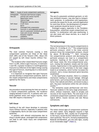
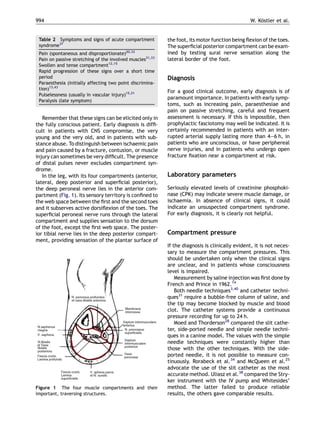
The document is a review article discussing acute compartment syndrome of the limb. It covers the aetiology, clinical signs, diagnosis, and treatment of compartment syndrome. Some key points:
- Compartment syndrome is a limb-threatening emergency caused by increased pressure within a limited muscle space, compromising circulation and tissue function.
- It most commonly results from orthopedic injuries like fractures but can also be caused by vascular injuries, soft tissue injuries, prolonged limb compression, and iatrogenic factors.
- Early symptoms are subtle like pain disproportionate to the injury but signs progress rapidly to include pain with passive stretching, tense compartments, paralysis.
- Diagnosis is clinical but compartment pressure measurements can help
This was a presentation done at Kanungo Institute of diabetic Specialitis Bhubaneswar . the audience included the students from Karolinkska Institute Sweden
Seminar clinical anatomy of upper limb joints and musclesQuan Fu Gan
This is not all, there are many more clinical anatomy in terms of condition such as Popeye Deformity with are not included here and Special Test such as Neer's Impingement and Hawkins Kennedy etc... with touches on the upper limb muscles and joints. Also not forgotten Long tendon test and so forth. In general, this is just a simplified slides. Tq
Seminar presentation by 4th year medical student of Lincoln University College, supervised by HRPZ Orthopedic's specialist.
Reference were from reliable medical websites and also from texttbook; Apley and Solomon's Concise System of Orthopaedics and Trauma, 4th Ed.
Acute Transverse Myelitis
Blockage of the Spinal Cord’s Blood Supply
Cervical Spondylosis
Compression of the Spinal Cord
Hereditary Spastic Paraparesis
Subacute Combined Degeneration
Syrinx of the Spinal Cord and Brain Stem
Shoulder pain is the third most common musculoskeletal symptom encountered in medical practice after back and neck pain, accounting for almost 3 million patient visits each year in the United States. A wide range of potential pathoanatomic entities can give rise to shoulder pain, from simple sprains to massive rotator cuff tears. The majority of these conditions are amenable to conservative treatment. Rotator cuff dysfunction is a particularly important entity because it occurs frequently and may necessitate surgical treatment. This report will provide a critical overview of current diagnostic and treatment techniques for rotator cuff disease. For more shoulder surgery and rotator cuff studies, visit Dr. Millett, shoulder surgeon, Greater Denver http://drmillett.com/shoulder-studies
Tom Selleck Health: A Comprehensive Look at the Iconic Actor’s Wellness Journeygreendigital
Tom Selleck, an enduring figure in Hollywood. has captivated audiences for decades with his rugged charm, iconic moustache. and memorable roles in television and film. From his breakout role as Thomas Magnum in Magnum P.I. to his current portrayal of Frank Reagan in Blue Bloods. Selleck's career has spanned over 50 years. But beyond his professional achievements. fans have often been curious about Tom Selleck Health. especially as he has aged in the public eye.
Follow us on: Pinterest
Introduction
Many have been interested in Tom Selleck health. not only because of his enduring presence on screen but also because of the challenges. and lifestyle choices he has faced and made over the years. This article delves into the various aspects of Tom Selleck health. exploring his fitness regimen, diet, mental health. and the challenges he has encountered as he ages. We'll look at how he maintains his well-being. the health issues he has faced, and his approach to ageing .
Early Life and Career
Childhood and Athletic Beginnings
Tom Selleck was born on January 29, 1945, in Detroit, Michigan, and grew up in Sherman Oaks, California. From an early age, he was involved in sports, particularly basketball. which played a significant role in his physical development. His athletic pursuits continued into college. where he attended the University of Southern California (USC) on a basketball scholarship. This early involvement in sports laid a strong foundation for his physical health and disciplined lifestyle.
Transition to Acting
Selleck's transition from an athlete to an actor came with its physical demands. His first significant role in "Magnum P.I." required him to perform various stunts and maintain a fit appearance. This role, which he played from 1980 to 1988. necessitated a rigorous fitness routine to meet the show's demands. setting the stage for his long-term commitment to health and wellness.
Fitness Regimen
Workout Routine
Tom Selleck health and fitness regimen has evolved. adapting to his changing roles and age. During his "Magnum, P.I." days. Selleck's workouts were intense and focused on building and maintaining muscle mass. His routine included weightlifting, cardiovascular exercises. and specific training for the stunts he performed on the show.
Selleck adjusted his fitness routine as he aged to suit his body's needs. Today, his workouts focus on maintaining flexibility, strength, and cardiovascular health. He incorporates low-impact exercises such as swimming, walking, and light weightlifting. This balanced approach helps him stay fit without putting undue strain on his joints and muscles.
Importance of Flexibility and Mobility
In recent years, Selleck has emphasized the importance of flexibility and mobility in his fitness regimen. Understanding the natural decline in muscle mass and joint flexibility with age. he includes stretching and yoga in his routine. These practices help prevent injuries, improve posture, and maintain mobilit
Ozempic: Preoperative Management of Patients on GLP-1 Receptor Agonists Saeid Safari
Preoperative Management of Patients on GLP-1 Receptor Agonists like Ozempic and Semiglutide
ASA GUIDELINE
NYSORA Guideline
2 Case Reports of Gastric Ultrasound
Flu Vaccine Alert in Bangalore Karnatakaaddon Scans
As flu season approaches, health officials in Bangalore, Karnataka, are urging residents to get their flu vaccinations. The seasonal flu, while common, can lead to severe health complications, particularly for vulnerable populations such as young children, the elderly, and those with underlying health conditions.
Dr. Vidisha Kumari, a leading epidemiologist in Bangalore, emphasizes the importance of getting vaccinated. "The flu vaccine is our best defense against the influenza virus. It not only protects individuals but also helps prevent the spread of the virus in our communities," he says.
This year, the flu season is expected to coincide with a potential increase in other respiratory illnesses. The Karnataka Health Department has launched an awareness campaign highlighting the significance of flu vaccinations. They have set up multiple vaccination centers across Bangalore, making it convenient for residents to receive their shots.
To encourage widespread vaccination, the government is also collaborating with local schools, workplaces, and community centers to facilitate vaccination drives. Special attention is being given to ensuring that the vaccine is accessible to all, including marginalized communities who may have limited access to healthcare.
Residents are reminded that the flu vaccine is safe and effective. Common side effects are mild and may include soreness at the injection site, mild fever, or muscle aches. These side effects are generally short-lived and far less severe than the flu itself.
Healthcare providers are also stressing the importance of continuing COVID-19 precautions. Wearing masks, practicing good hand hygiene, and maintaining social distancing are still crucial, especially in crowded places.
Protect yourself and your loved ones by getting vaccinated. Together, we can help keep Bangalore healthy and safe this flu season. For more information on vaccination centers and schedules, residents can visit the Karnataka Health Department’s official website or follow their social media pages.
Stay informed, stay safe, and get your flu shot today!
New Drug Discovery and Development .....NEHA GUPTA
The "New Drug Discovery and Development" process involves the identification, design, testing, and manufacturing of novel pharmaceutical compounds with the aim of introducing new and improved treatments for various medical conditions. This comprehensive endeavor encompasses various stages, including target identification, preclinical studies, clinical trials, regulatory approval, and post-market surveillance. It involves multidisciplinary collaboration among scientists, researchers, clinicians, regulatory experts, and pharmaceutical companies to bring innovative therapies to market and address unmet medical needs.
Ethanol (CH3CH2OH), or beverage alcohol, is a two-carbon alcohol
that is rapidly distributed in the body and brain. Ethanol alters many
neurochemical systems and has rewarding and addictive properties. It
is the oldest recreational drug and likely contributes to more morbidity,
mortality, and public health costs than all illicit drugs combined. The
5th edition of the Diagnostic and Statistical Manual of Mental Disorders
(DSM-5) integrates alcohol abuse and alcohol dependence into a single
disorder called alcohol use disorder (AUD), with mild, moderate,
and severe subclassifications (American Psychiatric Association, 2013).
In the DSM-5, all types of substance abuse and dependence have been
combined into a single substance use disorder (SUD) on a continuum
from mild to severe. A diagnosis of AUD requires that at least two of
the 11 DSM-5 behaviors be present within a 12-month period (mild
AUD: 2–3 criteria; moderate AUD: 4–5 criteria; severe AUD: 6–11 criteria).
The four main behavioral effects of AUD are impaired control over
drinking, negative social consequences, risky use, and altered physiological
effects (tolerance, withdrawal). This chapter presents an overview
of the prevalence and harmful consequences of AUD in the U.S.,
the systemic nature of the disease, neurocircuitry and stages of AUD,
comorbidities, fetal alcohol spectrum disorders, genetic risk factors, and
pharmacotherapies for AUD.
Recomendações da OMS sobre cuidados maternos e neonatais para uma experiência pós-natal positiva.
Em consonância com os ODS – Objetivos do Desenvolvimento Sustentável e a Estratégia Global para a Saúde das Mulheres, Crianças e Adolescentes, e aplicando uma abordagem baseada nos direitos humanos, os esforços de cuidados pós-natais devem expandir-se para além da cobertura e da simples sobrevivência, de modo a incluir cuidados de qualidade.
Estas diretrizes visam melhorar a qualidade dos cuidados pós-natais essenciais e de rotina prestados às mulheres e aos recém-nascidos, com o objetivo final de melhorar a saúde e o bem-estar materno e neonatal.
Uma “experiência pós-natal positiva” é um resultado importante para todas as mulheres que dão à luz e para os seus recém-nascidos, estabelecendo as bases para a melhoria da saúde e do bem-estar a curto e longo prazo. Uma experiência pós-natal positiva é definida como aquela em que as mulheres, pessoas que gestam, os recém-nascidos, os casais, os pais, os cuidadores e as famílias recebem informação consistente, garantia e apoio de profissionais de saúde motivados; e onde um sistema de saúde flexível e com recursos reconheça as necessidades das mulheres e dos bebês e respeite o seu contexto cultural.
Estas diretrizes consolidadas apresentam algumas recomendações novas e já bem fundamentadas sobre cuidados pós-natais de rotina para mulheres e neonatos que recebem cuidados no pós-parto em unidades de saúde ou na comunidade, independentemente dos recursos disponíveis.
É fornecido um conjunto abrangente de recomendações para cuidados durante o período puerperal, com ênfase nos cuidados essenciais que todas as mulheres e recém-nascidos devem receber, e com a devida atenção à qualidade dos cuidados; isto é, a entrega e a experiência do cuidado recebido. Estas diretrizes atualizam e ampliam as recomendações da OMS de 2014 sobre cuidados pós-natais da mãe e do recém-nascido e complementam as atuais diretrizes da OMS sobre a gestão de complicações pós-natais.
O estabelecimento da amamentação e o manejo das principais intercorrências é contemplada.
Recomendamos muito.
Vamos discutir essas recomendações no nosso curso de pós-graduação em Aleitamento no Instituto Ciclos.
Esta publicação só está disponível em inglês até o momento.
Prof. Marcus Renato de Carvalho
www.agostodourado.com
Knee anatomy and clinical tests 2024.pdfvimalpl1234
This includes all relevant anatomy and clinical tests compiled from standard textbooks, Campbell,netter etc..It is comprehensive and best suited for orthopaedicians and orthopaedic residents.
Report Back from SGO 2024: What’s the Latest in Cervical Cancer?bkling
Are you curious about what’s new in cervical cancer research or unsure what the findings mean? Join Dr. Emily Ko, a gynecologic oncologist at Penn Medicine, to learn about the latest updates from the Society of Gynecologic Oncology (SGO) 2024 Annual Meeting on Women’s Cancer. Dr. Ko will discuss what the research presented at the conference means for you and answer your questions about the new developments.
ARTIFICIAL INTELLIGENCE IN HEALTHCARE.pdfAnujkumaranit
Artificial intelligence (AI) refers to the simulation of human intelligence processes by machines, especially computer systems. It encompasses tasks such as learning, reasoning, problem-solving, perception, and language understanding. AI technologies are revolutionizing various fields, from healthcare to finance, by enabling machines to perform tasks that typically require human intelligence.
Explore natural remedies for syphilis treatment in Singapore. Discover alternative therapies, herbal remedies, and lifestyle changes that may complement conventional treatments. Learn about holistic approaches to managing syphilis symptoms and supporting overall health.
MANAGEMENT OF ATRIOVENTRICULAR CONDUCTION BLOCK.pdfJim Jacob Roy
Cardiac conduction defects can occur due to various causes.
Atrioventricular conduction blocks ( AV blocks ) are classified into 3 types.
This document describes the acute management of AV block.
MANAGEMENT OF ATRIOVENTRICULAR CONDUCTION BLOCK.pdf
Compartment syndrome
1. Injury, Int. J. Care Injured (2005) 36, 992—998
www.elsevier.com/locate/injury
ERRATUM
Acute compartment syndrome of the limb
W. Kostler *, P.C. Strohm, N.P. Sudkamp
¨ ¨
Department fur Orthopadie und Traumatologie, Klinik fur Traumatologie, Universitatsklinikum Freiburg,
¨ ¨ ¨ ¨
Hugstetterstr. 55, 79106 Freiburg im Breisgau, Germany
Accepted 15 April 2004
KEYWORDS Summary In this review the aetiology, clinical signs, diagnosis and therapy of the
Compartment; acute compartment syndrome of the limb is discussed. It is a limb- and untreated life
Compartment threatening emergency. For good results, early detection is necessary. It is important
syndrome; to educate those taking care of patients of risk, especially in the early symptoms and
Fracture complications signs. In uncooperative, unconscious and sedated patients pressure monitoring is
recommended. The critical level of the absolute intracompartmental pressure is
unclear. It is recommended to use a delta p pressure of 30 mmHg. Below this pressure
in the presence of clinical signs a fasciotomy of all compartments is the treatment of
choice.
# 2004 Elsevier Ltd. All rights reserved.
Introduction abdominal pressure alters cardiovascular haemody-
namics, respiratory mechanics and renal function.
Matsen defined the compartment syndrome as ‘‘a In this review, only the aetiology, clinical signs,
condition in which increased pressure within a lim- diagnosis and treatment of the limb compartment
ited space compromises the circulation and function syndromes are discussed.
of the tissues within that space.’’24
Malgaigne was the first to describe compartment
syndrome, and the first medical reference was by Aetiology and incidence
Volkmann39 in 1881. Jepsen reported successful
treatment by decompression18 in an experimental Most compartment syndromes are associated with
study. traumatic insults, but the condition also occurs after
The abdominal compartment syndrome was first reperfusion, following a period of ischaemia, burns,
described by Baggot in 1951.4 The increasing intra- prolonged limb compression after drug abuse or
poor positioning during prolonged surgical proce-
DOI of original article: 10.1016/j.injury.2004.04.009 dures (Table 1).
* Corresponding author. Tel.: +49 761 270 2401;
fax: +49 761 270 2520.
This classification, based on aetiology, is a sim-
E-mail address: koestler@ch11.ukl.uni-freiburg.de plification; in most cases, a combination of factors is
(W. Kostler).
¨ responsible.11,37
0020–1383/$ — see front matter # 2004 Elsevier Ltd. All rights reserved.
doi:10.1016/j.injury.2005.01.007
2. Acute compartment syndrome of the limb 993
Table 1 Causes of acute compartment syndromes Iatrogenic
Orthopaedic Fractures and fracture surgery
Vascular Arterial and venous injuries The use of a pneumatic antishock garment, or mili-
Reperfusion injury tary antishock trousers, may also lead to compart-
Haemorrhage ment syndrome. In combination with hypotension,
Phlegmasia caerulea dolens normally the reason for its use, and with application
Soft tissue Crush injury for more than 30 min, the development of compart-
Burns ment syndrome should be borne in mind.2
Prolonged limb compression
Prolonged surgery, especially in the Trendelen-
Iatrogenic Puncture in anticoagulated patients
burg position, sometimes causes compartment syn-
Use of a pneumatic antishock garment
Casts and circular dressings dromes.7 In combination with poor positioning, it
Pulsatile irrigation can also cause soft tissue necrosis, as a result of
Occasional Snakebite direct pressure.
Overuse of muscles
Pathophysiology
The normal pressure in the muscle compartments is
Orthopaedic below 10—12 mmHg (4—21).42 The compartmental
perfusion pressure, which is the mean arterial
The most common fractures causing a limb pressure minus the compartment pressure, should
compartment syndrome are those of the tibial be above 70—80 mmHg.17,23. Both increasing
shaft (40%) and of the forearm (18%). About 23% the compartmental pressure and decreasing the
are caused by soft tissue injuries without frac- perfusion pressure can lead to a compartment
ture.27,33 syndrome. Swelling of the injured muscle and
The incidence after closed tibial fractures ranges soft tissue raises the intra-compartmental pres-
from 1 to 29%, while in open fractures, it is between sure, closing the lymphatic vessels and the small
1 and 10%. A greater incidence in multifragmentary venules. Hypertension in the capillary bed and the
fractures is a reflection of the higher energy causing compression of the arterioles in the later stages
this type of fracture, resulting also in a more severe leads to an ischaemia, which further increases the
soft tissue injury. pressure, and a vicious downward spiral becomes
It is important to recognise that open fractures established.
can also develop a compartment syndrome, both in In the reperfusion situation, there is a complex
the closed compartments and those compartments mechanism leading to vascular leakage. Although
opened by the injury. some factors, such as leucotrienes, tumour necrosis
factor, free radicals and others, are known to play a
part, the exact mechanism within the cell remains
Vascular unclear.10
The ischaemia causes perifascicular and intra-
Any procedure revascularising the limb can result in fascicular oedema in the early stage presentation,
a muscle compartment syndrome, the incidence which, without treatment, leads to atrophy and
ranging between 0 and 21%. In patients with com- necrosis of the muscle fibres. The end result of an
bined popliteal artery and venous injuries, more unchecked compartment syndrome includes neuro-
than 50% require a fasciotomy.36 logical deficit, muscle necrosis and late fibrous
contractures.
Soft tissue
Symptoms and signs
Swelling of the soft tissue develops in contusion
injury without fracture, especially in patients with The initial clinical signs of compartment syndrome
a coagulopathy. Burns can also cause compartment are often subtle. Early diagnosis requires a high
syndrome. index of suspicion. Pain, usually out of proportion
In patients with altered consciousness due to to the injury, can be the first indication of compart-
drug or alcohol abuse, prolonged limb compression ment syndrome. The six P’s, formerly emphasised by
can lead to soft tissue injury and compartment Mubarak and Rorabeck, are very late signs, usually
syndrome. at an irreversible stage29 (Table 2).
3. 994 W. Kostler et al.
¨
Table 2 Symptoms and signs of acute compartment the foot, its motor function being flexion of the toes.
syndrome37 The superficial posterior compartment can be exam-
Pain (spontaneous and disproportionate)20,33 ined by testing sural nerve sensation along the
Pain on passive stretching of the involved muscles31,33 lateral border of the foot.
Swollen and tense compartment12,15
Rapid progression of these signs over a short time
period Diagnosis
Paraesthesia (initially affecting two point discrimina-
tion)13,43
For a good clinical outcome, early diagnosis is of
Pulselessness (usually in vascular injury)15,21
paramount importance. In patients with early symp-
Paralysis (late symptom)
toms, such as increasing pain, paraesthesiae and
pain on passive stretching, careful and frequent
Remember that these signs can be elicited only in assessment is necessary. If this is impossible, then
the fully conscious patient. Early diagnosis is diffi- prophylactic fasciotomy may well be indicated. It is
cult in patients with CNS compromise, the very certainly recommended in patients with an inter-
young and the very old, and in patients with sub- rupted arterial supply lasting more than 4—6 h, in
stance abuse. To distinguish between ischaemic pain patients who are unconscious, or have periphereal
and pain caused by a fracture, contusion, or muscle nerve injuries, and in patients who undergo open
injury can sometimes be very difficult. The presence fracture fixation near a compartment at risk.
of distal pulses never excludes compartment syn-
drome.
In the leg, with its four compartments (anterior, Laboratory parameters
lateral, deep posterior and superficial posterior),
the deep peroneal nerve lies in the anterior com- Seriously elevated levels of creatinine phosphoki-
partment (Fig. 1). Its sensory territory is confined to nase (CPK) may indicate severe muscle damage, or
the web space between the first and the second toes ischaemia. In absence of clinical signs, it could
and it subserves active dorsiflexion of the toes. The indicate an unsuspected compartment syndrome.
superficial peroneal nerve runs through the lateral For early diagnosis, it is clearly not helpful.
compartment and supplies sensation to the dorsum
of the foot, except the first web space. The poster-
ior tibial nerve lies in the deep posterior compart- Compartment pressure
ment, providing sensation of the plantar surface of
If the diagnosis is clinically evident, it is not neces-
sary to measure the compartment pressures. This
should be undertaken only when the clinical signs
are unclear, and in patients whose consciousness
level is impaired.
Measurement by saline injection was first done by
French and Prince in 1962.14
Both needle techniques3,40 and catheter techni-
ques31 require a bubble-free column of saline, and
the tip may become blocked by muscle and blood
clot. The catheter systems provide a continuous
pressure recording for up to 24 h.
Moed and Thorderson28 compared the slit cathe-
ter, side-ported needle and simple needle techni-
ques in a canine model. The values with the simple
needle techniques were constantly higher than
those with the other techniques. With the side-
ported needle, it is not possible to measure con-
tinuously. Rorabeck et al.34 and McQueen et al.25
advocate the use of the slit catheter as the most
accurate method. Uliasz et al.38 compared the Stry-
ker instrument with the IV pump and Whitesides’
Figure 1 The four muscle compartments and their method. The latter failed to produce reliable
important, traversing structures. results, the others gave comparable results.
4. Acute compartment syndrome of the limb 995
Despite most surgeons’ agreeing that pressure a big difference between individuals, when corre-
measurement is the standard method, only about lating absolute pressure levels, clinical signs, nerve
50% of the hospitals in the UK and Germany have the function (EMG) and oxygen levels in the muscle
technical equipment to do this, according to inves- tissue.42 At a level of 50 mmHg of intra-compart-
tigations of Williams and Sterk.41 It should be men- mental pressure, some healthy volunteers had no
tioned that it is important to measure all muscle decrease in oxygen saturation in the muscle; others
compartments in a limb suspected of acute com- showed a decrease to nearly zero. The decrease in
partment syndrome. the muscle response started at 30 mmHg, but at a
level of 50 mmHg there was still one volunteer with
no measurable muscular response. It is clear that
Other methods the definition of an absolute critical level of the
intra-compartmental pressure is not possible.
Willy et al.42 showed the easy and highly accurate Whitesides introduced the concept that the level
use of transducer tipped probes. They work without of intra-compartmental pressure which causes
the artefacts associated with saline-column sys- ischaemic compromise is related to the perfusion
tems. It is also possible to measure the partial pressure.40 This ‘‘delta p’’ pressure, comparable to
pressure of oxygen in the muscle with catheters, the CPP in brain injury, is the diastolic pressure
although this method is not routinely used clinically, minus the intra-compartmental pressure. The most
as the critical levels of tissue oxygenations have not commonly cited Dp is less than or equal to 30 mmHg.
yet been defined or validated, and its worth has to In studies using Dp, unnecessary fasciotomies were
be proven. Theoretically, it should be helpful to avoided and no significant complications
measure the oxygen saturation in the tissue, since occurred.26
the pressure is only an indirect measure of tissue The time factor is more critical for the neural
damage and perfusion. structures than it is for the muscle, but it is impor-
Near-infrared spectroscopy can measure the tant to remember that there is a dynamic relation-
levels of muscle haemoglobin and myoglobin. The ship between the level of the intra-compartmental
method is applied percutaneously, but the limited pressure and the duration of elevated pressure. The
measurement depth of about 40 mm is a problem, longer the delay to fasciotomy, the worse the out-
especially for the deep posterior compartment in come. If the delay is more than 12 h, bad results are
the leg. Most studies with this technique were done inevitable.
in chronic compartment syndromes; the value in
patients with an evolving acute compartment syn-
drome remains unclear. With further technical Treatment
improvements, it may offer a rapid, non-invasive
tool in the future.16,22 Surgical therapy
MRI can show the tissue changes in established
compartment syndrome, but fails to identify If compartment syndrome is suspected, all circum-
changes in an imminent compartment syndrome ferential dressings should be removed, normal blood
without neurological deficit. It is not possible to pressure should be achieved by dealing with any
decide if the swelling and oedema are correlated cause of hypotension. The extremity should not be
with a soft tissue injury itself, or represent signs of elevated, but kept at heart level, in order to main-
an evolving compartment syndrome.32 tain perfusion in the compartment. Supplementary
Laser Doppler flowmetry and scintigraphy have oxygen, to improve the tissue oxygenation, is help-
only been evaluated in chronic compartment syn- ful.
dromes. If the Dp is below 30 mmHg, and/or clinical signs
are present, fasciotomy of all relevant compart-
ments is the treatment of choice and should be
Critical pressure performed as an emergency. Prophylactic fasciot-
omy is recommended in patients with vascular inju-
More important than improving the technical ries, who have had a warm ischaemia time of more
devices is to improve the guidelines for interpreta- than 4—6 h, patients with ligation of the major veins
tion of the results and their clinical relevance. in the popliteal region or the distal thigh and
The critical level of the absolute intra-compart- patients with crush injuries.
mental pressure remains yet undecided. In the lit- In the lower leg, a four-compartment fasciotomy
erature, levels ranging from 30 to 50 mmHg are is recommended (Fig. 2). This can be performed by
proposed.1,29,31 Experimental studies have shown different techniques: with a single lateral incision,
5. 996 W. Kostler et al.
¨
Figure 2 Generous releases are necessary for complete decompression of all four lower leg muscle compartments.
with or without fibulectomy or by a double incision anterior and lateral compartments from the lateral
technique, using medial and lateral incisions.19,30 In side. Its advantage is the possible use of local
the acute situation, the skin incision must be long anaesthesia, even at the bedside; its disadvantage
enough to ensure that all underlying muscle is is the need for two separate incisions.
decompressed; so-called ‘‘percutaneous’’ fascio- For temporary wound coverage, hypoallogenic
tomies are dangerous and are not recommended. materials, such as Epigard or Vacuseal, can be used
The single incision technique allows adequate (Fig. 3a and b), followed by early definitive closure
exposure of all four compartments;8 fibulectomy using split skin grafting. Recently, in the authors’
should be avoided, especially in patients with com- department, since elastic bands have been used to
plex tibial fractures. close the wound incrementally, the necessity for
The skin incision is made directly over the fibula. skin grafting has decreased and has only been man-
A transverse incision is made in the fascia to identify dated in a few cases (Fig. 4).
the inter-muscular septum between the anterior In patients with fractures and muscle compart-
and lateral compartments and to identify the super- ment syndrome, osteosynthesis is recommended,
ficial peroneal nerve. Longitudinal fasciotomies of the technique depending on the fracture localisa-
the anterior, the lateral and the superficial dorsal tion, the patient’s status and the quality of the soft
compartments are then performed. The final step is tissue. If possible, definitive fracture surgery should
to divide the origins of the soleus from the fibula and be undertaken at the time of fasciotomy.
then, directly behind the fibula, release the deep In the thigh, all three compartments can be
posterior compartment. decompressed via a lateral incision. In order to
With the double incision technique, the posterior prevent muscle hernia, the authors favour two par-
compartments are opened from medially, and the allel incisions in the fascia lata, with an intervening
6. Acute compartment syndrome of the limb 997
Hyperbaric oxygen
In patients with crush injuries, Bouachour et al.
showed that adjunctive hyperbaric therapy was
associated with significantly better wound healing
after fasciotomy.5 There are also some experimental
data that show better functional results.35
It is our impression, in patients with clinically
borderline symptoms, for example, after intra-
medullary nailing, that immediate hyperbaric ther-
apy in the first 2 days often renders fasciotomy
unnecessary, but further studies are necessary.
Drug therapy
Mannitol, as a hyperosmotic diuretic, has been
shown to reduce the incidence of compartment
syndrome after revascularisation.6 Additionally,
free radical scavengers are thought to have bene-
ficial effects. In animal studies, the effects are
variable and their use in humans is not established.9
Figure 3 Before (a) and after (b) provisional tissue cover Summary
using a synthetic skin substitute.
The final clinical outcome of an untreated compart-
ment syndrome is the replacement of muscle with
bridge of at least 4—5 cm. The fracture causing the scar tissue. This produces a severe fibrous contrac-
compartment syndrome is fixed definitively at the ture and a neuropathy of any peripheral nerve
same time, provided the patient is physiologically traversing the compartment, leading to serious dys-
stable. function. Once this stage is reached, it is never
In the forearm, volar and dorsal incisions are possible to restore normal function.
necessary. A complete forearm fasciotomy requires For early detection of muscle compartment syn-
decompression of each nerve and muscle. When in drome, it is necessary to educate those taking care
combination with fractures, definitive stabilisation, of patients at risk, especially in the early symptoms
usually with plates, is recommended. The implants and signs.
should be covered by vital muscle. If the clinical Monitoring of intra-compartmental pressure
status of the hand remains unclear, the volar incision should be routine in unconscious, sedated and unco-
should be extended into the palm and the carpal operative patients. If the Dp remains less than
tunnel released. 30 mmHg, in the presence of clinical signs and
In patients who develop sepsis, it is important to despite conservative measures, fasciotomy should
remember that all necrotic tissue has to be excised. be performed as an emergency to preserve the
Renal function should be monitored and renal fail- function of the limb.
ure prevented.
References
1. Allen M, Stirling A, Crawshaw C, Barnes M. Intracompart-
mental pressure monitoring of leg injuries. An aid to manage-
ment. J Bone Joint Surg 1985;67Br:53—7.
2. Aprahamian T, Gessert G, Banoyk D. MAST-associated com-
partment syndrome (MAcompartment syndrome): a review. J
Trauma 1989;29:549.
3. Awbrey B, Stenkiewicz P, Mankin H. Chronic exercise induced
compartment pressure elevation measured with a miniatured
fluid pressure monitor: a laboratory and clinical study. Am J
Figure 4 Elastic closure technique. Sports Med 1988;16:610.
7. 998 W. Kostler et al.
¨
4. Baggot M. Abdominal blow-out: a concept. Anesth Analg 24. Matsen F, Winquist R, Krugmire R. Diagnosis and management
1951;30:295—9. of compartmental syndromes. J Bone Joint Surg 1980;62-
5. Bouachour G, Cronier P, Gouello J, Toulemonde J, Talha A, A:286.
Alquier P. Hyperbaric oxygen therapy in the management of 25. McQueen M, Christie J, Court-Brown C. Compartment pres-
crush injuries: a randomized double blind placebo-controlled sures after intramedullary nailing of the tibia. J Bone Joint
trial. J Trauma 1996;41:333—9. Surg 1990;72B:395—7.
6. Buchbinder D, Karmody A, Leather R, Shah D. Hypertonic 26. McQueen M, Court-Brown C. Compartment monitoring in
mannitol: its use in the prevention of revascularisation syn- tibial fractures. The presure threshold for decompressions.
drome after acute arterial ischaemia. Arch Surg 1981; J Bone Joint Surg 1996;72BR:99—104.
116:414—21. 27. McQueen M, Gaston P, Court-Brown C. Acute compartment
7. Chase J, Harford F, Pinzur M, Zussman M. Intraoperative lower syndrome: who’s at risk? J Bone Joint Surg 2000;82-B:200—3.
extremity compartment pressures. Dis Colon Rectum 2000; 28. Moed B, Thorderson P. Measurement of intracompartmental
43:678—80. pressure: a comparison of the slit catheter, sideported nee-
8. Cooper G. A method of single-incision, four compartment dle, simple needle. J Bone Joint Surg 1993;75A:231—5.
fasciotomy of the leg. Eur J Vasc Surg 1992;6:659—61. 29. Mubarak S, Hargens A. Acute compartment syndromes. Surg
9. Dabby D, Greif F, Yaniv M, Rubin M, Dekel S, Leleuk S. Clin North Am 1983;63:539—65.
Thromboxane A2 in postischaemic acute compartmental syn- 30. Mubarak S, Owen C. Double-incision fasciotomy of the leg for
drome. Arch Surg 1998;133:953—6. decompression in compartment syndromes. J Bone Joint Surg
10. Duran W, Takenaka H, Hobson R. Microvascular pathophysiol- 1977;59A:184—7.
ogy of skeletal muscle ischaemia-reperfusion. Semin Vasc 31. Mubarak S, Owen C, Hargens A, Garetto L, Akeson W. Acute
Surg 1998;11:203—14. compartment syndromes: diagnosis and treatment with the
11. Elliott K, Johnstone A. Diagnosing acute compartment syn- aid of the wick catheter. J Bone Joint Surg 1978;60A:1091—5.
drome. J Bone Joint Surg 2003;85:625—32. 32. Rominger M, Lukosch C, Bachmann G, Langer C, Schnettler R.
12. Fakhouri A, Manoli A. Acute foot compartment syndromes. J Compartment syndrome: value of MR imaging. Radiology
Orthop Trauma 1992;6:223—8. 1995;197:296.
13. Franc-Law J, Rossignol M, Vernec A, Somogyi D, Shrier J. 33. Rorabeck C. The treatment of compartment syndromes of the
Poisoning-induced acute atraumatic compartment syn- leg. J Bone Joint Surg 1984;66-B:93—7.
drome. Am J Emerg Med 2000;18:616—21. 34. Rorabeck C, Castle G, Hardie R, Logan J. Compartmental
14. French E, Price W. Anterior tibial pain. Br Med J 1962;ii: pressure measurements: an experimental investigation using
1291. the slit catheter. J Trauma 1980;21:446—9.
15. Gelbermann R, Garfin S, Hergenroeder P, Mubarak S, Menon J. 35. Strauss M, Hargens A, Gershuni D, Greenberg D, Grenshaw A,
Compartment syndromes of the forearm: diagnosis and treat- Hart G. Reduction of skeletal muscle necrosis using inter-
ment. Clin Orthop 1981;161:252—61. mittent hyperbaric oxygen in a model compartment syn-
16. Giannotti G, Cohn S, Brown M, Varela J, McKenney M, Wise- drome. J Bone Joint Surg 1986;68A:1218—24.
berg J. Utility of near-infrared spectroscopy in the diagnosis 36. Thomas DD, Wilson RF, Wiencek RG. Vascular injury about the
of lower extremity compartment syndrome. J Trauma knee: improved outcome. Am Surg 1989;55:370—7.
2000;48:396—9. 37. Tiwari A, Haq A, Myint F, Hamilton G. Acute compartment
17. Heppenstall R, Scott R, Sapega A. A comparative study of the syndromes. Br J Surg 2002;89:397—412.
tolerance of skeletal muscle to ischaemia. J Bone Joint Surg 38. Uliasz A, Ishida J, Fleming J, Yamamoto L. Comparing the
1986;68-A:820. methods of measuring compartment pressures in acute com-
18. Jepson P. Ischemic contracture, experimental study. Ann Surg partment syndrome. Am J Emerg Med 2003;21:143—5.
1926;84:785. 39. von Volkmann R. Die ischamischen Kontrakturen. Zentralbl
¨
19. Kelly R, Whitesides T. Transfibular route for fasciotomy of the Chir 1881;8:801.
leg. J Bone Joint Surg 1967;49A:1022—3. 40. Whitesides T, Haney T, Morimoto K, Hirada H. Tissue pressure
20. Lagerstrom C, Reed R, Rowlands B, Fischer R. Early fasciot- measurements as a determinant for the need of fasciotomy.
omy for acute clinically evident post-traumatic compartment Clin Orthop 1975;113:43.
syndrome. Am J Surg 1989;158:36—9. 41. Williams P, Russell I, Mintowt-Czyz W. Compartment pressure
21. Leach R, Hammond G, Stryker W. Anterior tibial compart- monitoring-current UK orthopeaedic practice. Injury 1998;
ment syndrome. Acute and chronic. J Bone Joint Surg 29:229—32.
1967;49A:451—62. 42. Willy C, Sterk J, Voelker H, et al. Das akute Kompartment-
22. Mancini D, Bolinger L, Li H, Kendrick K, Chance B, Wilson J. syndrom. Unfallchirurg 2001;104:381—91.
Validation of near-infrared spectroscopy in humans. J Appl 43. Zweifach S, Hargens A, Evans K, Smith R, Mubarak S, Akeson
Physiol 1994;77:2740—7. W. Skeletal muscle necrosis in pressurized compartments
23. Matsen F, Krugmire R. Compartmental syndromes. Surg Gyne- associated with hemorrhagic hypotension. J Trauma 1980;
col Obstet 1978;147:943—9. 20:941—7.