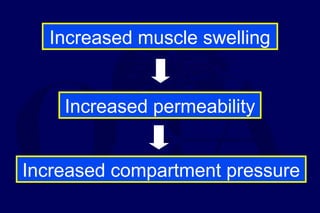
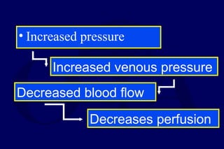
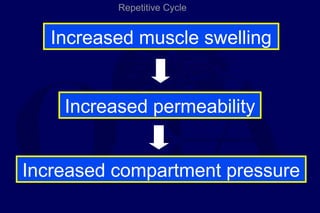
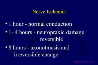
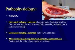
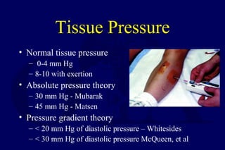
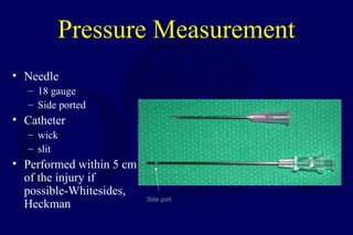
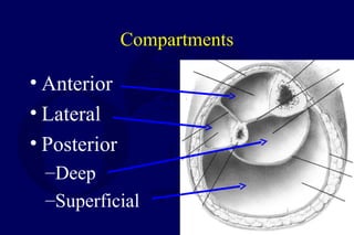
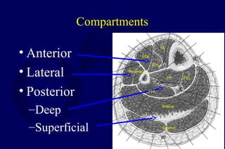
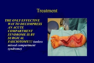
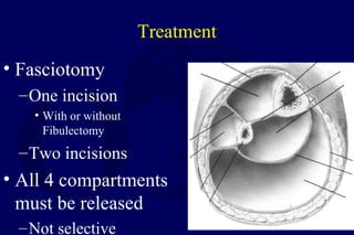
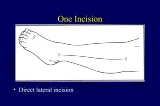
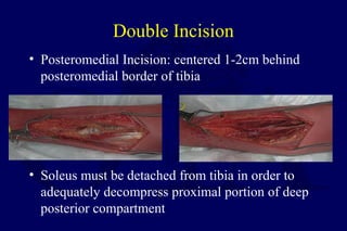
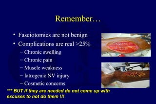
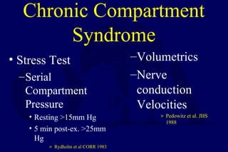
Compartment Syndromes
Dicky Aligheri ,MD
Dept of CardioThoracic & Vascular Surgery
National CardioVascular Centre Harapan Kita
Jakarta 2013
Today
• What is it
• Pathophysiology
• Diagnosis
• Treatment
Increase in hydrostatic pressure in closed osteofascial
space resulting in decreased perfusion of muscle
and nerves within compartment
• RAISED PRESSURERAISED PRESSURE
WITHIN A CLOSEDWITHIN A CLOSED
SPACESPACE with a potential to
cause irreversible damageirreversible damage
to the contents of the
closed space
Richard Von Volkmann, 1881
• “For many years I have noted on occasion, following the
use of bandages too tightly appliedbandages too tightly applied, the occurrence of
paralysis and contraction of the limb, NOT … due toparalysis and contraction of the limb, NOT … due to
thethe paralysis of the nerve by pressureparalysis of the nerve by pressure, but as a quick and
massive disintegration of the contractile substance and
the effect of the ensuing reaction and degeneration.”
Definition
• Symptoms resulting from
increased pressure within a
limited space
– compromising
• circulation
• function
Pathophysiology
• Local Blood Flow is reduced as a
consequence:
LBF=Pa-Pv / R (A-V Gradient)
Pathophysiology
• A continuous increase in
pressure within a
compartment occurs until
the low intramuscularlow intramuscular
arteriolar pressure isarteriolar pressure is
exceededexceeded and blood
cannot enter the
capillaries
Pathophysiology
• Increased compartment pressure
Increased venous pressure
Decreased blood flow
Decreases perfusion
Pathophysiology
• Autoregulatory mechanisms may compensate:
– Decrease in peripheral vascular resistance
– Increased extraction of oxygen
• As system becomes overwhelmed:
– Critical closing pressure is reached
– Oxygen perfusion of muscles and nerves decreases
Muscle Ischemia
• 4 hours - reversible damage
• 8 hours - irreversible changes
• 4-8 hours - variable
Hargens JBJS 1981
Muscle Ischemia
• Myoglobinuria after 4 hours
–Renal failure
–Maintain a high urinary output
–Alkalinize the urine
• Cell death initiates a “vicious cycle”
–increase capillary permeability
–increased muscle swelling
Increased muscle swelling
Increased permeability
Increased compartment pressure
• Increased pressure
Increased venous pressure
Decreased blood flow
Decreases perfusion
Increased muscle swelling
Increased permeability
Increased compartment pressure
Repetitive Cycle
Nerve Ischemia
• 1 hour - normal conduction
• 1- 4 hours - neuropraxic damage
reversible
• 8 hours - axonotmesis and
irreversible change
Hargens et al. JBJS 1979
Pathophysiology:
• CAUSES:
• Increased Volume - internal :Increased Volume - internal : hemmorhage, fractures, swelling
from traumatized tissue, increased fluid secondary to burns, post-
ischemic swelling
• Decreased volume - external:Decreased volume - external: tight casts, dressings
• Most common cause of hemmorhage into a compartment:Most common cause of hemmorhage into a compartment:
fractures of the tibia, elbow, forearm or femur
Etiology
• Fractures
• Soft Tissue Injury (Crush)
• Arterial Injury
– Post-ischemic swelling
– Reperfusion injury
• Drug Overdose (limb compression)
• Burns
Pathophysiology:
Most common causeMost common cause of compartment
syndrome is muscle injurymuscle injury that leads to
edema
Arterial Injuries
• Secondary toSecondary to
revascularizationrevascularization:
• Ischemia causes damage
to cellular basement
membrane that results in
edema
• With reestablishment of
flow, fluid leaks into the
compartment increasing
the pressure
Diagnosis
• Clinical diagnosis
– High index of suspicion
• Syndrome
– History
– Physical Exam
Difficult Diagnosis
• Classic signs of the 5 P’s - ARE NOT RELIABLE:
– pain
– pallor
– paralysis
– pulselessness
– paresthesias
• These are signs of an ESTABLISHED compartment
syndrome where ischemic injury has already taken place
• These signs may be present in the absence of
compartment syndrome.
Diagnosis
• Pain
• Compartment pressure
– Confirmatory test
– Don’t just measure
Diagnosis
• Palpable pulses are usually present in acute
compartment syndromes unless an arterial
injury occurs
• Sensory changes and paralysisSensory changes and paralysis do not occur
until ischemia has been present for about 11
hour or morehour or more
Diagnosis
• The most importantmost important
symptomsymptom of an impending
compartment syndrome is
PAINPAIN
DISPROPORTIONATEDISPROPORTIONATE
TO THAT EXPECTEDTO THAT EXPECTED
FOR THE INJURYFOR THE INJURY
Signs & Symptoms
• Pain
–Passive muscle stretching
–Out of proportion
–Progressive
–Not relieved by immobilization
Signs & Symptoms
• Pain
–May be worse with elevation
–Patient will not initiate motion on
own
• Be careful with coexisting nerve
injury
Signs & Symptoms
• Parasthesia
–Secondary to nerve ischemia
• Must be differentiated from nerve
injury
Signs & Symptoms
• Paralysis (Weakness)
– Ischemic muscles lose function
Signs & Symptoms
• Tense compartment on palpation
• Elevated compartment pressure
Tissue Pressure
• Normal tissue pressure
– 0-4 mm Hg
– 8-10 with exertion
• Absolute pressure theory
– 30 mm Hg - Mubarak
– 45 mm Hg - Matsen
• Pressure gradient theory
– < 20 mm Hg of diastolic pressure – Whitesides
– < 30 mm Hg of diastolic pressure McQueen, et al
Tissue-Pressure: Principles
• Originally, fasciotomies for tissue-pressures greater-than
30mmHg
• Whitesides et al in 1975Whitesides et al in 1975 was the first to suggest that the
significance of tissue pressures was in their relation torelation to
diastolic blood pressurediastolic blood pressure.
• McQueen et al: absolute compartment pressure is anabsolute compartment pressure is an
UNRELIABLE indication for the need for fasciotomies.UNRELIABLE indication for the need for fasciotomies. BUT,
pressures within 30mmHg of DP indicate compartment
syndrome
Tissue-Pressure: Principles
• Heckman et al demonstrated that
pressure within a givenpressure within a given
compartment is not uniformcompartment is not uniform
• They found tissue pressures to be
highest at the site or within 5cmhighest at the site or within 5cm
of the injuryof the injury
• 3 of their 5 patients requiring
fasciotomies had sub-critical
pressure values 5cm from the site
of highest pressure
Who is at high risk?
High energy fractures
• Severe
comminution
• Joint extension
• Segmental
injuries
• Widely
displaced
• Bilateral
• Floating knee
• Open fractures
Impaired Sensorium
• Alcohol
• Drug
• Decreased
GCS
• Unconscious
• Chemically
unconscious
• Neurologic
deficit
• Cognitively
challenged
Diagnosis
• The presence of an open fracture does NOT rule out the presenceopen fracture does NOT rule out the presence
of a compartment syndromeof a compartment syndrome
• 6-9% of open tibial fractures are associated with compartment
syndromes
• McQueen et al found no significant differences in compartmentno significant differences in compartment
pressures between open and closed tibial fracturespressures between open and closed tibial fractures
• No significant difference in pressures between tibial fracturesNo significant difference in pressures between tibial fractures
treated with IM Nails and those treated with Ex-Fixtreated with IM Nails and those treated with Ex-Fix
Criteria-Compartment Pressure
• Accurately examine
– Difference < 30mm Hg
• Impaired
– Absolute > than 30mm Hg
Needle Infusion Technique-Historical
• Needle inserted into muscle, tube
with air/saline interval kept at this
height, manometer indicates
pressure
• Air injected by syringe via 3-way
stopcock
• When the pressure of the injected
air exceeds the compartment
pressure pressure, the saline interval
moves in the tube
• AT this point, the second personthe second person
reads the pressure from thereads the pressure from the
manometermanometer
NEED 2 PEOPLE !NEED 2 PEOPLE !
saline
Pressure Measurement
• Infusion
– manometer
– saline
– 3-way stopcock
(Whitesides, CORR 1975)
• Catheter
– wick
– slit catheter
• Arterial line
– 16 - 18 ga. Needle
(5-19 mm Hg higher)
– transducer
– monitor
• Stryker device
– Side port needle
• Needle
– 18 gauge
– Side ported
• Catheter
– wick
– slit
• Performed within 5 cm
of the injury if
possible-Whitesides,
Heckman
Side port
Pressure Measurement
• Unit and needle set
• Assemble unit and prime
• Hold at angle to measure
• Zero machine
• Test each of 4 compartments
– Keep calf off of bed
Most Common Locations
• Leg: deep posterior and thedeep posterior and the
anterioranterior compartmentscompartments
• Forearm: volar compartmentvolar compartment,
especially in the deep flexor area
Where to Measure
Pressure
• Deeper muscles are initially involved
• Distance from fracture affects pressure
Heckmen et al. JBJS 1994
Compartments
• Anterior
• Lateral
• Posterior
–Deep
–Superficial
Compartments
• Anterior
• Lateral
• Posterior
–Deep
–Superficial
EDL
FDLTP
Gastroc
Soleus
TA
EHL
FHL
Peroneus
Treatment
• Remove restricting bandages
• Serial exams
• When diagnosis made
– Immediate surgery
• 4 compartment fasciotomy
Treatment
THE ONLY EFFECTIVE
WAY TO DECOMPRESS
AN ACUTE
COMPARTMENT
SYNDROME IS BY
SURGICAL
FASCIOTOMY!!! (unless
missed compartment
syndrome)
Treatment
• Fasciotomy
–One incision
• With or without
Fibulectomy
–Two incisions
• All 4 compartments
must be released
–Not selective
One Incision
• Direct lateral incision
Perifibular Fasciotomy
• One incision
• Head of fibula to proximal tip of lateral malleolus
• Incise fascia between soleus and FHL distally and
extended proximally to origin of soleus from fibula
• Deep posterior compartment released off of the
interosseous membrane, approached from the interval
between the lateral and superfical posterior
compartments
• Lateral compartment
• Anterior compartment
Alternative
Through
intermuscular
septum to reach
superficial
posterior
compartment
Two incisions
• Lateral • Medial
Double Incision
• 2 vertical incisions separated by a skin bridge of
at least 8 cm
• Anterolateral Incision: from knee to ankle,
centered over interval between anterior and lateral
compartments
Double Incision
• Posteromedial Incision: centered 1-2cm behind
posteromedial border of tibia
• Soleus must be detached from tibia in order to
adequately decompress proximal portion of deep
posterior compartment
Thigh
• Rare
• Crush injury with femur fracture
• Over distraction
– relative under distraction
Thigh
• Quadriceps
–Lateral
• Hamstrings
–Posterior
• Abductor
–Medial
Treatment
• Based upon involvement
• Usually Quadriceps and
Hamstrings
• Usually, a single lateral incision
will suffice
Compartments of the Forearm
• Forearm can be divided into 3
compartments: Dorsal, Volar and “Mobile
Wad”
• Mobile Wad: Brachioradialis, ECRL, ECRB
• Dorsal: EPB, EPL, ECU, EDC
• Volar: FPL, FCR, FCU, FDS, FDP, PQ
Henry Approach
• Incision begins proximal to antecubital
fossa and extends across carpal tunnel
• Begins lateral to biceps tendon, crosses
elbow crease and extends radially, then
it is extended distally along medial
aspect of brachioradialis and extends
across the palm along the thenar crease
• Alternatively, a straight incision from
lateral biceps to radial styloid can be
used.
Henry Approach
• Fascia over superficial muscles is
incised
• Care of NV structures
Henry Approach
• Brachioradialis and superficial
radial n. are retracted radially and
FCR and radial artery are retracted
ulnar to expose the deep volar
muscles
• Fascia of each of the deep muscles
is then incised
Post Fasciotomy…
• Must get bone stability
– IMN
– exfix
• ~48hrs after procedure patient should be
brought back to OR for further debridement
• Delayed skin closure or skin-grafting 3-7
days after the fasciotomies
Aftercare
• Xeroform
• VAC dressings
• Elevation of limb
• Delayed wound closure
– Split thickness skin graft
Remember…
• Fasciotomies are not benign
• Complications are real >25%
– Chronic swelling
– Chronic pain
– Muscle weakness
– Iatrogenic NV injury
– Cosmetic concerns
*** BUT if they are needed do not come up with*** BUT if they are needed do not come up with
excuses to not do them !!!excuses to not do them !!!
Chronic (Exertional)
Compartment Syndrome
• Transient rise in compartmental
pressure following activity
• Symptoms
–Pain
–Weakness
–Neurologic deficits
Chronic Compartment
Syndrome
• Stress Test
–Serial
Compartment
Pressure
• Resting >15mm Hg
• 5 min post-ex. >25mm
Hg
» Rydholm et al CORR 1983
–Volumetrics
–Nerve
conduction
Velocities
» Pedowitz et al. JHS
1988
Chronic Compartment
Syndrome
• Treatment
– Modification of activity
– Splinting
– Elective Fasciotomy
Conclusion
• Very important to make diagnosis
• Missed compartment is devastating
• Physical exam
• Re-examine patient!

compartment syndrome

  • 1.
    Compartment Syndromes Dicky Aligheri,MD Dept of CardioThoracic & Vascular Surgery National CardioVascular Centre Harapan Kita Jakarta 2013
  • 2.
    Today • What isit • Pathophysiology • Diagnosis • Treatment
  • 3.
    Increase in hydrostaticpressure in closed osteofascial space resulting in decreased perfusion of muscle and nerves within compartment
  • 4.
    • RAISED PRESSURERAISEDPRESSURE WITHIN A CLOSEDWITHIN A CLOSED SPACESPACE with a potential to cause irreversible damageirreversible damage to the contents of the closed space
  • 5.
    Richard Von Volkmann,1881 • “For many years I have noted on occasion, following the use of bandages too tightly appliedbandages too tightly applied, the occurrence of paralysis and contraction of the limb, NOT … due toparalysis and contraction of the limb, NOT … due to thethe paralysis of the nerve by pressureparalysis of the nerve by pressure, but as a quick and massive disintegration of the contractile substance and the effect of the ensuing reaction and degeneration.”
  • 6.
    Definition • Symptoms resultingfrom increased pressure within a limited space – compromising • circulation • function
  • 7.
    Pathophysiology • Local BloodFlow is reduced as a consequence: LBF=Pa-Pv / R (A-V Gradient)
  • 8.
    Pathophysiology • A continuousincrease in pressure within a compartment occurs until the low intramuscularlow intramuscular arteriolar pressure isarteriolar pressure is exceededexceeded and blood cannot enter the capillaries
  • 9.
    Pathophysiology • Increased compartmentpressure Increased venous pressure Decreased blood flow Decreases perfusion
  • 10.
    Pathophysiology • Autoregulatory mechanismsmay compensate: – Decrease in peripheral vascular resistance – Increased extraction of oxygen • As system becomes overwhelmed: – Critical closing pressure is reached – Oxygen perfusion of muscles and nerves decreases
  • 11.
    Muscle Ischemia • 4hours - reversible damage • 8 hours - irreversible changes • 4-8 hours - variable Hargens JBJS 1981
  • 12.
    Muscle Ischemia • Myoglobinuriaafter 4 hours –Renal failure –Maintain a high urinary output –Alkalinize the urine • Cell death initiates a “vicious cycle” –increase capillary permeability –increased muscle swelling
  • 13.
    Increased muscle swelling Increasedpermeability Increased compartment pressure
  • 14.
    • Increased pressure Increasedvenous pressure Decreased blood flow Decreases perfusion
  • 15.
    Increased muscle swelling Increasedpermeability Increased compartment pressure Repetitive Cycle
  • 16.
    Nerve Ischemia • 1hour - normal conduction • 1- 4 hours - neuropraxic damage reversible • 8 hours - axonotmesis and irreversible change Hargens et al. JBJS 1979
  • 17.
    Pathophysiology: • CAUSES: • IncreasedVolume - internal :Increased Volume - internal : hemmorhage, fractures, swelling from traumatized tissue, increased fluid secondary to burns, post- ischemic swelling • Decreased volume - external:Decreased volume - external: tight casts, dressings • Most common cause of hemmorhage into a compartment:Most common cause of hemmorhage into a compartment: fractures of the tibia, elbow, forearm or femur
  • 18.
    Etiology • Fractures • SoftTissue Injury (Crush) • Arterial Injury – Post-ischemic swelling – Reperfusion injury • Drug Overdose (limb compression) • Burns
  • 19.
    Pathophysiology: Most common causeMostcommon cause of compartment syndrome is muscle injurymuscle injury that leads to edema
  • 20.
    Arterial Injuries • SecondarytoSecondary to revascularizationrevascularization: • Ischemia causes damage to cellular basement membrane that results in edema • With reestablishment of flow, fluid leaks into the compartment increasing the pressure
  • 21.
    Diagnosis • Clinical diagnosis –High index of suspicion • Syndrome – History – Physical Exam
  • 22.
    Difficult Diagnosis • Classicsigns of the 5 P’s - ARE NOT RELIABLE: – pain – pallor – paralysis – pulselessness – paresthesias • These are signs of an ESTABLISHED compartment syndrome where ischemic injury has already taken place • These signs may be present in the absence of compartment syndrome.
  • 23.
    Diagnosis • Pain • Compartmentpressure – Confirmatory test – Don’t just measure
  • 24.
    Diagnosis • Palpable pulsesare usually present in acute compartment syndromes unless an arterial injury occurs • Sensory changes and paralysisSensory changes and paralysis do not occur until ischemia has been present for about 11 hour or morehour or more
  • 25.
    Diagnosis • The mostimportantmost important symptomsymptom of an impending compartment syndrome is PAINPAIN DISPROPORTIONATEDISPROPORTIONATE TO THAT EXPECTEDTO THAT EXPECTED FOR THE INJURYFOR THE INJURY
  • 26.
    Signs & Symptoms •Pain –Passive muscle stretching –Out of proportion –Progressive –Not relieved by immobilization
  • 27.
    Signs & Symptoms •Pain –May be worse with elevation –Patient will not initiate motion on own • Be careful with coexisting nerve injury
  • 28.
    Signs & Symptoms •Parasthesia –Secondary to nerve ischemia • Must be differentiated from nerve injury
  • 29.
    Signs & Symptoms •Paralysis (Weakness) – Ischemic muscles lose function
  • 30.
    Signs & Symptoms •Tense compartment on palpation • Elevated compartment pressure
  • 31.
    Tissue Pressure • Normaltissue pressure – 0-4 mm Hg – 8-10 with exertion • Absolute pressure theory – 30 mm Hg - Mubarak – 45 mm Hg - Matsen • Pressure gradient theory – < 20 mm Hg of diastolic pressure – Whitesides – < 30 mm Hg of diastolic pressure McQueen, et al
  • 32.
    Tissue-Pressure: Principles • Originally,fasciotomies for tissue-pressures greater-than 30mmHg • Whitesides et al in 1975Whitesides et al in 1975 was the first to suggest that the significance of tissue pressures was in their relation torelation to diastolic blood pressurediastolic blood pressure. • McQueen et al: absolute compartment pressure is anabsolute compartment pressure is an UNRELIABLE indication for the need for fasciotomies.UNRELIABLE indication for the need for fasciotomies. BUT, pressures within 30mmHg of DP indicate compartment syndrome
  • 33.
    Tissue-Pressure: Principles • Heckmanet al demonstrated that pressure within a givenpressure within a given compartment is not uniformcompartment is not uniform • They found tissue pressures to be highest at the site or within 5cmhighest at the site or within 5cm of the injuryof the injury • 3 of their 5 patients requiring fasciotomies had sub-critical pressure values 5cm from the site of highest pressure
  • 34.
    Who is athigh risk?
  • 35.
    High energy fractures •Severe comminution • Joint extension • Segmental injuries • Widely displaced • Bilateral • Floating knee • Open fractures
  • 36.
    Impaired Sensorium • Alcohol •Drug • Decreased GCS • Unconscious • Chemically unconscious • Neurologic deficit • Cognitively challenged
  • 37.
    Diagnosis • The presenceof an open fracture does NOT rule out the presenceopen fracture does NOT rule out the presence of a compartment syndromeof a compartment syndrome • 6-9% of open tibial fractures are associated with compartment syndromes • McQueen et al found no significant differences in compartmentno significant differences in compartment pressures between open and closed tibial fracturespressures between open and closed tibial fractures • No significant difference in pressures between tibial fracturesNo significant difference in pressures between tibial fractures treated with IM Nails and those treated with Ex-Fixtreated with IM Nails and those treated with Ex-Fix
  • 38.
    Criteria-Compartment Pressure • Accuratelyexamine – Difference < 30mm Hg • Impaired – Absolute > than 30mm Hg
  • 39.
    Needle Infusion Technique-Historical •Needle inserted into muscle, tube with air/saline interval kept at this height, manometer indicates pressure • Air injected by syringe via 3-way stopcock • When the pressure of the injected air exceeds the compartment pressure pressure, the saline interval moves in the tube • AT this point, the second personthe second person reads the pressure from thereads the pressure from the manometermanometer NEED 2 PEOPLE !NEED 2 PEOPLE ! saline
  • 40.
    Pressure Measurement • Infusion –manometer – saline – 3-way stopcock (Whitesides, CORR 1975) • Catheter – wick – slit catheter • Arterial line – 16 - 18 ga. Needle (5-19 mm Hg higher) – transducer – monitor • Stryker device – Side port needle
  • 41.
    • Needle – 18gauge – Side ported • Catheter – wick – slit • Performed within 5 cm of the injury if possible-Whitesides, Heckman Side port Pressure Measurement
  • 42.
    • Unit andneedle set • Assemble unit and prime • Hold at angle to measure • Zero machine • Test each of 4 compartments – Keep calf off of bed
  • 43.
    Most Common Locations •Leg: deep posterior and thedeep posterior and the anterioranterior compartmentscompartments • Forearm: volar compartmentvolar compartment, especially in the deep flexor area
  • 44.
  • 45.
    Pressure • Deeper musclesare initially involved • Distance from fracture affects pressure Heckmen et al. JBJS 1994
  • 46.
    Compartments • Anterior • Lateral •Posterior –Deep –Superficial
  • 47.
    Compartments • Anterior • Lateral •Posterior –Deep –Superficial EDL FDLTP Gastroc Soleus TA EHL FHL Peroneus
  • 48.
    Treatment • Remove restrictingbandages • Serial exams • When diagnosis made – Immediate surgery • 4 compartment fasciotomy
  • 49.
    Treatment THE ONLY EFFECTIVE WAYTO DECOMPRESS AN ACUTE COMPARTMENT SYNDROME IS BY SURGICAL FASCIOTOMY!!! (unless missed compartment syndrome)
  • 50.
    Treatment • Fasciotomy –One incision •With or without Fibulectomy –Two incisions • All 4 compartments must be released –Not selective
  • 51.
    One Incision • Directlateral incision
  • 52.
    Perifibular Fasciotomy • Oneincision • Head of fibula to proximal tip of lateral malleolus • Incise fascia between soleus and FHL distally and extended proximally to origin of soleus from fibula • Deep posterior compartment released off of the interosseous membrane, approached from the interval between the lateral and superfical posterior compartments
  • 53.
  • 54.
  • 55.
  • 56.
  • 58.
    Double Incision • 2vertical incisions separated by a skin bridge of at least 8 cm • Anterolateral Incision: from knee to ankle, centered over interval between anterior and lateral compartments
  • 59.
    Double Incision • PosteromedialIncision: centered 1-2cm behind posteromedial border of tibia • Soleus must be detached from tibia in order to adequately decompress proximal portion of deep posterior compartment
  • 60.
    Thigh • Rare • Crushinjury with femur fracture • Over distraction – relative under distraction
  • 61.
  • 62.
    Treatment • Based uponinvolvement • Usually Quadriceps and Hamstrings • Usually, a single lateral incision will suffice
  • 63.
    Compartments of theForearm • Forearm can be divided into 3 compartments: Dorsal, Volar and “Mobile Wad” • Mobile Wad: Brachioradialis, ECRL, ECRB • Dorsal: EPB, EPL, ECU, EDC • Volar: FPL, FCR, FCU, FDS, FDP, PQ
  • 64.
    Henry Approach • Incisionbegins proximal to antecubital fossa and extends across carpal tunnel • Begins lateral to biceps tendon, crosses elbow crease and extends radially, then it is extended distally along medial aspect of brachioradialis and extends across the palm along the thenar crease • Alternatively, a straight incision from lateral biceps to radial styloid can be used.
  • 65.
    Henry Approach • Fasciaover superficial muscles is incised • Care of NV structures
  • 66.
    Henry Approach • Brachioradialisand superficial radial n. are retracted radially and FCR and radial artery are retracted ulnar to expose the deep volar muscles • Fascia of each of the deep muscles is then incised
  • 67.
    Post Fasciotomy… • Mustget bone stability – IMN – exfix • ~48hrs after procedure patient should be brought back to OR for further debridement • Delayed skin closure or skin-grafting 3-7 days after the fasciotomies
  • 68.
    Aftercare • Xeroform • VACdressings • Elevation of limb • Delayed wound closure – Split thickness skin graft
  • 69.
    Remember… • Fasciotomies arenot benign • Complications are real >25% – Chronic swelling – Chronic pain – Muscle weakness – Iatrogenic NV injury – Cosmetic concerns *** BUT if they are needed do not come up with*** BUT if they are needed do not come up with excuses to not do them !!!excuses to not do them !!!
  • 70.
    Chronic (Exertional) Compartment Syndrome •Transient rise in compartmental pressure following activity • Symptoms –Pain –Weakness –Neurologic deficits
  • 71.
    Chronic Compartment Syndrome • StressTest –Serial Compartment Pressure • Resting >15mm Hg • 5 min post-ex. >25mm Hg » Rydholm et al CORR 1983 –Volumetrics –Nerve conduction Velocities » Pedowitz et al. JHS 1988
  • 72.
    Chronic Compartment Syndrome • Treatment –Modification of activity – Splinting – Elective Fasciotomy
  • 73.
    Conclusion • Very importantto make diagnosis • Missed compartment is devastating • Physical exam • Re-examine patient!

Editor's Notes

  • #32 Normal resting muscle tissue pressure is up to 4 mm Hg and 8-10 with exertion. Exercise induced CS may have a resting based line of 10-15 mmHg. Many studies utilizing clinical evaluations and animal models by Whitesides, Mubarak, Matsen, Heckman, Heppenstall and Matava have help to establish a better understanding of the pathophysiology and thresholds of ischemia. Two schools of though prevail: 1. The Absolute Pressure Theory of Murbarak and Matsen who suggest surgical decompression in CS with pressures that reach or exceed these thresholds. 2. The Perfusion Theory of Whitesides who demonstrated in animal models and human subjects the relationship of tissue perfusion and diastolic blood pressure. His group recommends surgical decompression when the tissue pressure is within 20mm Hg of the DBP. McQueen suggested a differential &amp;lt;30mmHg of the diastolic pressure and the intramuscular pressure as a threshold for release as being more reliable. Mean arterial pressure can also serve as a benchmark with a release suggested when intramuscular pressure is within 45 mm Hg. Caution must be exercised in traumatized tissue and especially in hypotensive patients
  • #41 Whitesides described the use of a 3-way stop cock connected to a mercury manometer(now against JCAH rules-biohazard) An arterial line using a large bore needle hooked up to a transducer and monitor in any ICU, OR or the recovery room will work. Remember that a standard needle will give higher results than a side port (Srtyker) or wick catheter. (Moed and Thorderson, JBJS(A), 1993) The stryker device is one of the more commonly used portable hand-held devices used for the tissue pressure measurements and since the redesign of the side port needle is very accurate. All devices must have the transducer at the level of the needle to be zeroed for an accurate reading.
  • #42 Whitesides et al have demonstrated that the pressure measurements should be done within 5 cm of the fracture (tibia) to obtain a true pressure reading within the suspected compartment.