Downloaded 1,026 times
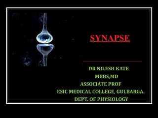
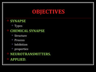
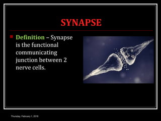
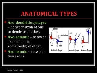
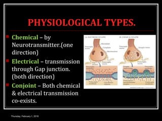
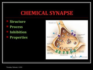
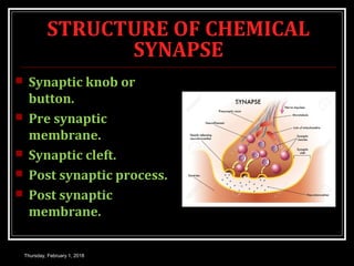
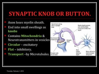
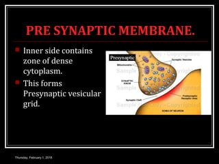
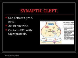
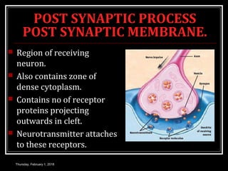
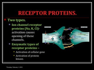
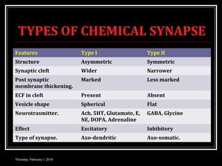
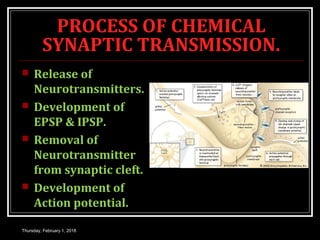
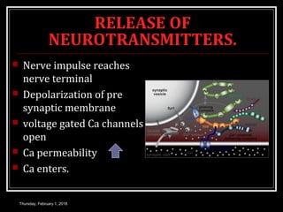
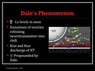
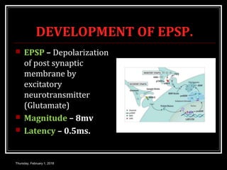
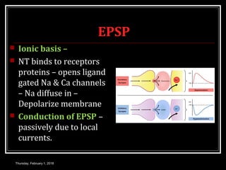
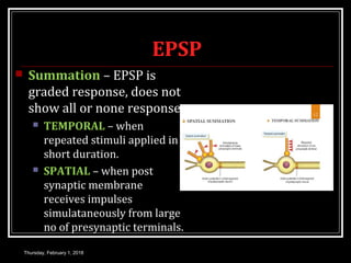
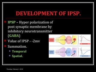
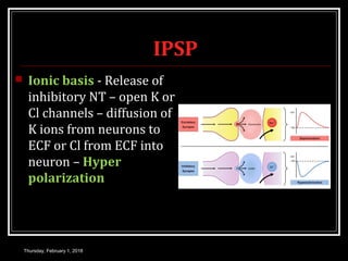
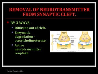
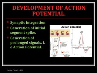
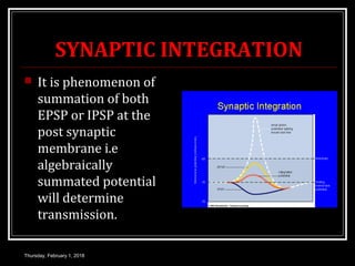
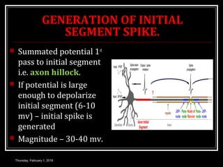
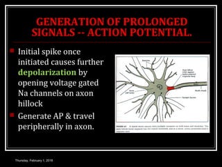
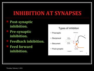
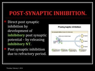
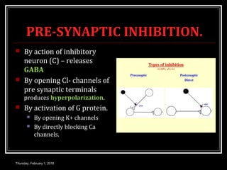
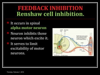
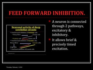
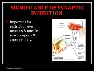
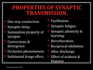
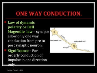
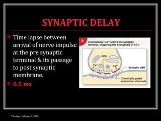
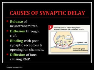
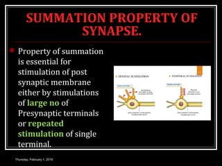
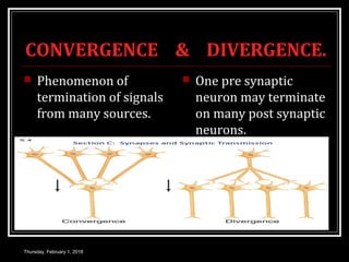
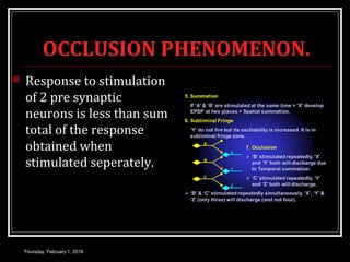

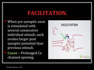
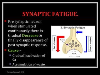
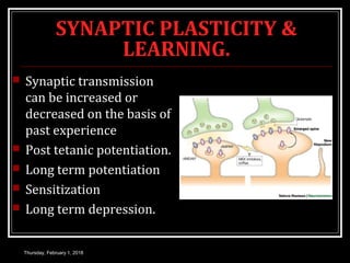
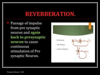
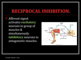
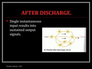

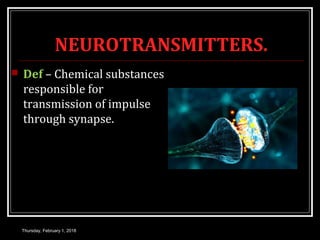
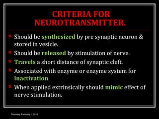
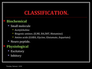
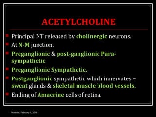
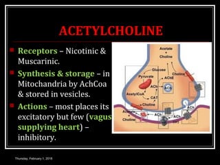
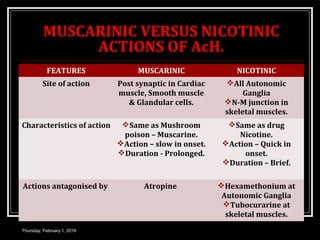
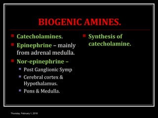
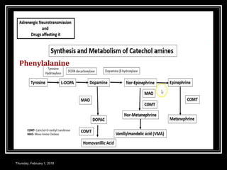
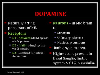
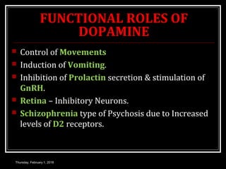
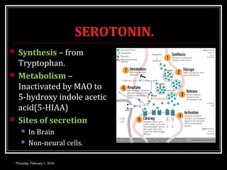
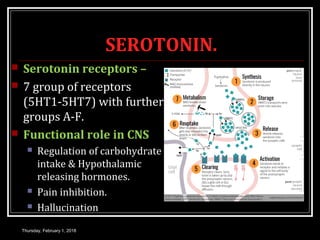
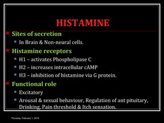
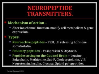


The document discusses the structure and function of chemical synapses. It begins by defining a synapse as the junction between two nerve cells. It then describes the key anatomical components of a chemical synapse, including the presynaptic knob, synaptic cleft, and postsynaptic membrane. It explains the process of neurotransmission, including the release of neurotransmitters into the synaptic cleft, their binding to receptors on the postsynaptic membrane, and the resulting postsynaptic potentials. The document also discusses inhibition at synapses, the properties of synaptic transmission, and examples of neurotransmitters.


![PERI-PROSTHETIC FRACTURE NAIL-PLATE CONSTRUCT [NPC].pptx](https://cdn.slidesharecdn.com/ss_thumbnails/drarunkumardrmohamedashrafperiprostheticfrasturenail-plateconstructnpc-260209164459-7e9d15a1-thumbnail.jpg?width=640&height=640&fit=bounds)


