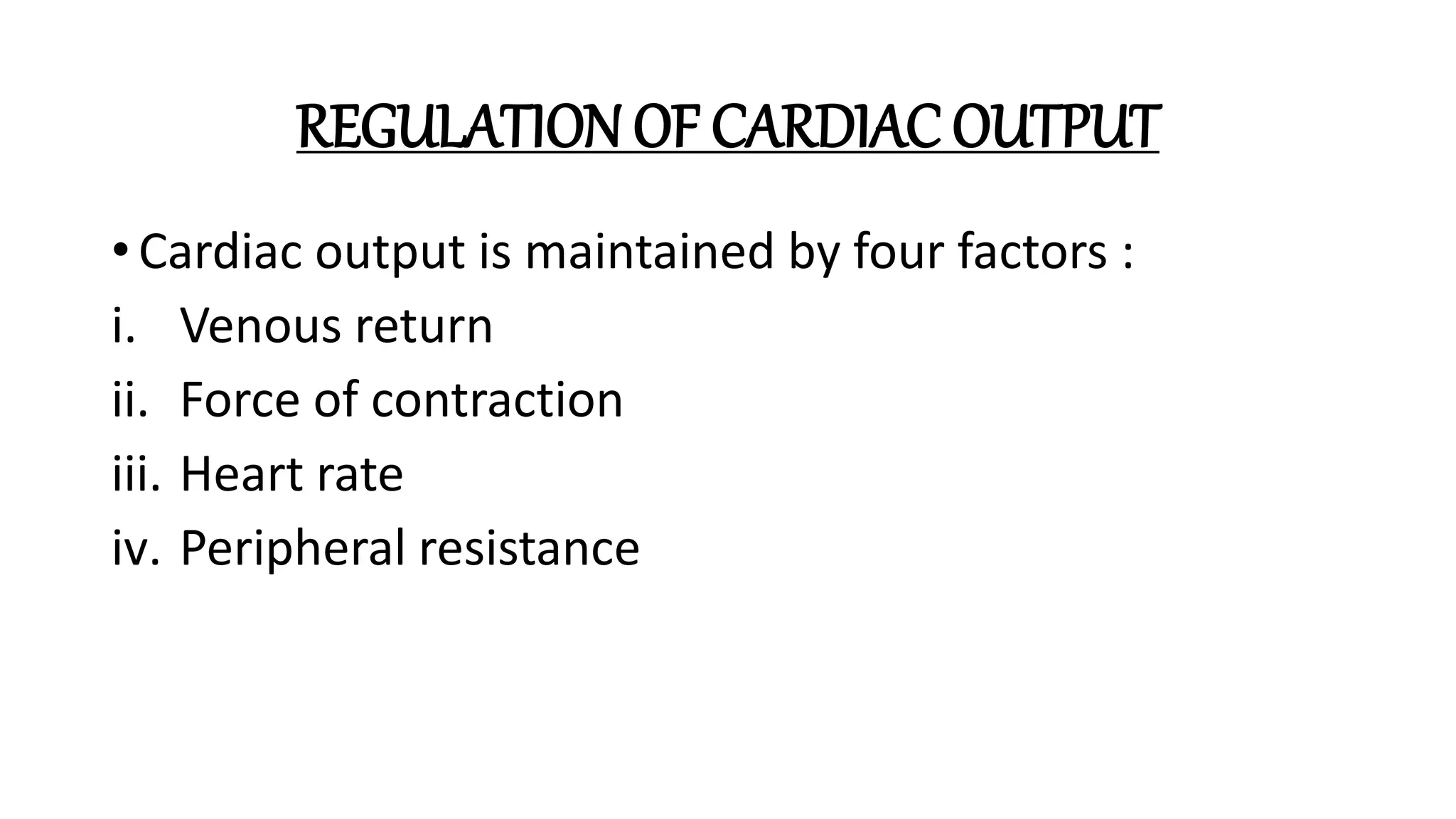
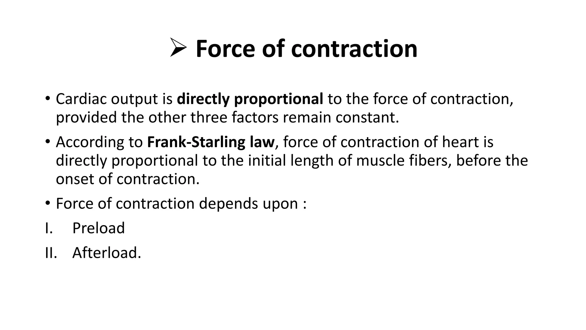
Cardiac Output and its
Regulation
Submitted By:- Sakshi Upadhyay
MPT Sports
INTRODUCTION
• Cardiac output is the amount of blood pumped from each ventricle.
Usually, it refers to left ventricular output through aorta.
• Cardiac output is the most important factor in cardiovascular system,
because rate of blood flow through different parts of the body
depends upon cardiac output.
• Cardiac Output(mL/minute) = Heart Rate(beats/minute) x Stoke
Volume(mL/beat)
• Cardiac Output is expressed in three ways:
1. Stoke Volume
2. Minute Volume
3. Cardiac Index
Stoke Volume
• The amount of blood pumped out by each ventricles during each
beat.
• Normal value :- 70mL (60-80mL) when the heart rate is normal (72
per minute).
• As the stroke volume increases the cardiac output also increases.
SV = EDV – ESV
• On average, Men have higher stroke volumes than women due to the
larger size of their hearts.
Minute Volume
• The amount of blood pumped out by each ventricle in 1 minutes.
• Minute volume = stoke volume x heart rate
• Normal value :- 5L/ventricle/min.
Cardiac Index
• The amount of blood pumped out per ventricle/minute/square meter
of the body surface area.
• Normal value :- 2.8+ 0.3L/sq. m of body surface area/min.
DISTRIBUTION
EJECTION FRACTION
• The fraction of end diastolic volume that is ejected out by each
ventricle.
• Normal ejection fraction is 60-65%.
CARDIAC RESERVE
• The maximum amount of blood that can be pumped out by heart
above the normal value.
• Plays an important role in increasing the cardiac output during the
conditions like exercise.
Factors affecting
Heart Rate (HR)
•Autonomic innervation
•Hormones
•Fitness levels
•Age
HR
Factors affecting
Stoke Volume
(SV)
Heart size
Fitness levels
Gender
Contractility
Duration of contraction
Preload (EDV)
Afterload (resistance)
SV=EDV-ESV
CO=HR X SV
REGULATION OF CARDIAC OUTPUT
• Cardiac output is maintained by four factors :
i. Venous return
ii. Force of contraction
iii. Heart rate
iv. Peripheral resistance
 VENOUS RETURN
• The amount of blood which is returned to heart from different parts of the body.
• Cardiac output is directly proportional to venous return.
• Factors of Venous Return :-
i. Respiratory pump - During inspiration, increase in venous return.
ii. Muscle pump - During exercise, increase in venous return.
iii. Gravity - When a person stands for a long period, gravity causes poling of
blood in the legs, which is called Venous Pooling. During standing, decrease in
venous return.
iv. Venous pressure - Driving force for venous return.
v. Sympathetic tone – vasomotor tone causes constriction of venules.
Venoconstriction pushes the blood towards heart.
 Force of contraction
• Cardiac output is directly proportional to the force of contraction,
provided the other three factors remain constant.
• According to Frank-Starling law, force of contraction of heart is
directly proportional to the initial length of muscle fibers, before the
onset of contraction.
• Force of contraction depends upon :
I. Preload
II. Afterload.
Preload
• Increase in ventricular pressure caused by filling of blood during
diastole.
• The force of contraction of heart and cardiac output are directly
proportional to preload.
Afterload
• The pressure increases in these two vessels. Now, the ventricles have
to work against this pressure for further ejection.
• Force of contraction of heart and cardiac output are inversely
proportional to afterload.
Heart Rate
• Cardiac output is directly proportional to heart rate.
• Change in heart rate does not alter the cardiac output.
• If increase in heart rate, cardiac output is increased and decrease in
heart rate, cardiac output is decreased.
Peripheral Resistance
• Peripheral resistance is the resistance offered to blood flow at the
peripheral blood vessels.
• The cardiac output is inversely proportional to peripheral resistance.
MEASUREMENT OF CARDIAC OUTPUT
• Methods based on Fick’s principle
• Indicator or dye dilution method
• Echocardiography
DIRECT FICK METHOD
• It states that the amount of a substance taken up by an organ per unit
of time is equal to the arterial level of the substance minus the
venous level (A-V difference) times the blood flow, i.e. amount of
substance taken per min = (A-V) difference of the substance x blood
flow / min.
• Blood flow/ min = amount of substance taken
(A- V) difference of the substance
INDICATOR (or DYE) DILUTION METHOD
• A amount of the dye is injected into an arm vein. It will first pass through the heart,
then the pulmonary circulation and finally will be evenly distributed in the blood
stream. Its mean concentration during its 1st passage through an artery can be
determined from successive samples of blood taken from the artery.
• The blood flow (F) is given by the formula:
• F = 1
c.t
• where,
I = total amount of dye injected
c = mean concentration of the dye
t = duration in sec of the 1st passage of the dye through the artery
ECHOCARDIOGRAPHY
• A non-invasive technique that
does not involve injection or
insertion of a catheter.
• In echo-cardiography, pulses of
ultrasonic waves, at a frequency of
2.25 mega-hertz (MHz), are emitted
from a transducer that also functions
as a receiver to detect waves reflected
back from various parts of the heart.
References
• Textbook of Physiology by AK Jain
• Essentials of Medical Physiology by K Sembulingam, Prema
Sembulingam
Cardiac Output and its Regulation.pptx

Cardiac Output and its Regulation.pptx

  • 1.
    Cardiac Output andits Regulation Submitted By:- Sakshi Upadhyay MPT Sports
  • 2.
    INTRODUCTION • Cardiac outputis the amount of blood pumped from each ventricle. Usually, it refers to left ventricular output through aorta. • Cardiac output is the most important factor in cardiovascular system, because rate of blood flow through different parts of the body depends upon cardiac output. • Cardiac Output(mL/minute) = Heart Rate(beats/minute) x Stoke Volume(mL/beat)
  • 3.
    • Cardiac Outputis expressed in three ways: 1. Stoke Volume 2. Minute Volume 3. Cardiac Index Stoke Volume • The amount of blood pumped out by each ventricles during each beat. • Normal value :- 70mL (60-80mL) when the heart rate is normal (72 per minute). • As the stroke volume increases the cardiac output also increases.
  • 4.
    SV = EDV– ESV • On average, Men have higher stroke volumes than women due to the larger size of their hearts. Minute Volume • The amount of blood pumped out by each ventricle in 1 minutes. • Minute volume = stoke volume x heart rate • Normal value :- 5L/ventricle/min.
  • 5.
    Cardiac Index • Theamount of blood pumped out per ventricle/minute/square meter of the body surface area. • Normal value :- 2.8+ 0.3L/sq. m of body surface area/min.
  • 6.
  • 7.
    EJECTION FRACTION • Thefraction of end diastolic volume that is ejected out by each ventricle. • Normal ejection fraction is 60-65%. CARDIAC RESERVE • The maximum amount of blood that can be pumped out by heart above the normal value. • Plays an important role in increasing the cardiac output during the conditions like exercise.
  • 8.
    Factors affecting Heart Rate(HR) •Autonomic innervation •Hormones •Fitness levels •Age HR Factors affecting Stoke Volume (SV) Heart size Fitness levels Gender Contractility Duration of contraction Preload (EDV) Afterload (resistance) SV=EDV-ESV CO=HR X SV
  • 9.
    REGULATION OF CARDIACOUTPUT • Cardiac output is maintained by four factors : i. Venous return ii. Force of contraction iii. Heart rate iv. Peripheral resistance
  • 10.
     VENOUS RETURN •The amount of blood which is returned to heart from different parts of the body. • Cardiac output is directly proportional to venous return. • Factors of Venous Return :- i. Respiratory pump - During inspiration, increase in venous return. ii. Muscle pump - During exercise, increase in venous return. iii. Gravity - When a person stands for a long period, gravity causes poling of blood in the legs, which is called Venous Pooling. During standing, decrease in venous return. iv. Venous pressure - Driving force for venous return. v. Sympathetic tone – vasomotor tone causes constriction of venules. Venoconstriction pushes the blood towards heart.
  • 11.
     Force ofcontraction • Cardiac output is directly proportional to the force of contraction, provided the other three factors remain constant. • According to Frank-Starling law, force of contraction of heart is directly proportional to the initial length of muscle fibers, before the onset of contraction. • Force of contraction depends upon : I. Preload II. Afterload.
  • 12.
    Preload • Increase inventricular pressure caused by filling of blood during diastole. • The force of contraction of heart and cardiac output are directly proportional to preload. Afterload • The pressure increases in these two vessels. Now, the ventricles have to work against this pressure for further ejection. • Force of contraction of heart and cardiac output are inversely proportional to afterload.
  • 13.
    Heart Rate • Cardiacoutput is directly proportional to heart rate. • Change in heart rate does not alter the cardiac output. • If increase in heart rate, cardiac output is increased and decrease in heart rate, cardiac output is decreased. Peripheral Resistance • Peripheral resistance is the resistance offered to blood flow at the peripheral blood vessels. • The cardiac output is inversely proportional to peripheral resistance.
  • 14.
    MEASUREMENT OF CARDIACOUTPUT • Methods based on Fick’s principle • Indicator or dye dilution method • Echocardiography
  • 15.
    DIRECT FICK METHOD •It states that the amount of a substance taken up by an organ per unit of time is equal to the arterial level of the substance minus the venous level (A-V difference) times the blood flow, i.e. amount of substance taken per min = (A-V) difference of the substance x blood flow / min. • Blood flow/ min = amount of substance taken (A- V) difference of the substance
  • 16.
    INDICATOR (or DYE)DILUTION METHOD • A amount of the dye is injected into an arm vein. It will first pass through the heart, then the pulmonary circulation and finally will be evenly distributed in the blood stream. Its mean concentration during its 1st passage through an artery can be determined from successive samples of blood taken from the artery. • The blood flow (F) is given by the formula: • F = 1 c.t • where, I = total amount of dye injected c = mean concentration of the dye t = duration in sec of the 1st passage of the dye through the artery
  • 17.
    ECHOCARDIOGRAPHY • A non-invasivetechnique that does not involve injection or insertion of a catheter. • In echo-cardiography, pulses of ultrasonic waves, at a frequency of 2.25 mega-hertz (MHz), are emitted from a transducer that also functions as a receiver to detect waves reflected back from various parts of the heart.
  • 18.
    References • Textbook ofPhysiology by AK Jain • Essentials of Medical Physiology by K Sembulingam, Prema Sembulingam