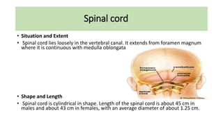
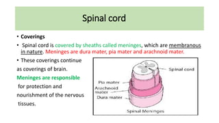
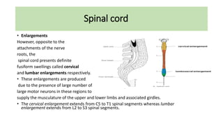
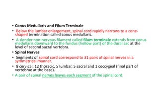
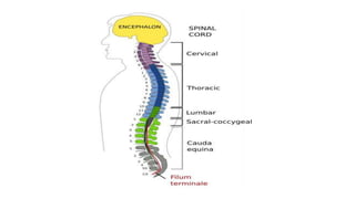
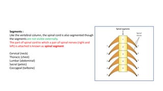
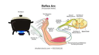
understanding spinal cord, its bransches, lesions, functions and anatomy.
hope to give you better knowledge of spinal cord by the end of it.
plese review ans comment for my future updates and corrections that iw ill be needing in this.
cerebrum, sulci and gyri of cerebrum, lobes of cerebrum, frontal lobe , parietal lobe, temporal lobe and occipital lobe, sulci and gyri presnet in each lobes, and the functional areas , of cerebrum, brodmann areas of cerebrum, borders and surfaces of cerebrum, insula,
Peripheral Nervous System, Audumbar MaliAudumbar Mali
Peripheral Nervous System,
Types of PNS,
Spinal nerves,
Types of neuron (3 basic types),
Plexus,
Cranial nerves,
Autonomic nervous system,
Structure of Neuron,
Human Anatomy and Physiology-I,
Syllabus As per PCI,
B. Pharm-I
The detail description about peripheral nervous system, neuron, its covering, types of neuron, synapses, spinal nerves, plexus, and more about cranial nerves at last not the least about somatic and autonomic nervous system. you may also find the information about types of peripheral nervous system in detail.
The spinal cord is a long, thin, tubular structure made up of nervous tissue, which extends from the medulla oblongata in the brainstem to the lumbar region of the vertebral column. It encloses the central canal of the spinal cord, which contains cerebrospinal fluid. The brain and spinal cord together make up the central nervous system (CNS). In humans, the spinal cord begins at the occipital bone, passing through the foramen magnum and entering the spinal canal at the beginning of the cervical vertebrae.
Largest part of hind brain.
Called “ silent area/Little Brain ”
Weight- 150 gms.
Cerebellar cortex is a large folded sheet, each fold is called Folium.
Connected to brain stem by 3 pairs of peduncles- Superior (Brachium conjunctiva), Middle (Brachium Pontis) & Inferior (Restiform body) peduncle.
The all the content in this profile is completed by the teachers, students as well as other health care peoples.
thank you, all the respected peoples, for giving the information to complete this presentation.
this information is free to use by anyone.
1.Organization of the Nervous System.pptxSana67616
CNS physiology general description and introduction
Brain and spinal cord
Brain is covered by meninges, CSF is fluid present in subarachnoid space
It has a protective function
Peripheral nervous system consists of cranial nerves and spinal nerves
There are 12 pairs of cranial and 31 pairs of spinal nerves Spinal cord is present in the vertebral canal of vertebral column, surrounded by the meninges and there is CSF present in subarachnoid space.
- It is almost cylindrical but in cervical and lower thoracic and lumbar regions it contains fusiform shaped enlargements called cervical enlargements and lumbar enlargements.
In the cervical segments there is more gray matter and also there is more gray matter in the lower thoracic and lumbar region. Cervical segments supply the upper limb i.e. it supplies more muscles, so more gray matter is present. Similarly the lumbar region supplies the lower limb so more gray matter is present in this region.
Just below the lumbar enlargements, spinal cord abruptly tapers, this tapering part is called conus medularis, and it gives attachment to a fibrous thread called filum terminale, which is attached to the back of coccvx.
In the spinal cord on the anterior surface there is anterior median fissure and on the posterior surface is posterior median sulcus. Fissure is much deeper than the sulcus.
Spinal nerves arise from the spinal cord and each spinal nerve has two roots.
1. Ventral (anterior root); which is motor and it contains both somatic and autonomic fibers.
2. Dorsal (Posterior root); is sensory and in the dorsal root there is dorsal root ganglia. It contains sensory neurons which give rise to cell fibers.
Title: Sense of Taste
Presenter: Dr. Faiza, Assistant Professor of Physiology
Qualifications:
MBBS (Best Graduate, AIMC Lahore)
FCPS Physiology
ICMT, CHPE, DHPE (STMU)
MPH (GC University, Faisalabad)
MBA (Virtual University of Pakistan)
Learning Objectives:
Describe the structure and function of taste buds.
Describe the relationship between the taste threshold and taste index of common substances.
Explain the chemical basis and signal transduction of taste perception for each type of primary taste sensation.
Recognize different abnormalities of taste perception and their causes.
Key Topics:
Significance of Taste Sensation:
Differentiation between pleasant and harmful food
Influence on behavior
Selection of food based on metabolic needs
Receptors of Taste:
Taste buds on the tongue
Influence of sense of smell, texture of food, and pain stimulation (e.g., by pepper)
Primary and Secondary Taste Sensations:
Primary taste sensations: Sweet, Sour, Salty, Bitter, Umami
Chemical basis and signal transduction mechanisms for each taste
Taste Threshold and Index:
Taste threshold values for Sweet (sucrose), Salty (NaCl), Sour (HCl), and Bitter (Quinine)
Taste index relationship: Inversely proportional to taste threshold
Taste Blindness:
Inability to taste certain substances, particularly thiourea compounds
Example: Phenylthiocarbamide
Structure and Function of Taste Buds:
Composition: Epithelial cells, Sustentacular/Supporting cells, Taste cells, Basal cells
Features: Taste pores, Taste hairs/microvilli, and Taste nerve fibers
Location of Taste Buds:
Found in papillae of the tongue (Fungiform, Circumvallate, Foliate)
Also present on the palate, tonsillar pillars, epiglottis, and proximal esophagus
Mechanism of Taste Stimulation:
Interaction of taste substances with receptors on microvilli
Signal transduction pathways for Umami, Sweet, Bitter, Sour, and Salty tastes
Taste Sensitivity and Adaptation:
Decrease in sensitivity with age
Rapid adaptation of taste sensation
Role of Saliva in Taste:
Dissolution of tastants to reach receptors
Washing away the stimulus
Taste Preferences and Aversions:
Mechanisms behind taste preference and aversion
Influence of receptors and neural pathways
Impact of Sensory Nerve Damage:
Degeneration of taste buds if the sensory nerve fiber is cut
Abnormalities of Taste Detection:
Conditions: Ageusia, Hypogeusia, Dysgeusia (parageusia)
Causes: Nerve damage, neurological disorders, infections, poor oral hygiene, adverse drug effects, deficiencies, aging, tobacco use, altered neurotransmitter levels
Neurotransmitters and Taste Threshold:
Effects of serotonin (5-HT) and norepinephrine (NE) on taste sensitivity
Supertasters:
25% of the population with heightened sensitivity to taste, especially bitterness
Increased number of fungiform papillae
The prostate is an exocrine gland of the male mammalian reproductive system
It is a walnut-sized gland that forms part of the male reproductive system and is located in front of the rectum and just below the urinary bladder
Function is to store and secrete a clear, slightly alkaline fluid that constitutes 10-30% of the volume of the seminal fluid that along with the spermatozoa, constitutes semen
A healthy human prostate measures (4cm-vertical, by 3cm-horizontal, 2cm ant-post ).
It surrounds the urethra just below the urinary bladder. It has anterior, median, posterior and two lateral lobes
It’s work is regulated by androgens which are responsible for male sex characteristics
Generalised disease of the prostate due to hormonal derangement which leads to non malignant enlargement of the gland (increase in the number of epithelial cells and stromal tissue)to cause compression of the urethra leading to symptoms (LUTS
Explore natural remedies for syphilis treatment in Singapore. Discover alternative therapies, herbal remedies, and lifestyle changes that may complement conventional treatments. Learn about holistic approaches to managing syphilis symptoms and supporting overall health.
Title: Sense of Smell
Presenter: Dr. Faiza, Assistant Professor of Physiology
Qualifications:
MBBS (Best Graduate, AIMC Lahore)
FCPS Physiology
ICMT, CHPE, DHPE (STMU)
MPH (GC University, Faisalabad)
MBA (Virtual University of Pakistan)
Learning Objectives:
Describe the primary categories of smells and the concept of odor blindness.
Explain the structure and location of the olfactory membrane and mucosa, including the types and roles of cells involved in olfaction.
Describe the pathway and mechanisms of olfactory signal transmission from the olfactory receptors to the brain.
Illustrate the biochemical cascade triggered by odorant binding to olfactory receptors, including the role of G-proteins and second messengers in generating an action potential.
Identify different types of olfactory disorders such as anosmia, hyposmia, hyperosmia, and dysosmia, including their potential causes.
Key Topics:
Olfactory Genes:
3% of the human genome accounts for olfactory genes.
400 genes for odorant receptors.
Olfactory Membrane:
Located in the superior part of the nasal cavity.
Medially: Folds downward along the superior septum.
Laterally: Folds over the superior turbinate and upper surface of the middle turbinate.
Total surface area: 5-10 square centimeters.
Olfactory Mucosa:
Olfactory Cells: Bipolar nerve cells derived from the CNS (100 million), with 4-25 olfactory cilia per cell.
Sustentacular Cells: Produce mucus and maintain ionic and molecular environment.
Basal Cells: Replace worn-out olfactory cells with an average lifespan of 1-2 months.
Bowman’s Gland: Secretes mucus.
Stimulation of Olfactory Cells:
Odorant dissolves in mucus and attaches to receptors on olfactory cilia.
Involves a cascade effect through G-proteins and second messengers, leading to depolarization and action potential generation in the olfactory nerve.
Quality of a Good Odorant:
Small (3-20 Carbon atoms), volatile, water-soluble, and lipid-soluble.
Facilitated by odorant-binding proteins in mucus.
Membrane Potential and Action Potential:
Resting membrane potential: -55mV.
Action potential frequency in the olfactory nerve increases with odorant strength.
Adaptation Towards the Sense of Smell:
Rapid adaptation within the first second, with further slow adaptation.
Psychological adaptation greater than receptor adaptation, involving feedback inhibition from the central nervous system.
Primary Sensations of Smell:
Camphoraceous, Musky, Floral, Pepperminty, Ethereal, Pungent, Putrid.
Odor Detection Threshold:
Examples: Hydrogen sulfide (0.0005 ppm), Methyl-mercaptan (0.002 ppm).
Some toxic substances are odorless at lethal concentrations.
Characteristics of Smell:
Odor blindness for single substances due to lack of appropriate receptor protein.
Behavioral and emotional influences of smell.
Transmission of Olfactory Signals:
From olfactory cells to glomeruli in the olfactory bulb, involving lateral inhibition.
Primitive, less old, and new olfactory systems with different path
Factory Supply Best Quality Pmk Oil CAS 28578–16–7 PMK Powder in Stockrebeccabio
Factory Supply Best Quality Pmk Oil CAS 28578–16–7 PMK Powder in Stock
Telegram: bmksupplier
signal: +85264872720
threema: TUD4A6YC
You can contact me on Telegram or Threema
Communicate promptly and reply
Free of customs clearance, Double Clearance 100% pass delivery to USA, Canada, Spain, Germany, Netherland, Poland, Italy, Sweden, UK, Czech Republic, Australia, Mexico, Russia, Ukraine, Kazakhstan.Door to door service
Hot Selling Organic intermediates
New Directions in Targeted Therapeutic Approaches for Older Adults With Mantl...i3 Health
i3 Health is pleased to make the speaker slides from this activity available for use as a non-accredited self-study or teaching resource.
This slide deck presented by Dr. Kami Maddocks, Professor-Clinical in the Division of Hematology and
Associate Division Director for Ambulatory Operations
The Ohio State University Comprehensive Cancer Center, will provide insight into new directions in targeted therapeutic approaches for older adults with mantle cell lymphoma.
STATEMENT OF NEED
Mantle cell lymphoma (MCL) is a rare, aggressive B-cell non-Hodgkin lymphoma (NHL) accounting for 5% to 7% of all lymphomas. Its prognosis ranges from indolent disease that does not require treatment for years to very aggressive disease, which is associated with poor survival (Silkenstedt et al, 2021). Typically, MCL is diagnosed at advanced stage and in older patients who cannot tolerate intensive therapy (NCCN, 2022). Although recent advances have slightly increased remission rates, recurrence and relapse remain very common, leading to a median overall survival between 3 and 6 years (LLS, 2021). Though there are several effective options, progress is still needed towards establishing an accepted frontline approach for MCL (Castellino et al, 2022). Treatment selection and management of MCL are complicated by the heterogeneity of prognosis, advanced age and comorbidities of patients, and lack of an established standard approach for treatment, making it vital that clinicians be familiar with the latest research and advances in this area. In this activity chaired by Michael Wang, MD, Professor in the Department of Lymphoma & Myeloma at MD Anderson Cancer Center, expert faculty will discuss prognostic factors informing treatment, the promising results of recent trials in new therapeutic approaches, and the implications of treatment resistance in therapeutic selection for MCL.
Target Audience
Hematology/oncology fellows, attending faculty, and other health care professionals involved in the treatment of patients with mantle cell lymphoma (MCL).
Learning Objectives
1.) Identify clinical and biological prognostic factors that can guide treatment decision making for older adults with MCL
2.) Evaluate emerging data on targeted therapeutic approaches for treatment-naive and relapsed/refractory MCL and their applicability to older adults
3.) Assess mechanisms of resistance to targeted therapies for MCL and their implications for treatment selection
These simplified slides by Dr. Sidra Arshad present an overview of the non-respiratory functions of the respiratory tract.
Learning objectives:
1. Enlist the non-respiratory functions of the respiratory tract
2. Briefly explain how these functions are carried out
3. Discuss the significance of dead space
4. Differentiate between minute ventilation and alveolar ventilation
5. Describe the cough and sneeze reflexes
Study Resources:
1. Chapter 39, Guyton and Hall Textbook of Medical Physiology, 14th edition
2. Chapter 34, Ganong’s Review of Medical Physiology, 26th edition
3. Chapter 17, Human Physiology by Lauralee Sherwood, 9th edition
4. Non-respiratory functions of the lungs https://academic.oup.com/bjaed/article/13/3/98/278874
Ethanol (CH3CH2OH), or beverage alcohol, is a two-carbon alcohol
that is rapidly distributed in the body and brain. Ethanol alters many
neurochemical systems and has rewarding and addictive properties. It
is the oldest recreational drug and likely contributes to more morbidity,
mortality, and public health costs than all illicit drugs combined. The
5th edition of the Diagnostic and Statistical Manual of Mental Disorders
(DSM-5) integrates alcohol abuse and alcohol dependence into a single
disorder called alcohol use disorder (AUD), with mild, moderate,
and severe subclassifications (American Psychiatric Association, 2013).
In the DSM-5, all types of substance abuse and dependence have been
combined into a single substance use disorder (SUD) on a continuum
from mild to severe. A diagnosis of AUD requires that at least two of
the 11 DSM-5 behaviors be present within a 12-month period (mild
AUD: 2–3 criteria; moderate AUD: 4–5 criteria; severe AUD: 6–11 criteria).
The four main behavioral effects of AUD are impaired control over
drinking, negative social consequences, risky use, and altered physiological
effects (tolerance, withdrawal). This chapter presents an overview
of the prevalence and harmful consequences of AUD in the U.S.,
the systemic nature of the disease, neurocircuitry and stages of AUD,
comorbidities, fetal alcohol spectrum disorders, genetic risk factors, and
pharmacotherapies for AUD.
Tom Selleck Health: A Comprehensive Look at the Iconic Actor’s Wellness Journeygreendigital
Tom Selleck, an enduring figure in Hollywood. has captivated audiences for decades with his rugged charm, iconic moustache. and memorable roles in television and film. From his breakout role as Thomas Magnum in Magnum P.I. to his current portrayal of Frank Reagan in Blue Bloods. Selleck's career has spanned over 50 years. But beyond his professional achievements. fans have often been curious about Tom Selleck Health. especially as he has aged in the public eye.
Follow us on: Pinterest
Introduction
Many have been interested in Tom Selleck health. not only because of his enduring presence on screen but also because of the challenges. and lifestyle choices he has faced and made over the years. This article delves into the various aspects of Tom Selleck health. exploring his fitness regimen, diet, mental health. and the challenges he has encountered as he ages. We'll look at how he maintains his well-being. the health issues he has faced, and his approach to ageing .
Early Life and Career
Childhood and Athletic Beginnings
Tom Selleck was born on January 29, 1945, in Detroit, Michigan, and grew up in Sherman Oaks, California. From an early age, he was involved in sports, particularly basketball. which played a significant role in his physical development. His athletic pursuits continued into college. where he attended the University of Southern California (USC) on a basketball scholarship. This early involvement in sports laid a strong foundation for his physical health and disciplined lifestyle.
Transition to Acting
Selleck's transition from an athlete to an actor came with its physical demands. His first significant role in "Magnum P.I." required him to perform various stunts and maintain a fit appearance. This role, which he played from 1980 to 1988. necessitated a rigorous fitness routine to meet the show's demands. setting the stage for his long-term commitment to health and wellness.
Fitness Regimen
Workout Routine
Tom Selleck health and fitness regimen has evolved. adapting to his changing roles and age. During his "Magnum, P.I." days. Selleck's workouts were intense and focused on building and maintaining muscle mass. His routine included weightlifting, cardiovascular exercises. and specific training for the stunts he performed on the show.
Selleck adjusted his fitness routine as he aged to suit his body's needs. Today, his workouts focus on maintaining flexibility, strength, and cardiovascular health. He incorporates low-impact exercises such as swimming, walking, and light weightlifting. This balanced approach helps him stay fit without putting undue strain on his joints and muscles.
Importance of Flexibility and Mobility
In recent years, Selleck has emphasized the importance of flexibility and mobility in his fitness regimen. Understanding the natural decline in muscle mass and joint flexibility with age. he includes stretching and yoga in his routine. These practices help prevent injuries, improve posture, and maintain mobilit
These lecture slides, by Dr Sidra Arshad, offer a quick overview of physiological basis of a normal electrocardiogram.
Learning objectives:
1. Define an electrocardiogram (ECG) and electrocardiography
2. Describe how dipoles generated by the heart produce the waveforms of the ECG
3. Describe the components of a normal electrocardiogram of a typical bipolar leads (limb II)
4. Differentiate between intervals and segments
5. Enlist some common indications for obtaining an ECG
Study Resources:
1. Chapter 11, Guyton and Hall Textbook of Medical Physiology, 14th edition
2. Chapter 9, Human Physiology - From Cells to Systems, Lauralee Sherwood, 9th edition
3. Chapter 29, Ganong’s Review of Medical Physiology, 26th edition
4. Electrocardiogram, StatPearls - https://www.ncbi.nlm.nih.gov/books/NBK549803/
5. ECG in Medical Practice by ABM Abdullah, 4th edition
6. ECG Basics, http://www.nataliescasebook.com/tag/e-c-g-basics
2. Spinal cord
• Situation and Extent
• Spinal cord lies loosely in the vertebral canal. It extends from foramen magnum
where it is continuous with medulla oblongata
• Shape and Length
• Spinal cord is cylindrical in shape. Length of the spinal cord is about 45 cm in
males and about 43 cm in females, with an average diameter of about 1.25 cm.
3. Spinal cord
• Coverings
• Spinal cord is covered by sheaths called meninges, which are membranous
in nature. Meninges are dura mater, pia mater and arachnoid mater.
• These coverings continue
as coverings of brain.
Meninges are responsible
for protection and
nourishment of the nervous
tissues.
4. Spinal cord
• Enlargements
However, opposite to the
attachments of the nerve
roots, the
spinal cord presents definite
fusiform swellings called cervical
and lumbar enlargements respectively.
• These enlargements are produced
due to the presence of large number of
large motor neurons in these regions to
supply the musculature of the upper and lower limbs and associated girdles.
• The cervical enlargement extends from C5 to T1 spinal segments whereas lumbar
enlargement extends from L2 to S3 spinal segments.
5. • Conus Medullaris and Filum Terminale
• Below the lumbar enlargement, spinal cord rapidly narrows to a cone-
shaped termination called conus medullaris.
• A slender non-nervous filament called filum terminale extends from conus
medullaris downward to the fundus (hollow part) of the dural sac at the
level of second sacral vertebra.
• Spinal Nerves
• Segments of spinal cord correspond to 31 pairs of spinal nerves in a
symmetrical manner.
• 8 cervical, 12 thoracic, 5 lumbar, 5 sacral and 1 coccygeal (final part of
vertebrae at the base).
• A pair of spinal nerves leaves each segment of the spinal cord.
6.
7. Segments :
Like the vertebral column, the spinal cord is also segmented though
the segments are not visible externally.
The part of spinal cord to which a pair of spinal nerves (right and
left) is attached is known as spinal segment
Cervical (neck)
Thoracic (chest)
Lumbar (abdominal)
Sacral (pelvic)
Coccygeal (tailbone)
8. Spinal cord
• Internal Structure of the Spinal Cord
• The cross-section of the spinal cord shows that it consists of an inner
core of grey matter, and a peripheral zone of white matter.
9. • Grey Matter
• In cross-section of cord the grey matter is seen as H-shaped (or butterfly-
shaped) fluted column, extending throughout the length of the spinal cord.
• It is divided into symmetrical right and left comma-shaped masses which
are connected across the midline by a transverse grey commissure.
• The central canal of the cord passes through the centre of grey
commissure.
• The canal is surrounded by substantia gelatinosa centralis.
• The lateral comma-shaped mass of grey matter is further divided by a
transverse grey commissure into a narrow elongated posterior horn, and a
broad anterior horn.
10. Transverse sections of the spinal cord at
different levels showing the arrangement of
grey and white matters.
The amount of the grey matter seen at a particular
level is well correlated with the mass of tissue it
supplies.
• It is, therefore, maximum in the regions of
cervical and lumbar enlargements, which supply
the limbs and their associated girdles.
• The horns are thus largest in the regions
cervical and lumbar enlargements.
• The amount of white matter in the spinal
cord undergoes progressive increase from
below upwards.
11. • Structure of the grey matter
• Like in other regions of the CNS, the grey matter of spinal cord consists of
(a) nerve cells, (b) neuroglia (glial cells), and (c) blood vessels.
• Neurons in the grey matter of the spinal cord
• The nerve cells in the grey matter of spinal cord are multi-polar and can be
classified into the following two ways:
• Structural classification
• • Golgi type I, having long axons, which leave the grey matter and either
join the anterior nerve roots or form the nerve tracts.
• • Golgi type II, having short axons, which do not leave the grey matter and
remain intrasegmental or intersegmental in position.
12. • Functional classification
• • Motor neurons: These are present in the anterior and lateral horns.
• Types of motor neurons in the anterior grey columns:
• 1. Alpha (α) neurons: They are large multipolar cells (25 µm or more in diameter) and
supply the extrafusal skeletal muscle fibres.
• 2. Gamma (γ) neurons: They are small multipolar cells (15–25 µm in diameter) and
supply the intrafusal muscle fibres of the neuromuscular spindles in skeletal muscles.
• Sensory neurons: These are present in the posterior horn and involved in relay of
sensory information to the different parts of the brain, forming ascending tracts; or to
the other segments of spinal cord forming intersegmental tracts.
• Interneurons: These are small neurons present throughout the grey matter of the spinal
cord. They connect different types of neurons, hence also called association neurons.
These are either inhibitory or excitatory, and concerned with integration of segmental
activities.
13. • White Matter
• The white matter of the spinal cord surrounds the central ‘H-shaped mass of grey matter, and mainly
consists of nerve fibers, the large proportion of them being myelinated, give it a white appearance’.
• Types of fibers in the white matter
• Functionally, the fibers in the white matter of spinal cord are divided into following three types:
• 1. Sensory fibers: These include:
• – The central processes of primary sensory neurons of the posterior root ganglia which enter the spinal
cord and ascend or descend for varying lengths, and
• – The ascending fibers from the nuclei of spinal grey columns that convey sensory modalities to the higher
centers.
• 2. Motor fibers: These include:
• – The descending fibers from higher centers (supraspinal levels) to the spinal cord, and
• – The nerve fibers of anterior and lateral horn cells that go to the motor roots of the spinal nerves.
• 3. Association fibers: These fibers originate and end within the spinal cord, interconnecting the neurons of
the same segment or of different segmental levels.
14. Function of spinal cord
• The central nervous system (CNS) controls most functions of the body
and mind. It consists of two parts: the brain and the spinal cord.
• The brain is the center of our thoughts, the interpreter of our external
environment, and the origin of control over body movement. Like a
central computer, it interprets information from our eyes (sight),
ears (sound), nose (smell), tongue (taste), and skin (touch), as well
as from internal organs such as the stomach.
• The spinal cord is the highway for communication between the body
and the brain. When the spinal cord is injured, the exchange of
information between the brain and other parts of the body is
disrupted.
15. • The brain is the command center for your body, and the spinal cord is the
pathway for messages sent by the brain to the body and from the body to the
brain.
• the peripheral nervous system is the network of nerves strands that branch off
from the left and right sides of the spinal cord through openings between each
vertebra on the spinal canal. These nerve pairs spread throughout your body to
deliver commands from your brain and spinal cord to and from parts of your body
• The spinal cord is a complex cylinder of nerves that starts at the base of your
brain and runs down the vertebral canal to the backbone. It is part of the body’s
collection of nerves, called the central nervous system, along with the brain
• In each of the spinal cord’s many segments lives a pair of roots that are made up
of nerve fibers. These roots are referred to as the dorsal (which is towards the
back) and the ventral (which is away from the back) roots.
16. • We depend on the spinal column to be the main support of our body. It allows us to
stand upright, bend, and twist while protecting the spinal cord from injury. If the spinal
cord is injured, it often causes issues like:
• Permanent changes in the body’s strength;
• Loss of sensation; and
• Loss of motor control or other functions.
• The spinal cord conducts sensory information from the peripheral nervous system (both
somatic and autonomic) to the brain
• conducts motor information from the brain to our various effectors
• skeletal muscles
• cardiac muscle
• smooth muscle
• glands
• So it serves as a minor reflex center
17. Functions of spinal cord
• The Major Functions of the Spinal Cord
• Electrochemical communication. Electrical currents travel up and down the
spinal cord and across nerves, sending signals which allow different segments of
the body to communicate with the brain.
• Walking. While a person walks, a collection of muscle groups in the legs are
constantly contracting and relaxing. The action of taking step after step may seem
incredibly simple to us since we have been doing it all of our lives, but there are
actually a lot of factors that have to be coordinated properly to allow this to
happen. This central pattern generators in the spinal cord are made up of
neurons which send signals to the muscles in the legs, making them relax or
contract, and produce the alternating movements which occur when a person
walks.
• Reflexes. Reflexes are involuntary responses resulting from stimuli involving the
brain, spinal cord, and nerves of the peripheral nervous system.
18. Tracts of spinal cord
• Tracts of the Spinal Cord
• The tracts are defined as collections of nerve fibres within the central nervous system,
which have same origin, course and termination. They are sometimes referred to
as fasciculus (= bundle) or lemniscus (= ribbon).
• The spinal cord has numerous groups of nerve fibers going towards and coming from the
brain. These have been collectively called the ascending and descending tracts of the
spinal cord, respectively. The tracts are responsible for carrying sensory and motor
stimuli to and from the periphery (respectively).
• Ascending tracts of the spinal cord
• Growing up, the impression was given that there were only five senses that humans can detect. These
were sight, smell, sound, taste, and touch. However, it is clear that touch can be further expanded to include pain,
thermal changes, pressure, light (crude) touch, vibration, two-point discrimination, and proprioception. Sight,
sound, smell, and taste are special afferent stimuli that are conveyed through their respective cranial nerves.
However, the other tactile modalities are transmitted through the ascending tracts of the spinal cord. There are
eight known ascending tracts conveying a variety of sensory stimuli that are discussed below.
19. TRACTS IN SPINAL CORD
• Groups of nerve fibers passing through spinal cord are known as tracts of the spinal cord. The
spinal tracts are divided into two main groups.
• They are: 1. Short tracts 2. Long tracts.
• 1. Short Tracts
• Fibers of the short tracts connect different parts of spinal cord itself. Short tracts are of two types:
• i. Association or intrinsic tracts, which connect adjacent segments of spinal cord on the same
side
• ii. Commissural tracts, which connect opposite halves of same segment of spinal cord.
• 2. Long Tracts
• Long tracts of spinal cord, which are also called projection tracts, connect the spinal cord with
other parts of central nervous system. Long tracts are of two types:
• i. Ascending tracts, which carry sensory impulses from the spinal cord to brain
• ii. Descending tracts, which carry motor impulses from brain to the spinal cord.
20. Naming of tracts
• Transverse section of spinal cord at mid-cervical (neck) region showing main
descending (motor) tracts in the left half and ascending (sensory) tracts in the
right half of the spinal cord.
• The tracts are named after the names of masses of grey matter connected by
them.
• The name usually consists of two components (or terms), the first term denotes
the origin and second the termination of the tract.
• For example,
• a tract arising in cerebral cortex and terminating in the spinal cord is called
corticospinal tract, similarly a tract arising in the spinal cord and terminating in
the thalamus is called spinothalamic tract.
• he lateral spinothalamic tract refers to a cluster of nerve fibers traveling within
the lateral funiculus of the spinal cord, which originated within the cord and will
terminate within the thalamus.
21.
22.
23. Function of ascending tract
• Ascending Tracts
• The ascending tracts conduct the impulses from the periphery to the
brain through the cord.
• The important ascending tracts fall into the following three types:
• 1. Those concerned with pain and temperature sensations and crude
touch, e.g. lateral and anterior spinothalamic tracts.
• 2. Those concerned with fine touch and conscious proprioceptive
sensations, e.g. fasciculus gracilis and fasciculus cuneatus.
• 3. Those concerned with unconscious proprioception and muscular
coordination, e.g. anterior and posterior spinocerebellar tracts.
24.
25. Reflex action and reflexes
• DEFINITION AND SIGNIFICANCE OF REFLEXES
• Reflex: A reflex, or reflex action, is an involuntary and nearly
instantaneous movement in response to a stimulus
• Reflex activity is the response to a peripheral nervous stimulation
that occurs without our consciousness. It is a type of protective
mechanism and it protects the body from irreparable damages.
• For example, when hand is placed on a hot object, it is withdrawn
immediately. When a bright light is thrown into the eyes, eyelids are
closed and pupil is constricted to prevent the damage of retina by
entrance of excessive light into the eyes.
26. Reflex arc
• REFLEX ARC
• Reflex arc is the anatomical nervous pathway for a reflex action. A simple reflex arc includes five
components.
• 1. Receptor: Receptor is the end organ, which receives the stimulus. When receptor is stimulated, impulses
are generated in afferent nerve.
• 2. Afferent Nerve: Afferent or sensory nerve transmits sensory impulses from the receptor to center.
• 3. Center: Center receives the sensory impulses via afferent nerve fibers and in turn, it generates
appropriate motor impulses. Center is located in the brain or spinal cord.
• 4. Efferent Nerve: Efferent or motor nerve transmits motor impulses from the center to the effector organ.
• 5. Effector Organ: Effector organ is the structure such as muscle or gland where the activity occurs in
response to stimulus.
• (Afferent and efferent nerve fibers may be connected directly to the center. In some places, one or more
neurons are interposed between these nerve fibers and the center. Such neurons are called(connector
neurons or internuncial neurons or interneurons.)
27.
28.
29. Reflexes
• CLASSIFICATION OF REFLEXES
• Reflexes are classified by six different methods depending upon various factors.
• 1. DEPENDING UPON WHETHER INBORN OR ACQUIRED REFLEXES
• i. Inborn Reflexes or Unconditioned Reflexes Unconditioned reflexes are the
natural reflexes, which are present since the time of birth, hence the name
inborn reflexes. Such reflexes do not require previous learning, training or
conditioning. Best example is the secretion of saliva when a drop of honey is kept
in the mouth of a newborn baby for the first time. The baby does not know the
taste of honey, but still saliva is secreted.
• ii. Acquired Reflexes or Conditioned Reflexes Conditioned or acquired reflexes
are the reflexes that are developed after conditioning or training. These reflexes
are not inborn but, acquired after birth. Such reflexes need previous learning,
training or conditioning. Example is the secretion of saliva by sight, smell, thought
or hearing of a known edible substance.
30. • 2. DEPENDING UPON SITUATION – ANATOMICAL CLASSIFICATION
• In this method, reflexes are classified depending upon the situation of the center.
• i. Cerebellar Reflexes: Cerebellar reflexes are the reflexes which have their center in cerebellum.
• ii. Cortical Reflexes: Cortical reflexes are the reflexes that have their center in cerebral cortex.
• iii. Midbrain Reflexes: Midbrain reflexes are the reflexes which have their center in midbrain.
• iv. Bulbar or Medullary Reflexes: Bulbar or medullary reflexes are the reflexes which have their
center in medulla oblongata.
• v. Spinal Reflexes: Reflexes having their center in the spinal cord are called spinal reflexes.
• Depending upon the segments involved, spinal reflexes are divided into three groups:
• a. Segmental spinal reflexes
• b. Intrasegmental spinal reflexes
• c. Suprasegmental spinal reflexes.
31. • DEPENDING UPON PURPOSE – PHYSIOLOGICAL CLASSIFICATION
• In this method, reflexes are classified depending upon the purpose
(functional significance).
• i. Protective Reflexes or Flexor Reflexes
• Protective reflexes are the reflexes which protect the body from nociceptic
(harmful) stimuli. These reflexes are also called withdrawal reflexes or
flexor reflexes. Protective reflexes involve flexion at different joints hence
the name flexor reflexes.
• ii. Antigravity Reflexes or Extensor Reflexes
• Antigravity reflexes are the reflexes that protect the body against
gravitational force. These reflexes are also called the extensor reflexes
because, the extensor muscles contract during these reflexes resulting in
extension at joints.
32. • 4. DEPENDING UPON THE NUMBER OF SYNAPSE
• Depending upon the number of synapse in reflex arc, reflexes are classified
into two types:
• i. Monosynaptic Reflexes:
• Reflexes having only one synapse in the reflex arc are called monosynaptic
reflexes. Knee jerk is the best example for monosynaptic reflex and it is
elicited due to the stimulation of muscle spindle.
• ii. Polysynaptic Reflexes
• Reflexes having more than one synapse in the reflex arc are called
polysynaptic reflexes. Flexor reflexes (withdrawal reflexes) are the
polysynaptic reflexes
33. • 5. DEPENDING UPON WHETHER SOMATIC OR VISCERAL REFLEXES
• i. Somatic Reflexes
• Somatic reflexes are the reflexes, for which the reflex arc is formed by somatic
nerve fibers. These reflexes involve the participation of skeletal muscles. And
there may be flexion or extension at different joints during these reflexes.
• ii. Visceral or Autonomic Reflexes
• Visceral or autonomic reflexes are the reflexes, for which at least a part of reflex
arc is formed by autonomic nerve fibers. These reflexes involve participation of
smooth muscle or cardiac muscle. Visceral reflexes include pupillary reflexes,
gastrointestinal reflexes, cardiovascular reflexes, respiratory reflexes, etc.
• Some reflexes like swallowing, coughing or vomiting are considered as visceral
reflexes. However, these reflexes involve some participation of skeletal muscles
also.
34. • 6. DEPENDING UPON CLINICAL BASIS
• Depending upon the clinical basis, reflexes are classified into four types:
• i. Superficial reflexes ii. Deep reflexes iii. Visceral reflexes
iv. Pathological reflexes.
• SUPERFICIAL REFLEXES
• Superficial reflexes are the reflexes, which are elicited from the surface of
the body. Superficial reflexes are of two types:
• mucus membrane reflexes and skin reflexes.
• 1. MUCOUS MEMBRANE REFLEXES Mucous membrane reflexes arise from
the mucus membrane
35. Superficial mucous membrane reflexes
REFLEX STIMULUS RESPONSE AFFERENT
NERVE
CENTER EFFERENT
NERVE
Corneal reflex Irritation of
cornea
Blinking eye
(eyelid closure)
5th cranial nerve pons 7th cranial nerve
Conjunctival
reflex
Irritation of
conjunctiva
Blinking of eye 5th cranial nerve pons 7th cranial nerve
Nasal reflex Irritation of
nasal mucus
membrane
sneezing 5th cranial nerve Motor nucleus
of V cranial
nerve
10th cranial
nerve and upper
cervical nerves
Superficial mucous membrane reflexes
36. • 2. CUTANEOUS REFLEXES OR SKIN REFLEXES
• Cutaneous reflexes are elicited from skin by the stimulation of cutaneous receptors. Details of these reflexes are given in Table
• DEEP REFLEXES
• Deep reflexes are elicited from deeper structures beneath the skin like tendon. These reflexes are otherwise known as tendon
reflexes. Details of these are given in Table
• VISCERAL REFLEXES
• Visceral reflexes are the reflexes arising from pupil and visceral organs.
• Following are the visceral reflexes:
• 1. Pupillary reflexes 2. Oculocardiac reflex 3. Carotid sinus reflex.
• PUPILLARY REFLEXES: Pupillary reflexes are the reflexes in which, the size of pupil is altered.
• EXAMPLE OF PUPILLARY REFLEX IS:
• Light Reflex When retina of the eye is stimulated by a sudden flash of light, constriction of pupil occurs. It is called light reflex.
• ii. Accommodation Reflex While eyes are fixed on a distant object and if another object is brought in front of the eye (near the
eye) the vision shifts form far object to near object. During that time some changes occur in the eyes. Changes during
accommodation reflex are: a. Constriction of pupil b. Convergence of eyeball c. Increase in anterior curvature of lens.
37. Superficial cutaneous reflexes
Reflex Stimulus Response Central spinal
segment involved
Scapular (shoulder
blade)reflex
Irritation of skin at
the interscapular
space
Contraction of
scapular muscles
and drawing in of
scapula
C5 to T1
Lower abdominal
reflex
Stroking the
abdominal wall at
umbilical and iliac
level
contraction of
abdominal muscle
and movement of
umbilicus towards
the site of stroke
T10 to T12
Gluteal reflex Stroking the skin
over glutei
Contraction of glutei L4 to S1,2
38. Deep reflexes
REFLEX STIMULUS RESPONSE Center – spinal segments
involved
Jaw jerk Tapping middle of the
chin with slightly opened
mouth
Closure of mouth Pons – V cranial nerve
Biceps jerk Percussion of biceps
tendon
Flexion of forearm C5, C6
Triceps jerk Percussion of triceps
tendon
Extension of forearm C6 to C8
39. • OCULOCARDIAC REFLEX: Oculocardiac reflex is the reflex, in which
heart rate decreases due to the pressure applied over eyeball.
• CAROTID SINUS REFLEX: Carotid sinus reflex is the decrease in heart
rate and blood pressure caused by pressure over carotid sinus in neck
due to tight collar.
Carotid artery supplies blood to heart, neck & face
Carotid sinus- consist of baroreceptor, which monitor blood pressure.
40. PROPERTIES OF REFLEXES
• 1. ONE WAY CONDUCTION (BELL-MAGENDIE LAW) During any reflex activity, impulses
are transmitted in only one direction through the reflex arc as per Bell-Magendie law.
The impulses pass from receptors to center and then from center to effector organ.
• 2. REACTION TIME Reaction time is the time interval between application of stimulus
and the onset of reflex. It depends upon the length of afferent and efferent nerve fibers,
velocity of impulse through these fibers and central delay. Central delay is the delay at
the synapse. It is also called synaptic delay.
• 3. SUMMATION : Summation in reflex action is of two types:
• i. Spatial Summation When two afferent nerve fibers supplying a muscle are stimulated
separately with subliminal stimulus, there is no response. But the muscle contracts when
both the nerve fibers are stimulated together with same strength of stimulus. It is called
spatial summation.
• ii. Temporal Summation When one nerve fiber is stimulated repeatedly with subliminal
stimuli, these stimuli are summed up to give response in the muscle. It is called temporal
summation.
41. • RECRUITMENT
• Recruitment is defined as the successive activation of additional
motor units with pr When an excitatory nerve is stimulated for a long
time, there is a gradual increase in the response of reflex activities. It
is due to the activation of more and more motor neurons.
Recruitment is similar to the effect of temporal summation.
• Indefinite increase in response does not produce unlimited
recruitment. A plateau is reached. Thus, there is a limit to the number
of motor neurons, which are recruited. So, beyond certain limit, the
prolongation of stimulation does not increase the response
Pogressive increase in force of muscular contraction
42. • REBOUND PHENOMENON
• Reflex activities can be forcefully inhibited for some time. But, when
the inhibition is suddenly removed, the reflex activity becomes more
forceful than before inhibition. It is called rebound phenomenon.
Reason for this state of over excitation is not known
• FATIGUE
• When a reflex activity is continuously elicited for a long time, the
response is reduced slowly and at one stage, the response does not
occur. This type of failure to give response to the stimulus is called
fatigue. Center or the synapse of the reflex arc is the first seat of
fatigue.
43. Production of CSF
• CSF: Cerebrospinal fluid is a clear, watery fluid that surrounds the
brain and the spinal cord.
• It is an ultra-filtrate of blood
plasma and is contained within
the subarachnoid space and the
central canal of the spinal cord.
45. Cerebrospinal Fluid Circulation and Absorption
• CSF is formed within the ventricles by small, delicate tufts of specialized tissue called the
choroid plexus.
• The solid arrows in the drawing, Cerebrospinal fluid (CSF) Circulatory Pathway, show the
major pathway of CSF flow.
• Beginning in the lateral ventricles, CSF flows through two passageways into the third
ventricle.
• From the third ventricle it flows down a long, narrow passageway (the aqueduct of
Sylvius) into the fourth ventricle.
• From the fourth ventricle it passes through three small openings (foramina) into the
subarachnoid space surrounding the brain and spinal cord.
• CSF is absorbed through blood vessels over the surface of the brain back into the
bloodstream. Some absorption also occurs through the lymphatic system.
• Once in the bloodstream, it is carried away and filtered by our kidneys and liver in the
same way as are our other body fluids.
46.
47. • The ventricular system is the major pathway for the flow of CSF.
• CSF also flows directly from the ventricles into the brain tissue surrounding them. This is
shown by the broken arrows.
• Here the CSF passes through the spaces between the cells to where it eventually enters
the subarachnoid space.
• Our bodies produce approximately a pint (500 ml) of CSF daily, continuously replacing
CSF as it is absorbed. Under normal conditions there is a delicate balance between the
amount of CSF that is produced and the rate at which it is absorbed.
• Hydrocephalus occurs when this balance is disrupted.
• Hydrocephalus: is a pathological condition of abnormal accumulation of CSF caused by
increased CSF production, blockage of flow, or decreased absorption. The ventricles
distend to accommodate elevated CSF volumes, potentially causing damage to the brain
by pressing its tissue against the boney skull. Hydrocephalus may be congenital or
acquired.
48.
49.
50. • when compared to plasma, CSF has a higher concentration of sodium,
chloride, and magnesium but a lower concentration of potassium and
calcium.
• Unlike plasma, CSF has only trace amounts of cells, protein, and
immunoglobulins.
• No cells can pass through the blood-CSF barrier, although small numbers of
white blood cells are usually introduced to the CSF indirectly.
• The normal cell count of CSF is generally lower than 5 cells/ml.
• Despite changes in blood composition and flow, the composition of CSF is
kept constant, which provides a stable intraventricular environment, critical
for maintaining normal neuronal function
51. • CSF Clearance
• CSF gets drained into the superior sagittal venous sinus through the
arachnoid villi. The pressure gradient between the subarachnoid
space and the venous sinus results in the fluid moving through the
arachnoid villi.
• because there is no appreciable barrier between the CSF and the
extracellular space of the brain (ECSB), the blood-CSF barrier also
serves to regulate the environment of the brain. Larger substances
such as cells, protein, and glucose are not allowed passage, whereas
ions and small molecules such as vitamins and nutrients can pass into
the CSF relatively easily.
52. Functions of CSF
• The CSF has many functions:
• Buoyancy – the brain weighs ~1400g, but due to the presence of CSF
creating a bath, it only has a net weight of 50g. The brain otherwise is only
supported within the arachnoid space by blood vessels and nerve roots
which are fragile structures.
• Protection – CSF acts as a shock absorber preventing damage from
occurring to the brain when the cranium is jolted/hit.
• Homeostasis – regulates the distribution of metabolites surrounding the
brain keeping the environment ideal to prevent any damage to the nervous
system.
• Clearing waste – waste products produced by the brain move into the CSF
which then clears out through the arachnoid granulations into the venous
sinus so it can be absorbed into the bloodstream.