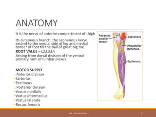
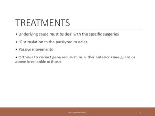
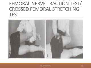
FEMORAL
NEUROPATHY
P/B :- DR NIYATI PATEL 1
P/B :- DR NIYATI PATEL 2
ANATOMY
It is the nerve of anterior compartment of thigh
Its cutaneous branch, the saphenous nerve
extend to the medial side of leg and medial
border of foot till the ball of great big toe
ROOT VALUE – L2,L3,L4
Arising from dorsal division of the ventral
primary rami of lumbar plexus
MOTOR SUPPLY
oAnterior division
•Sartorius
•Pectineus
oPosterior division
•Vastus medialis
•Vastus intermedius
•Vastus lateralis
•Rectus femoris
P/B :- DR NIYATI PATEL 3
P/B :- DR NIYATI PATEL 4
CAUSES
• Psoas abscess
• Pelvic neoplasm
• Fracture of the pelvis or femur
• Hip dislocation
• Inguinal hernia
• Complication of spinal anesthesia
• Prolapse intervertebral disc
• Lumbar spondylosis or stenosis
• Neuropathy secondary to diabetes mellitus known as diabetic
Amyotrophy
• Neurapraxia after hysterectomy or gynecological surgery
• Penetrating wounds of lower abdomen.
P/B :- DR NIYATI PATEL 5
SIGN & SYMPTOMS
Sensory
There is loss of sensation over the following areas:
a. Anterior division - anterior and medial aspect of the thigh
b. Posterior division – Continuous as the saphenous nerve involving the
medial aspect of the leg and foot right up to the ball of the great toe.
Motor
The muscles which are paralyzed due to the lesion of the anterior
division are Sartorius and pectineus.
The muscles which are paralyzed due to the lesion to the posterior
division are rectus femoris, vastus medialis, vastus lateralis and vastus
intermedius.
P/B :- DR NIYATI PATEL 6
Reflexes
Quadriceps jerk is lost.
Deformity
Genu recurvatum is seen because as
the quadriceps is paralyzed the
patient will try to lock the knee into
hyperextension to get the center of
gravity well in front of the knee joint
to keep it stable.
P/B :- DR NIYATI PATEL 7
SPECIAL TESTS
oSLUMP TEST
oPRONE KNEE BENDING TEST
oFEMORAL NERVE TRACTION TEST
P/B :- DR NIYATI PATEL 8
INVESTIGATION
RADIOGRAPH :- shows whether there is presence of fracture
MRI :- To delineate complete avulsion of nerve roots
SD CURVE:- abnormality in conduction can be verified. Sharp curve, long
chronaxie, low rheobase and the absence of contraction with repetitive
stimuli indicates denervation. If it is done 2-3 weeks after injury, it
shows the sign of denervation and to find out whether it is moderate or
severe injury
NCV:- To find out the severance of nerve fibers with wallerian
degeneration.
EMG:- it will help to find out reversible and irreversible nerve damage
and will help map out whether it pre ganglionic/ post ganglionic lesion
P/B :- DR NIYATI PATEL 9
TYPES OF INJURIES
In Neuropraxia  pain, numbness, muscle weakness,
minimal muscle wasting is present. Recovery occurs within
minutes to days
In Axonotmesis  there is pain, evident muscle wasting,
complete loss of motor, sensory and sympathetic functions.
Recovery time– months (axon regeneration at 1-1.5
mm/day)
In Neurotmesis  no pain, complete loss of motor, sensory
and sympathetic functions. Recovery time – months and
only with surgery
P/B :- DR NIYATI PATEL 10
TREATMENTS
• Underlying cause must be deal with the specific surgeries
• IG stimulation to the paralyzed muscles
• Passive movements
• Orthosis to correct genu recurvatum. Either anterior knee guard or
above knee ankle orthosis
P/B :- DR NIYATI PATEL 11
MERALGIA PARESTHETICA
P/B :- DR NIYATI PATEL 12
P/B :- DR NIYATI PATEL 13
It is also known as ‘’lateral femoral cutaneous
nerve entrapment”
Causes:-
•Tight clothing, such as belts, corsets and tight pants
•Obesity or weight gain
•Wearing a heavy tool belt
•Pregnancy
•Fluid accumulation in the abdomen causing increased
abdominal pressure
•Scar tissue near the inguinal ligament due to injury or past
surgery
P/B :- DR NIYATI PATEL 14
Sign and symptoms:-
•Tingling, numbness and burning pain in your lateral
aspect of thigh
•Decreased sensation
•Increased sensitivity and pain to even a light touch
P/B :- DR NIYATI PATEL 15
SLUMP TEST
P/B :- DR NIYATI PATEL 16
P/B :- DR NIYATI PATEL 17
P/B :- DR NIYATI PATEL 18
SLUMP1
P/B :- DR NIYATI PATEL 19
SLUMP2
P/B :- DR NIYATI PATEL 20
SLUMP3
P/B :- DR NIYATI PATEL 21
SLUMP4
P/B :- DR NIYATI PATEL 22
PRONE KNEE
BENDING
P/B :- DR NIYATI PATEL 23
P/B :- DR NIYATI PATEL 24
PKB1
P/B :- DR NIYATI PATEL 25
PKB2
P/B :- DR NIYATI PATEL 26
PKExtension
P/B :- DR NIYATI PATEL 27
FEMORAL NERVE TRACTION TEST/
CROSSED FEMORAL STRETCHING
TEST
P/B :- DR NIYATI PATEL 28

5.FEMORAL NERVE.pdf

  • 1.
  • 2.
    P/B :- DRNIYATI PATEL 2
  • 3.
    ANATOMY It is thenerve of anterior compartment of thigh Its cutaneous branch, the saphenous nerve extend to the medial side of leg and medial border of foot till the ball of great big toe ROOT VALUE – L2,L3,L4 Arising from dorsal division of the ventral primary rami of lumbar plexus MOTOR SUPPLY oAnterior division •Sartorius •Pectineus oPosterior division •Vastus medialis •Vastus intermedius •Vastus lateralis •Rectus femoris P/B :- DR NIYATI PATEL 3
  • 4.
    P/B :- DRNIYATI PATEL 4
  • 5.
    CAUSES • Psoas abscess •Pelvic neoplasm • Fracture of the pelvis or femur • Hip dislocation • Inguinal hernia • Complication of spinal anesthesia • Prolapse intervertebral disc • Lumbar spondylosis or stenosis • Neuropathy secondary to diabetes mellitus known as diabetic Amyotrophy • Neurapraxia after hysterectomy or gynecological surgery • Penetrating wounds of lower abdomen. P/B :- DR NIYATI PATEL 5
  • 6.
    SIGN & SYMPTOMS Sensory Thereis loss of sensation over the following areas: a. Anterior division - anterior and medial aspect of the thigh b. Posterior division – Continuous as the saphenous nerve involving the medial aspect of the leg and foot right up to the ball of the great toe. Motor The muscles which are paralyzed due to the lesion of the anterior division are Sartorius and pectineus. The muscles which are paralyzed due to the lesion to the posterior division are rectus femoris, vastus medialis, vastus lateralis and vastus intermedius. P/B :- DR NIYATI PATEL 6
  • 7.
    Reflexes Quadriceps jerk islost. Deformity Genu recurvatum is seen because as the quadriceps is paralyzed the patient will try to lock the knee into hyperextension to get the center of gravity well in front of the knee joint to keep it stable. P/B :- DR NIYATI PATEL 7
  • 8.
    SPECIAL TESTS oSLUMP TEST oPRONEKNEE BENDING TEST oFEMORAL NERVE TRACTION TEST P/B :- DR NIYATI PATEL 8
  • 9.
    INVESTIGATION RADIOGRAPH :- showswhether there is presence of fracture MRI :- To delineate complete avulsion of nerve roots SD CURVE:- abnormality in conduction can be verified. Sharp curve, long chronaxie, low rheobase and the absence of contraction with repetitive stimuli indicates denervation. If it is done 2-3 weeks after injury, it shows the sign of denervation and to find out whether it is moderate or severe injury NCV:- To find out the severance of nerve fibers with wallerian degeneration. EMG:- it will help to find out reversible and irreversible nerve damage and will help map out whether it pre ganglionic/ post ganglionic lesion P/B :- DR NIYATI PATEL 9
  • 10.
    TYPES OF INJURIES InNeuropraxia  pain, numbness, muscle weakness, minimal muscle wasting is present. Recovery occurs within minutes to days In Axonotmesis  there is pain, evident muscle wasting, complete loss of motor, sensory and sympathetic functions. Recovery time– months (axon regeneration at 1-1.5 mm/day) In Neurotmesis  no pain, complete loss of motor, sensory and sympathetic functions. Recovery time – months and only with surgery P/B :- DR NIYATI PATEL 10
  • 11.
    TREATMENTS • Underlying causemust be deal with the specific surgeries • IG stimulation to the paralyzed muscles • Passive movements • Orthosis to correct genu recurvatum. Either anterior knee guard or above knee ankle orthosis P/B :- DR NIYATI PATEL 11
  • 12.
    MERALGIA PARESTHETICA P/B :-DR NIYATI PATEL 12
  • 13.
    P/B :- DRNIYATI PATEL 13
  • 14.
    It is alsoknown as ‘’lateral femoral cutaneous nerve entrapment” Causes:- •Tight clothing, such as belts, corsets and tight pants •Obesity or weight gain •Wearing a heavy tool belt •Pregnancy •Fluid accumulation in the abdomen causing increased abdominal pressure •Scar tissue near the inguinal ligament due to injury or past surgery P/B :- DR NIYATI PATEL 14
  • 15.
    Sign and symptoms:- •Tingling,numbness and burning pain in your lateral aspect of thigh •Decreased sensation •Increased sensitivity and pain to even a light touch P/B :- DR NIYATI PATEL 15
  • 16.
    SLUMP TEST P/B :-DR NIYATI PATEL 16
  • 17.
    P/B :- DRNIYATI PATEL 17
  • 18.
    P/B :- DRNIYATI PATEL 18
  • 19.
    SLUMP1 P/B :- DRNIYATI PATEL 19
  • 20.
    SLUMP2 P/B :- DRNIYATI PATEL 20
  • 21.
    SLUMP3 P/B :- DRNIYATI PATEL 21
  • 22.
    SLUMP4 P/B :- DRNIYATI PATEL 22
  • 23.
    PRONE KNEE BENDING P/B :-DR NIYATI PATEL 23
  • 24.
    P/B :- DRNIYATI PATEL 24
  • 25.
    PKB1 P/B :- DRNIYATI PATEL 25
  • 26.
    PKB2 P/B :- DRNIYATI PATEL 26
  • 27.
    PKExtension P/B :- DRNIYATI PATEL 27
  • 28.
    FEMORAL NERVE TRACTIONTEST/ CROSSED FEMORAL STRETCHING TEST P/B :- DR NIYATI PATEL 28