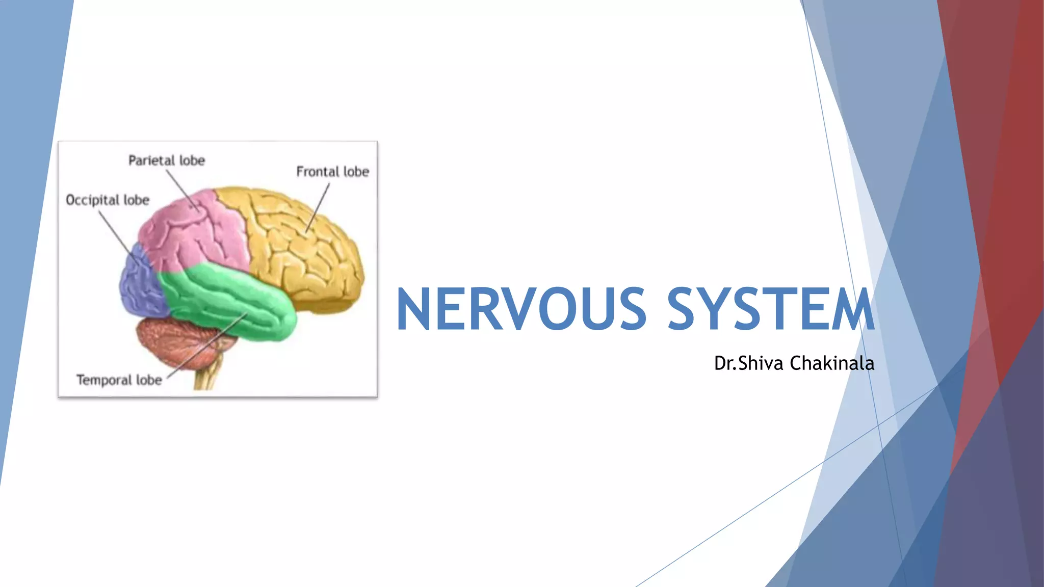
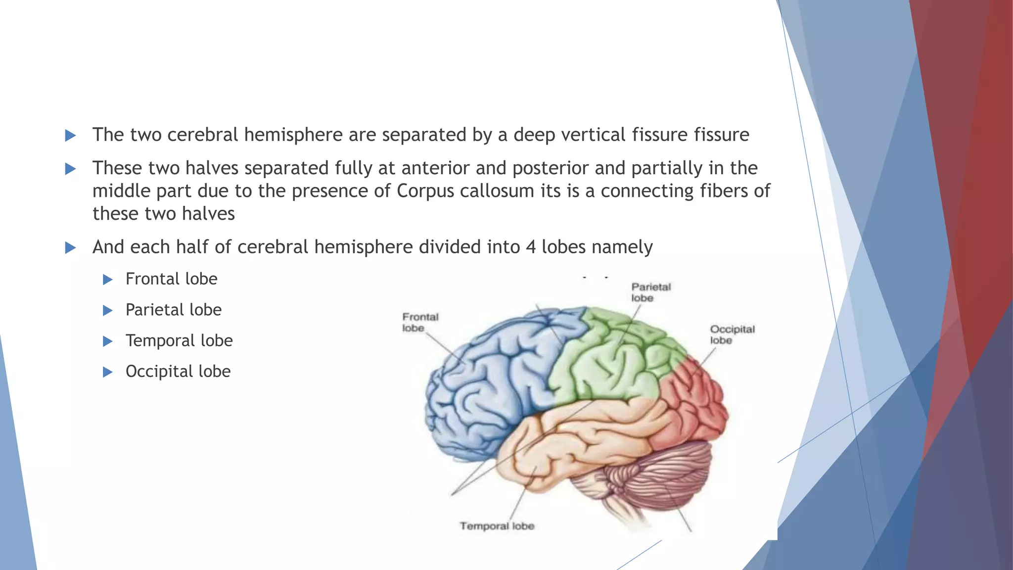
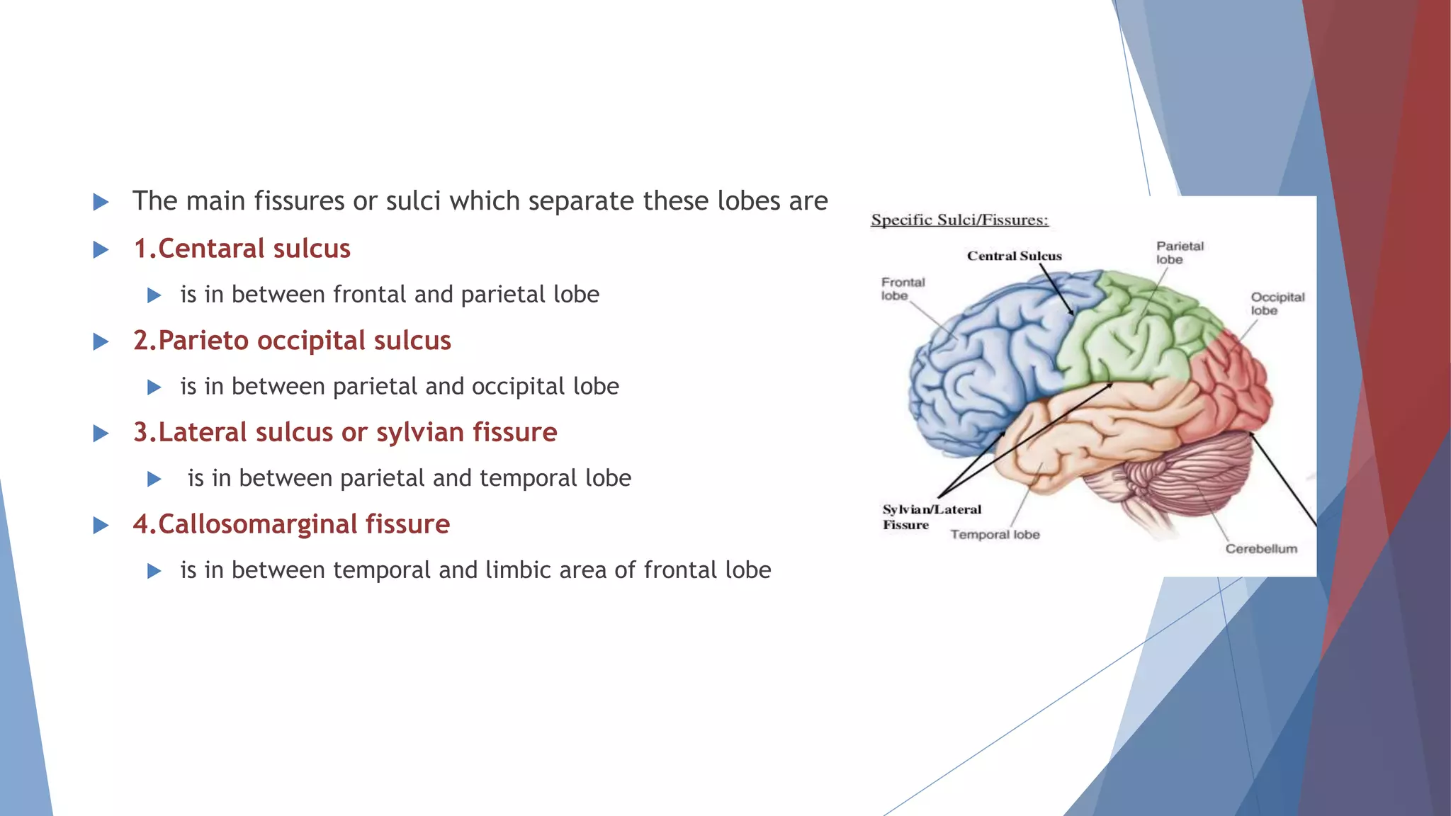
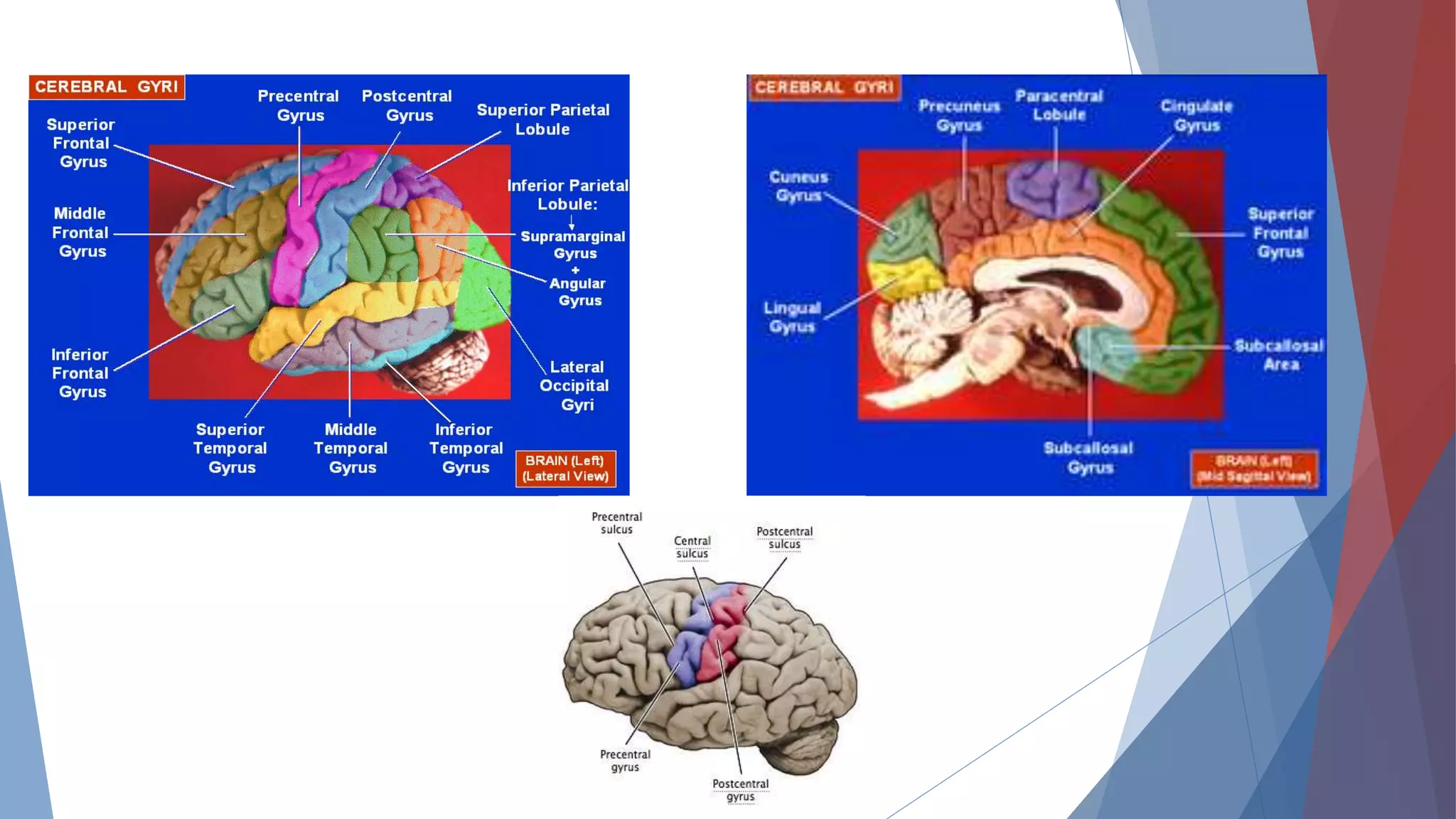
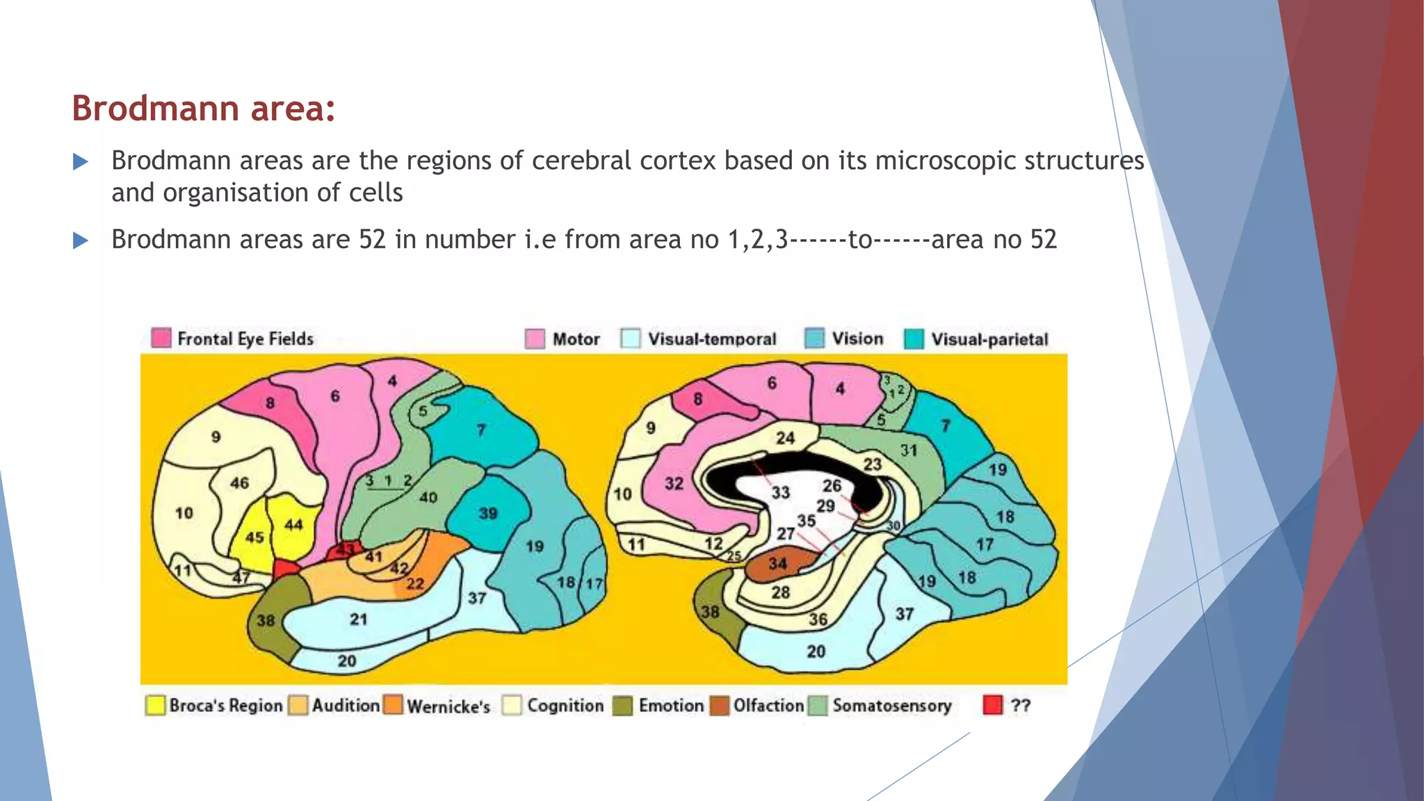
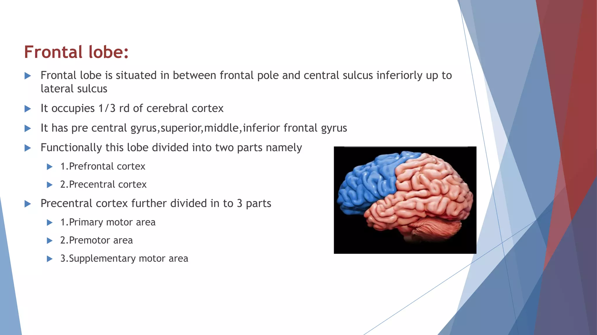
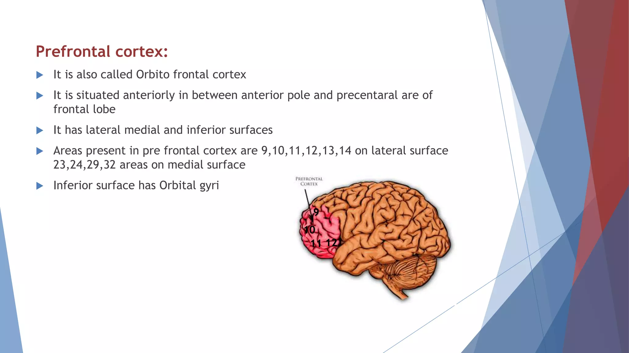
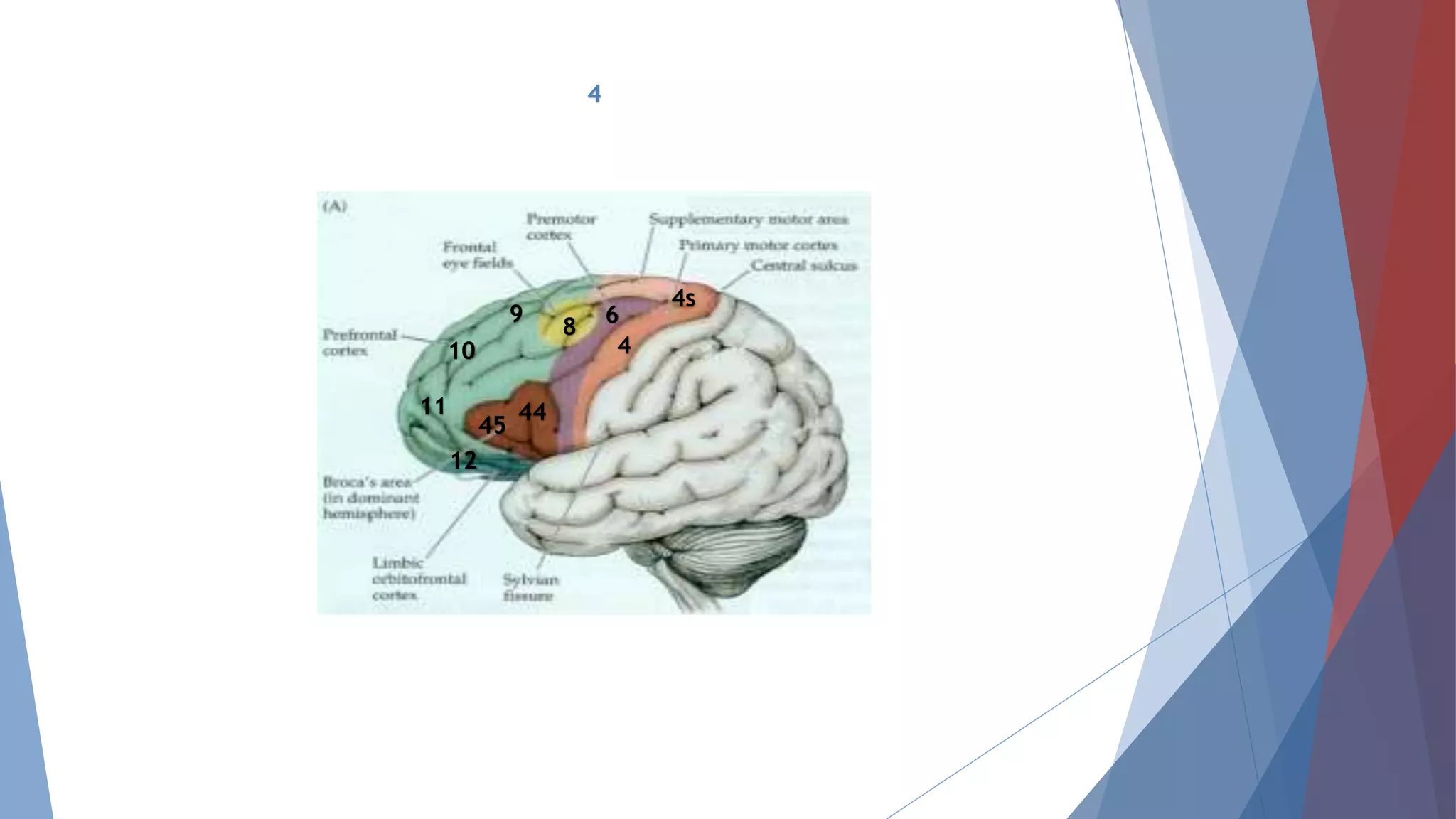
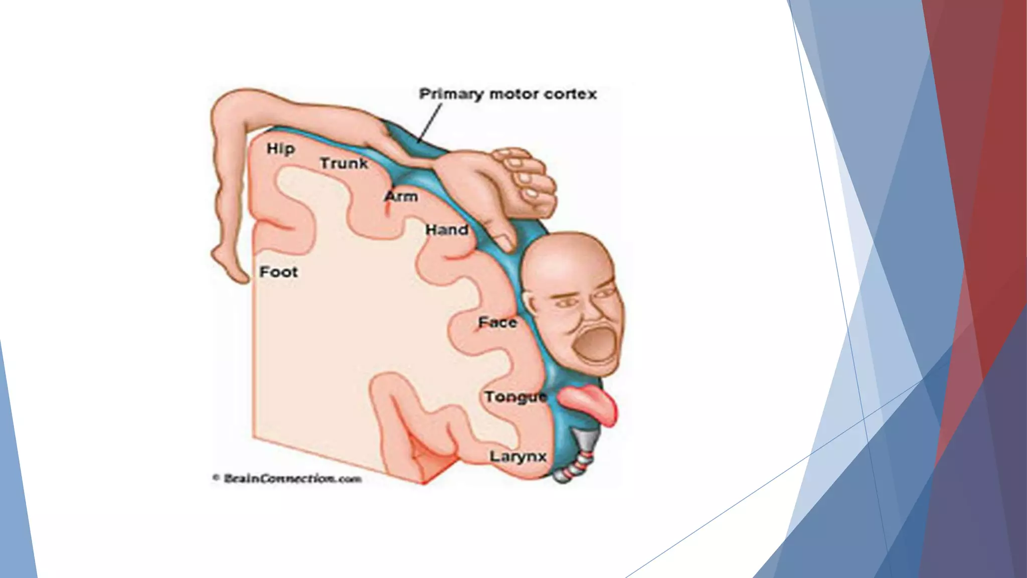
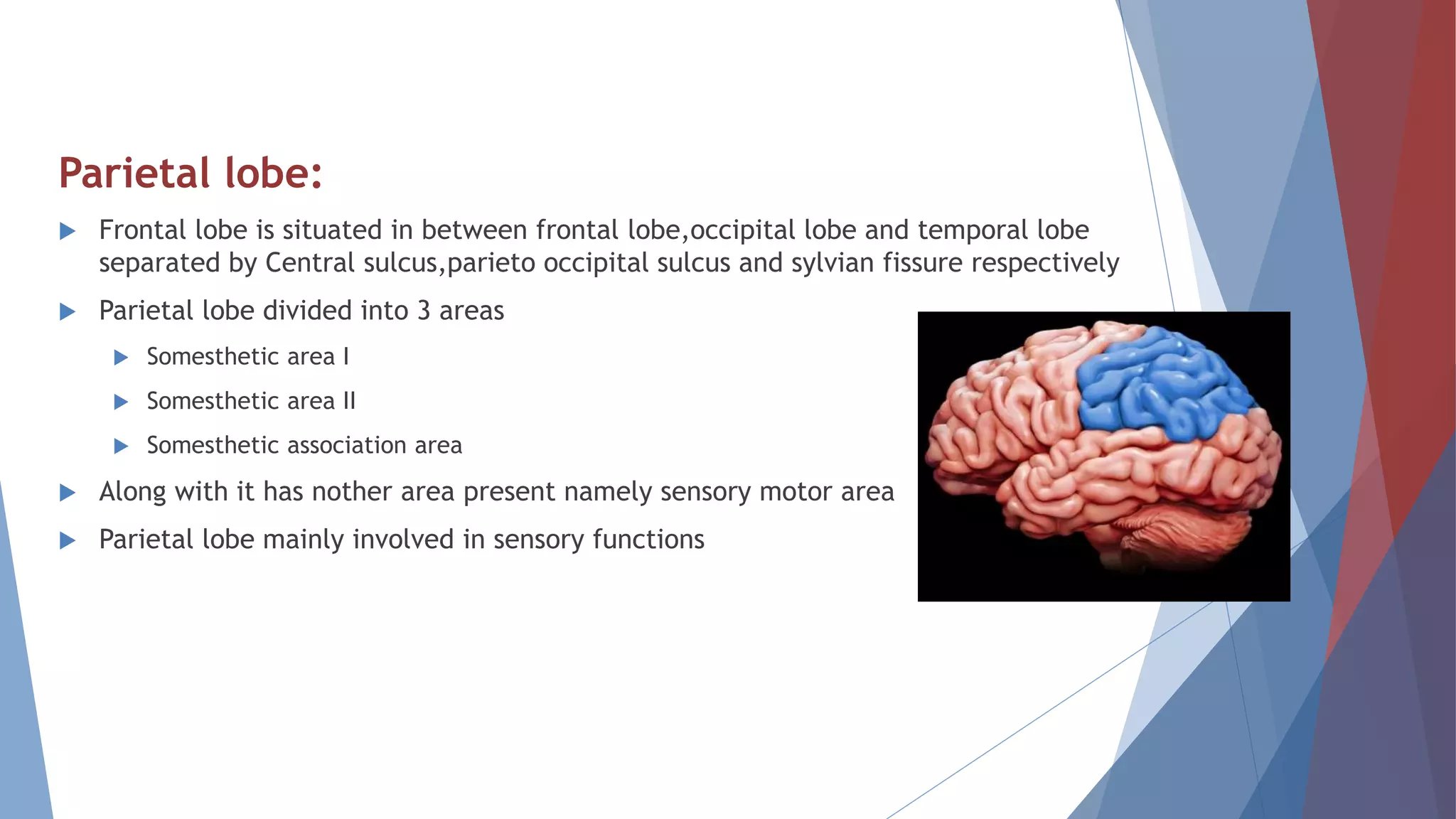
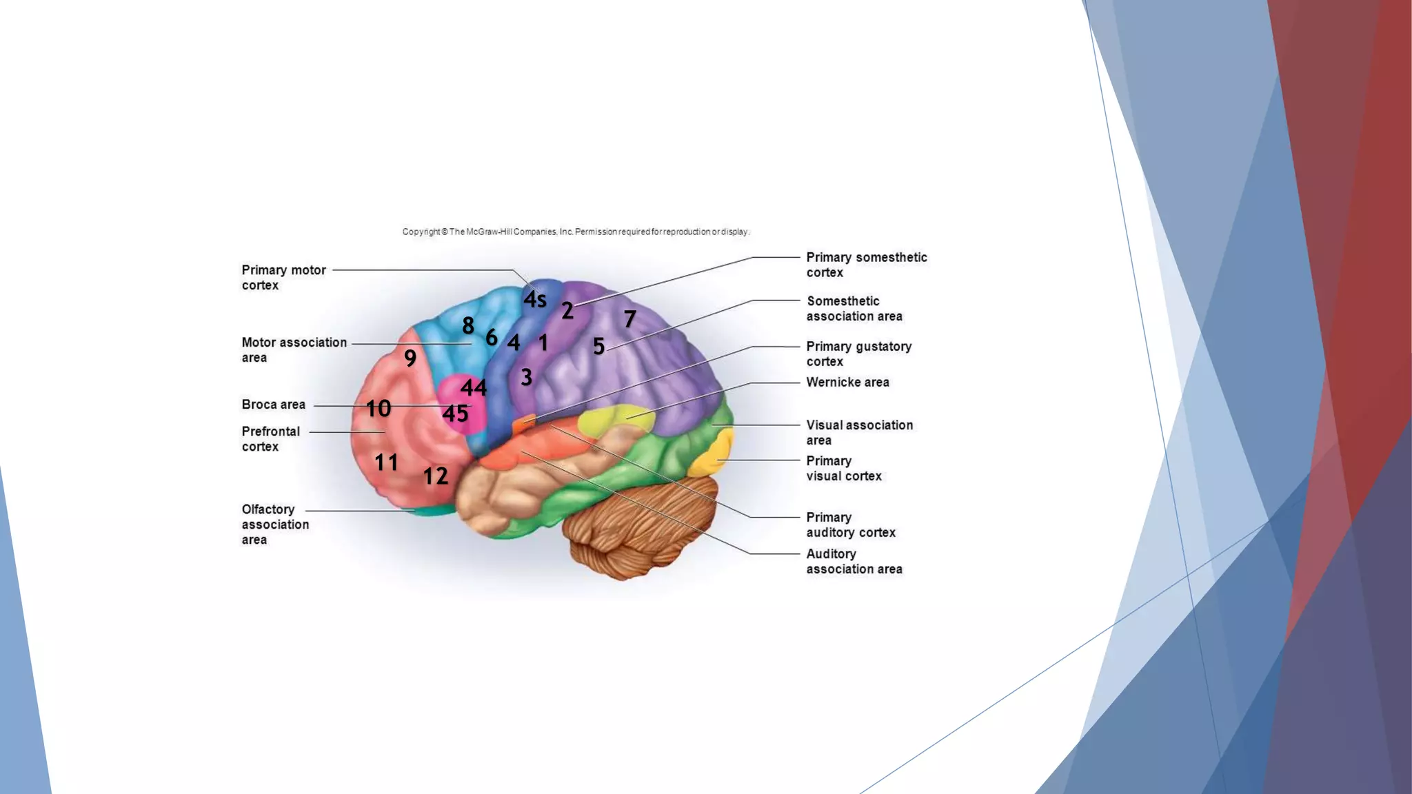
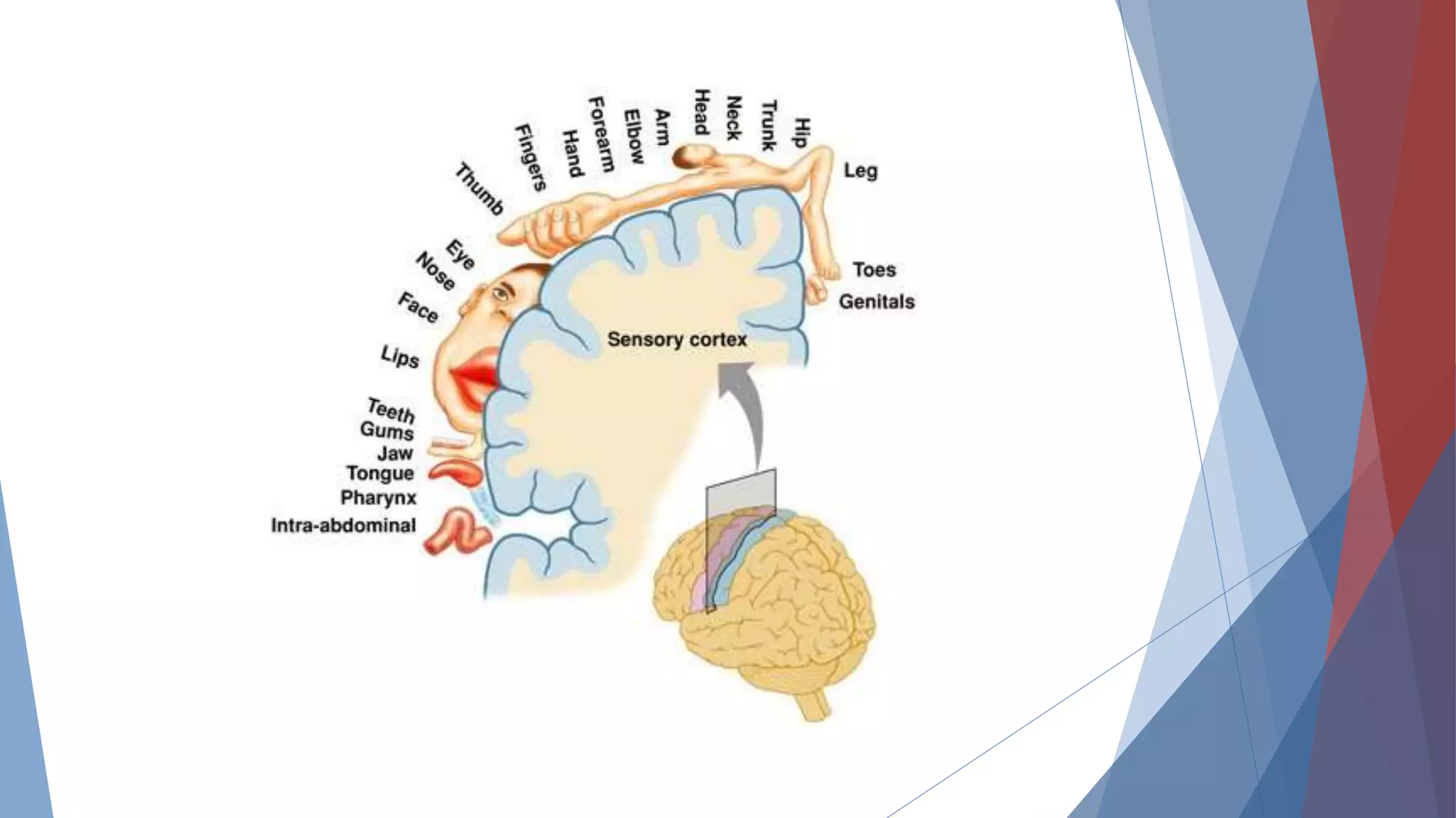
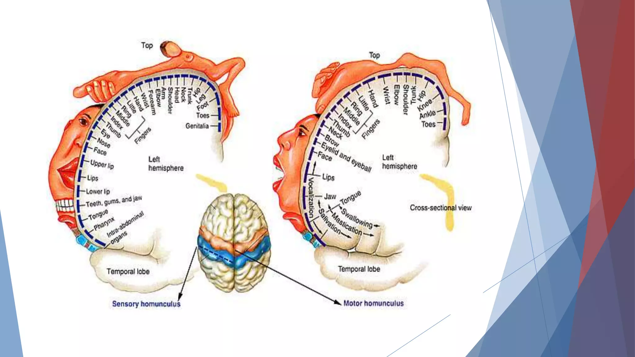
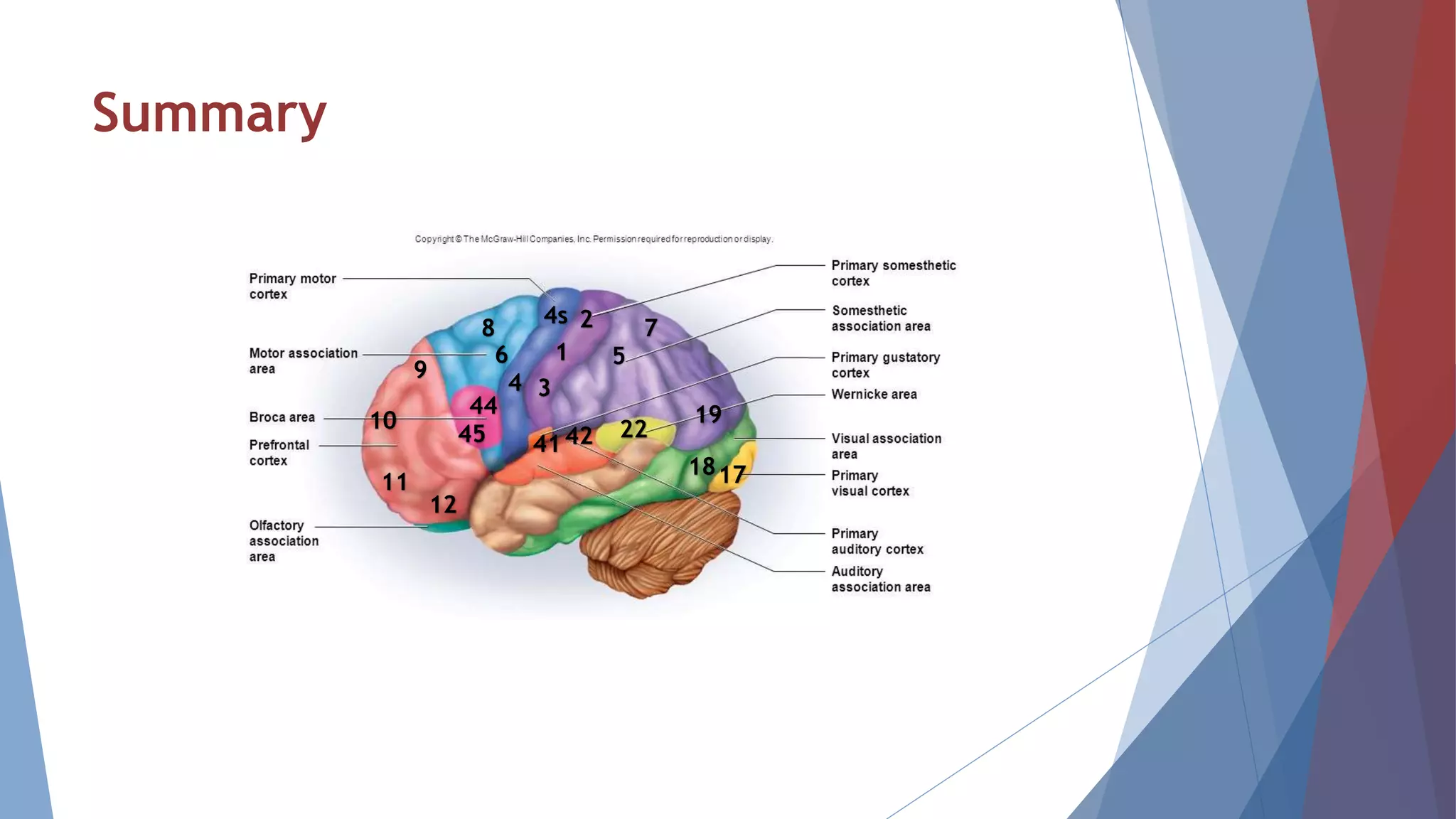
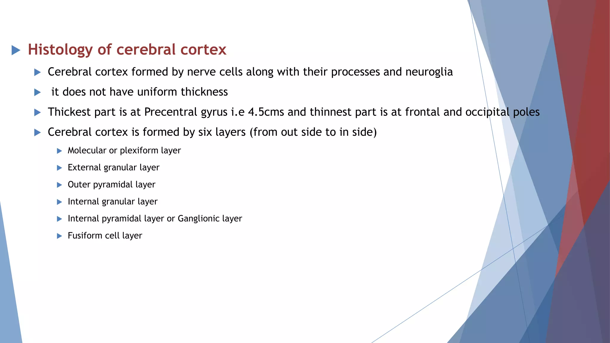
NERVOUS SYSTEM
Dr.Shiva Chakinala
CEREBRAL CORTEX
Introduction:
 Cerebral cortex is involved in different motor and sensory function
 Cerebral cortex area is about 2.2 square meter
 It has two cerebral hemisphere right and left
 The characteristic feature of cerebral cortex is Sulci and Gyri
 Sulci are the depression present on cerebral cortex
 Gyri are the elevations present on cerebral cortex
 The two cerebral hemisphere are separated by a deep vertical fissure fissure
 These two halves separated fully at anterior and posterior and partially in the
middle part due to the presence of Corpus callosum its is a connecting fibers of
these two halves
 And each half of cerebral hemisphere divided into 4 lobes namely
 Frontal lobe
 Parietal lobe
 Temporal lobe
 Occipital lobe
 The main fissures or sulci which separate these lobes are
 1.Centaral sulcus
 is in between frontal and parietal lobe
 2.Parieto occipital sulcus
 is in between parietal and occipital lobe
 3.Lateral sulcus or sylvian fissure
 is in between parietal and temporal lobe
 4.Callosomarginal fissure
 is in between temporal and limbic area of frontal lobe
Brodmann area:
 Brodmann areas are the regions of cerebral cortex based on its microscopic structures
and organisation of cells
 Brodmann areas are 52 in number i.e from area no 1,2,3------to------area no 52
Frontal lobe:
 Frontal lobe is situated in between frontal pole and central sulcus inferiorly up to
lateral sulcus
 It occupies 1/3 rd of cerebral cortex
 It has pre central gyrus,superior,middle,inferior frontal gyrus
 Functionally this lobe divided into two parts namely
 1.Prefrontal cortex
 2.Precentral cortex
 Precentral cortex further divided in to 3 parts
 1.Primary motor area
 2.Premotor area
 3.Supplementary motor area
Prefrontal cortex:
 It is also called Orbito frontal cortex
 It is situated anteriorly in between anterior pole and precentaral are of
frontal lobe
 It has lateral medial and inferior surfaces
 Areas present in pre frontal cortex are 9,10,11,12,13,14 on lateral surface
23,24,29,32 areas on medial surface
 Inferior surface has Orbital gyri
9
10
11 12
Connections of Prefrontal cortex:
Afferent connections
 Receives fibers mainly from
 Dorsomedial nucleus of thalamus
 Hypothalamus
 Corpus striatum
 amygdala
 Mid brain
Efferent connections
 Send fibers to
 Thalamus
 Hypothalamus
 Tegmentum
 Caudate nucleus
 Pons
 Temporal lobe
 Area 13 along with hippocampus send fibers to mammillary body via fornix this area related with
Emotions
Functions of Prefrontal cortex:
 This area is center for higher functions like Emotions,learning,memory,social
behaviour
 Short term memories stored
 Center for planned actions
 Center for intelligence
 Bilateral removal of these area leads to lack of mental alertness and lack of
intiation very little changes in memory judgement and intelligence
4
4s
4
68
44
45
9
10
11
12
Precentral cortex:
 It is posterior part of frontal lobe
 It is situated anteriorly in between central sulcus and prefrontal cortex ,posterior
portion of middle,superior and inferior frontal gyri
 It extend to medial surface also
 It is also called excito motor cortex
 Precentral cortex again divided into
 Primary motor area
 Pre motor area
 Supplementary motor area
Primary motor area:
 This area present through out the area of pre central gyrus adjoining area of
central sulcus
 Area no. 4 and 4s are present in it
Connections of Primary motor area:
Afferent connections
 Receives fibers from
 Dentate nucleus via red nucleus
 Thalamus
Efferent connections
 Send fibers to
 Corticospinal tract to spinal cord of opposite side
 Frontopontine fibers to pontine nuclei of same side
 Corpus striatum
 Red nucleus
 Thalamus
 Subthalamus
 Reticular formation
 Association fibers to this area to other area of cortex
Functions of Primary motor area:
 This area is involved in voluntary movements and speech
 Area 4:
 Area situated just in front of central sulcus on pre central gyrus as a strip
 Area has broad end at superiorly
 Most of efferent fibers arised from primary motor area and these fibers involved in voluntary movements
of opposite side on stimulation
 Area 4S:
 This is called suppressor area
 It is present in front of area no.4 on precentral gyrus
 It inhibited the movements created by the area no 4 so that it control exaggerated movements
• Lesion in this area leads to paralysis of contralateral side(opposite side of body)
• Lesion of this area both side leads to total paralysis
• Recovery is late while recovering upper limb recovered first than lower limbs
4
4s
6
8
44
45
Pre motor area:
 This area present anterior to primary motor area
 Area involved in postural movements of body sent efferent fibers to axial muscles of body
 Areas involved are 6,8,44 and 45
Area 6:
 Area no 6 divided in to 6a and 6b on frontal gyri, infront of area no 4
 This area also send efferent fibers through corticospinal tract along with area 4
Functions of Area 6:
Coardinate the movements intiated by area 4,that is it involved in skilled movements to perform
accurate
 lesion of this area leads to loss of skilled movements opposite side
 Lesion along with area 4 leads to severe hemiplegia
Area 8:
 This area also called Frontal eye field
 Present anterior to the area no 6
 It is concerned with eye ball movements
 Send fibers to i.e efferent connection with Occulomotor nuclei of midbrain
 Receive fibers from i.e afferent connection with Thalamus and Occipital lobe
Functions of Area 8:
this area involved in eye ball movements,open and closure of eyelids,dilatation of
pupil and lacrimation of opposite side of eye
 lesion of this area leads to loss of eye ball movements but not effect eyelids and pupil
Area 44 and 45:
 It is also called as Broca’s area
 It is a Motor area of speech
 Situated on inferior frontal gyrus of frontal lobe
Functions of Broca’s area:
area responsible for movements of tongue,lips,and larynx which area
involved in speech
 lesion of this area leads to Motor aphasia or Broca’s aphasia
 In this person has idea about his statement but he is unable to talk fluently
Supplementary motor area:
 This area present medial surface of frontal lobe
 Exact function of this area not understood clearly
 It suggested that it is concerned with coordinate skilled movements
4
4s
6
8
44
45
 Homunculus arrangement of motor areas in cerebral cortex
 The muscles of various parts of body represent in motor area 4 from medial to lateral
surface
 In this the lower part of body present at lateral surface and upper parts of body present
at medial surface
 The order of representation from medial to laeral cortex is
 Toes-->ankle-->knee-->hip-->trunk-->shoulder-->arm-->elbow-->wrist-->hand-->fingers-->face
 From their respective are the efferent fibers send to respective muscles of opposite side
except face
 Face receives efferents from both side cortex
Parietal lobe:
 Frontal lobe is situated in between frontal lobe,occipital lobe and temporal lobe
separated by Central sulcus,parieto occipital sulcus and sylvian fissure respectively
 Parietal lobe divided into 3 areas
 Somesthetic area I
 Somesthetic area II
 Somesthetic association area
 Along with it has nother area present namely sensory motor area
 Parietal lobe mainly involved in sensory functions
Somesthetic area I:
 It is also called Primary somesthetic area or Primary sensory area
 It is situated posterior to the central sulcus on post central gyrus and on paracentral lobule
of medial side
Areas:
 It has three areas 3,1 and 2
 Anteriorly on post central gyrus has area 3 and posteriorly has area 1 and 2
Connections:
 It receives afferent fibers from Thalamus as thalamic radiation
Functions of Somesthetic area I :
 It receives sensory impulses like pain,touch,temperature,pressure and proprioception from
opposite side
 Area 1 involved in sensory perception and area 2 and 3 in integration of sensory impulses
 These area send feedback to pre motor area
 Apart from this it has also involved in
 Spatial recognition:
 Tactile localization and two point discrimination
 Recognition of intensity of stimuli
 Recognition of similarities and difference between stimuli
 If lesion in this area without involvement of thalamus leads to loss of discrimination but
patient able perceive the stimuli
 If lesion in this area along with thalamus leads to loss of sensations opposite side
1
2
3
7
5
Somesthetic area II
Somesthetic area I
Somesthetic Association
area
Somesthetic area II:
 It is situated on post central gyrus below the somesthtic area I some part of
this area present in sylvian fissure
 Connections:
 It receives afferent fibers from Thalamus and somesthetic area directly
 Exact role of this area not clear but it concerned with perception of sensation
1
2
3
Somesthetic association area:
 It is situated posterior to post central gyrus infront of visual cortex and above auditory
cortexmedial side
Areas:
 It has areas 5 and 7
 Anteriorly on post central gyrus has area 3 and posteriorly has area 1 and 2
Function:
 This area center for combined sesation like Stereognosis
 Lesion of this area leads to Astereognosis
1
2
3
7
54
4s
68
44
45
9
10
11
12
Sensory motor area:
 Sensory area not limited to parietal lobe it extends in to motor area of pre central
gyrus
 Similarly motor area not limited to frontal lobe it extends In to sensory area post
central gyrus
 This combined area called as sensory motor area
 Timing and programming of skilled movents involved in Corticocerebellum are stored
in sensory motor area
 Homunculus arrangement of motor areas in cerebral cortex
 The various parts of body represent in primary sensory area from medial to lateral
surface
 In this the lower part of body present at lateral surface and upper parts of body
present at medial surface
 The order of representation from medial to laeral cortex is
 Toes-->ankle-->knee-->hip-->trunk-->shoulder-->arm-->elbow-->wrist-->hand-->fingers--
>face(eyelids-->nose-->cheek-->upper lip-->lower lip)
Temporal lobe:
 Temporal lobe is situated below the fronto parietal lobe
 Temporal includes 3 areas
 Primary auditory area
 Auditopsychic area
 Area for equilibrium
Primary auditory area:
 This area include area no 41,42
 41’42areas are present on anterior transvers gyrus and anterior part of
superiortemporal gyrus
Connections:
 This area receives afferent fibers from Medial geniculate body and Pulvinar
 Send efferent fibers to medial geniculate body,inferior colliculus of mid brain
and pulvinar
Functions of primary auditory area:
 primary auditory area involved in perception of auditory stimulus
 but interpretation of sound i.e analysis of speech source of sound is done with
the help of Auditopsychic area
Auditory psychic area:
 This area include area no 22
 This is also called wernick’s area
 It is situated on superior temporal gyrus posterior to area no 41,42
 It is helpful in interpretation of sound
 It is a sensory area of speech
 Lesion of this area leads to Sensory aphasia or Wernick’s aphasia
 In this patient able to talk clearly but not related
Area for equilibrium:
 It is situated posterior to superior temporal gyrus
 It is helpful maintainence of equilibrium
 Stimulation of this area leads to dizziness,swaying,falling and feeling of rotation of
body
22
22
4241
Occipital lobe:
 It is posterior part of cerebral cortex
 Temporal include visual cortex
 Visual cortex includes
 Primary visual area----- area no.17
 Visual association area--area no.18
 Occipital eye field-------area no.19
 Connections:
 It receives afferent fibers from latearal geniculate body
 Send efferents to superior colliculus and lateral geniculate body
Functions of Visual cortex :
 Primary visual are involved in perception of visual impulses
 Visual association area involved in interpretation of visual impulses
 Occipital eye field concerned with movement of eyes
17
18
19
Summary
4
4s
6
8
44
45
9
10
11
12
1
2
3
7
5
224241
1718
19
 Histology of cerebral cortex
 Cerebral cortex formed by nerve cells along with their processes and neuroglia
 it does not have uniform thickness
 Thickest part is at Precentral gyrus i.e 4.5cms and thinnest part is at frontal and occipital poles
 Cerebral cortex is formed by six layers (from out side to in side)
 Molecular or plexiform layer
 External granular layer
 Outer pyramidal layer
 Internal granular layer
 Internal pyramidal layer or Ganglionic layer
 Fusiform cell layer
THANK YOU..!

cerebrum(cerebral cortex) physiology

  • 1.
  • 2.
    CEREBRAL CORTEX Introduction:  Cerebralcortex is involved in different motor and sensory function  Cerebral cortex area is about 2.2 square meter  It has two cerebral hemisphere right and left  The characteristic feature of cerebral cortex is Sulci and Gyri  Sulci are the depression present on cerebral cortex  Gyri are the elevations present on cerebral cortex
  • 3.
     The twocerebral hemisphere are separated by a deep vertical fissure fissure  These two halves separated fully at anterior and posterior and partially in the middle part due to the presence of Corpus callosum its is a connecting fibers of these two halves  And each half of cerebral hemisphere divided into 4 lobes namely  Frontal lobe  Parietal lobe  Temporal lobe  Occipital lobe
  • 4.
     The mainfissures or sulci which separate these lobes are  1.Centaral sulcus  is in between frontal and parietal lobe  2.Parieto occipital sulcus  is in between parietal and occipital lobe  3.Lateral sulcus or sylvian fissure  is in between parietal and temporal lobe  4.Callosomarginal fissure  is in between temporal and limbic area of frontal lobe
  • 6.
    Brodmann area:  Brodmannareas are the regions of cerebral cortex based on its microscopic structures and organisation of cells  Brodmann areas are 52 in number i.e from area no 1,2,3------to------area no 52
  • 7.
    Frontal lobe:  Frontallobe is situated in between frontal pole and central sulcus inferiorly up to lateral sulcus  It occupies 1/3 rd of cerebral cortex  It has pre central gyrus,superior,middle,inferior frontal gyrus  Functionally this lobe divided into two parts namely  1.Prefrontal cortex  2.Precentral cortex  Precentral cortex further divided in to 3 parts  1.Primary motor area  2.Premotor area  3.Supplementary motor area
  • 8.
    Prefrontal cortex:  Itis also called Orbito frontal cortex  It is situated anteriorly in between anterior pole and precentaral are of frontal lobe  It has lateral medial and inferior surfaces  Areas present in pre frontal cortex are 9,10,11,12,13,14 on lateral surface 23,24,29,32 areas on medial surface  Inferior surface has Orbital gyri 9 10 11 12
  • 9.
    Connections of Prefrontalcortex: Afferent connections  Receives fibers mainly from  Dorsomedial nucleus of thalamus  Hypothalamus  Corpus striatum  amygdala  Mid brain Efferent connections  Send fibers to  Thalamus  Hypothalamus  Tegmentum  Caudate nucleus  Pons  Temporal lobe  Area 13 along with hippocampus send fibers to mammillary body via fornix this area related with Emotions
  • 10.
    Functions of Prefrontalcortex:  This area is center for higher functions like Emotions,learning,memory,social behaviour  Short term memories stored  Center for planned actions  Center for intelligence  Bilateral removal of these area leads to lack of mental alertness and lack of intiation very little changes in memory judgement and intelligence
  • 11.
  • 12.
    Precentral cortex:  Itis posterior part of frontal lobe  It is situated anteriorly in between central sulcus and prefrontal cortex ,posterior portion of middle,superior and inferior frontal gyri  It extend to medial surface also  It is also called excito motor cortex  Precentral cortex again divided into  Primary motor area  Pre motor area  Supplementary motor area
  • 13.
    Primary motor area: This area present through out the area of pre central gyrus adjoining area of central sulcus  Area no. 4 and 4s are present in it Connections of Primary motor area: Afferent connections  Receives fibers from  Dentate nucleus via red nucleus  Thalamus Efferent connections  Send fibers to  Corticospinal tract to spinal cord of opposite side  Frontopontine fibers to pontine nuclei of same side  Corpus striatum  Red nucleus  Thalamus  Subthalamus  Reticular formation  Association fibers to this area to other area of cortex
  • 14.
    Functions of Primarymotor area:  This area is involved in voluntary movements and speech  Area 4:  Area situated just in front of central sulcus on pre central gyrus as a strip  Area has broad end at superiorly  Most of efferent fibers arised from primary motor area and these fibers involved in voluntary movements of opposite side on stimulation  Area 4S:  This is called suppressor area  It is present in front of area no.4 on precentral gyrus  It inhibited the movements created by the area no 4 so that it control exaggerated movements • Lesion in this area leads to paralysis of contralateral side(opposite side of body) • Lesion of this area both side leads to total paralysis • Recovery is late while recovering upper limb recovered first than lower limbs
  • 15.
  • 16.
    Pre motor area: This area present anterior to primary motor area  Area involved in postural movements of body sent efferent fibers to axial muscles of body  Areas involved are 6,8,44 and 45 Area 6:  Area no 6 divided in to 6a and 6b on frontal gyri, infront of area no 4  This area also send efferent fibers through corticospinal tract along with area 4 Functions of Area 6: Coardinate the movements intiated by area 4,that is it involved in skilled movements to perform accurate  lesion of this area leads to loss of skilled movements opposite side  Lesion along with area 4 leads to severe hemiplegia
  • 17.
    Area 8:  Thisarea also called Frontal eye field  Present anterior to the area no 6  It is concerned with eye ball movements  Send fibers to i.e efferent connection with Occulomotor nuclei of midbrain  Receive fibers from i.e afferent connection with Thalamus and Occipital lobe Functions of Area 8: this area involved in eye ball movements,open and closure of eyelids,dilatation of pupil and lacrimation of opposite side of eye  lesion of this area leads to loss of eye ball movements but not effect eyelids and pupil
  • 18.
    Area 44 and45:  It is also called as Broca’s area  It is a Motor area of speech  Situated on inferior frontal gyrus of frontal lobe Functions of Broca’s area: area responsible for movements of tongue,lips,and larynx which area involved in speech  lesion of this area leads to Motor aphasia or Broca’s aphasia  In this person has idea about his statement but he is unable to talk fluently Supplementary motor area:  This area present medial surface of frontal lobe  Exact function of this area not understood clearly  It suggested that it is concerned with coordinate skilled movements
  • 19.
  • 20.
     Homunculus arrangementof motor areas in cerebral cortex  The muscles of various parts of body represent in motor area 4 from medial to lateral surface  In this the lower part of body present at lateral surface and upper parts of body present at medial surface  The order of representation from medial to laeral cortex is  Toes-->ankle-->knee-->hip-->trunk-->shoulder-->arm-->elbow-->wrist-->hand-->fingers-->face  From their respective are the efferent fibers send to respective muscles of opposite side except face  Face receives efferents from both side cortex
  • 22.
    Parietal lobe:  Frontallobe is situated in between frontal lobe,occipital lobe and temporal lobe separated by Central sulcus,parieto occipital sulcus and sylvian fissure respectively  Parietal lobe divided into 3 areas  Somesthetic area I  Somesthetic area II  Somesthetic association area  Along with it has nother area present namely sensory motor area  Parietal lobe mainly involved in sensory functions
  • 23.
    Somesthetic area I: It is also called Primary somesthetic area or Primary sensory area  It is situated posterior to the central sulcus on post central gyrus and on paracentral lobule of medial side Areas:  It has three areas 3,1 and 2  Anteriorly on post central gyrus has area 3 and posteriorly has area 1 and 2 Connections:  It receives afferent fibers from Thalamus as thalamic radiation
  • 24.
    Functions of Somestheticarea I :  It receives sensory impulses like pain,touch,temperature,pressure and proprioception from opposite side  Area 1 involved in sensory perception and area 2 and 3 in integration of sensory impulses  These area send feedback to pre motor area  Apart from this it has also involved in  Spatial recognition:  Tactile localization and two point discrimination  Recognition of intensity of stimuli  Recognition of similarities and difference between stimuli  If lesion in this area without involvement of thalamus leads to loss of discrimination but patient able perceive the stimuli  If lesion in this area along with thalamus leads to loss of sensations opposite side
  • 25.
    1 2 3 7 5 Somesthetic area II Somestheticarea I Somesthetic Association area
  • 26.
    Somesthetic area II: It is situated on post central gyrus below the somesthtic area I some part of this area present in sylvian fissure  Connections:  It receives afferent fibers from Thalamus and somesthetic area directly  Exact role of this area not clear but it concerned with perception of sensation
  • 27.
  • 28.
    Somesthetic association area: It is situated posterior to post central gyrus infront of visual cortex and above auditory cortexmedial side Areas:  It has areas 5 and 7  Anteriorly on post central gyrus has area 3 and posteriorly has area 1 and 2 Function:  This area center for combined sesation like Stereognosis  Lesion of this area leads to Astereognosis
  • 29.
  • 30.
    Sensory motor area: Sensory area not limited to parietal lobe it extends in to motor area of pre central gyrus  Similarly motor area not limited to frontal lobe it extends In to sensory area post central gyrus  This combined area called as sensory motor area  Timing and programming of skilled movents involved in Corticocerebellum are stored in sensory motor area
  • 31.
     Homunculus arrangementof motor areas in cerebral cortex  The various parts of body represent in primary sensory area from medial to lateral surface  In this the lower part of body present at lateral surface and upper parts of body present at medial surface  The order of representation from medial to laeral cortex is  Toes-->ankle-->knee-->hip-->trunk-->shoulder-->arm-->elbow-->wrist-->hand-->fingers-- >face(eyelids-->nose-->cheek-->upper lip-->lower lip)
  • 34.
    Temporal lobe:  Temporallobe is situated below the fronto parietal lobe  Temporal includes 3 areas  Primary auditory area  Auditopsychic area  Area for equilibrium
  • 35.
    Primary auditory area: This area include area no 41,42  41’42areas are present on anterior transvers gyrus and anterior part of superiortemporal gyrus Connections:  This area receives afferent fibers from Medial geniculate body and Pulvinar  Send efferent fibers to medial geniculate body,inferior colliculus of mid brain and pulvinar Functions of primary auditory area:  primary auditory area involved in perception of auditory stimulus  but interpretation of sound i.e analysis of speech source of sound is done with the help of Auditopsychic area
  • 36.
    Auditory psychic area: This area include area no 22  This is also called wernick’s area  It is situated on superior temporal gyrus posterior to area no 41,42  It is helpful in interpretation of sound  It is a sensory area of speech  Lesion of this area leads to Sensory aphasia or Wernick’s aphasia  In this patient able to talk clearly but not related Area for equilibrium:  It is situated posterior to superior temporal gyrus  It is helpful maintainence of equilibrium  Stimulation of this area leads to dizziness,swaying,falling and feeling of rotation of body 22
  • 37.
  • 38.
    Occipital lobe:  Itis posterior part of cerebral cortex  Temporal include visual cortex  Visual cortex includes  Primary visual area----- area no.17  Visual association area--area no.18  Occipital eye field-------area no.19  Connections:  It receives afferent fibers from latearal geniculate body  Send efferents to superior colliculus and lateral geniculate body
  • 39.
    Functions of Visualcortex :  Primary visual are involved in perception of visual impulses  Visual association area involved in interpretation of visual impulses  Occipital eye field concerned with movement of eyes 17 18 19
  • 40.
  • 41.
     Histology ofcerebral cortex  Cerebral cortex formed by nerve cells along with their processes and neuroglia  it does not have uniform thickness  Thickest part is at Precentral gyrus i.e 4.5cms and thinnest part is at frontal and occipital poles  Cerebral cortex is formed by six layers (from out side to in side)  Molecular or plexiform layer  External granular layer  Outer pyramidal layer  Internal granular layer  Internal pyramidal layer or Ganglionic layer  Fusiform cell layer
  • 43.