2. Organization of the
Nervous System
• We have only one nervous
system, but because of its
complexity, it is difficult to
consider all its parts at the
same time.
• So, to simplify its study, we
divide it in terms of its
structures (structural
classification) or in terms of its
activities (functional
classification).
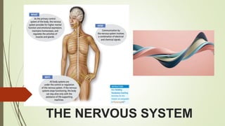
3. THE NERVOUS SYSTEM
Master control and communication system of the
body. Every thought, action, and emotion reflects
its activity.
It communicates with body cells using electrical
impulses which are rapid and specific and cause
almost immediate responses.
Does not work alone to regulate and maintain
body homeostasis; the endocrine system is a
second important regulating system.
Controls with rapid electrical nerve impulses, the
endocrine system produces hormones that are
released into the blood. Thus, the endocrine
system acts in a more leisurely way.
4. Three overlapping Functions of
the Nervous System
Sensory input –
Uses millions of sensory receptors to monitor changes
occurring both inside and outside the body.
These changes are called stimuli
Gathered information
Integration
Toprocess and interpret sensory input and decide if action
is needed
Motor output
It then causes a response, or effect, by activating muscles
or glands (effectors) via the motor output.
5. Structural Classification of the Nervous System
• Central nervous system (CNS)
Brain and Spinal cord
Acts as integrating and command
center – interpret incoming sensory
information and issue instructions
based on past experiences and
current conditions
• Peripheral nervous system (PNS)
Nerves outside the brain and spinal
cord
Link all parts of the body by carrying
impulses to the CNS and back
6. Functional Classification of the
Peripheral Nervous System
Motor
(efferent)
division
Nerve fibers that carry
impulses away from the
central nervous system
Sensory
(afferent)
division
Nerve fibers that carry
information to the central
nervous system
7. Functional Classification of the
Peripheral Nervous System
Motor
(efferent)
division
• Two subdivisions
Somatic nervous system =
voluntary nervous system
• Skeletal muscle reflexes
such as stretch
• reflex are initiated
involuntarily by same
fibers
Autonomic nervous system =
involuntary nervous system
• regulates events that are
automatic, or involuntary,
such as the activity of
smooth muscle, cardiac
muscle, and glands
• Sympathetic and
parasympathetic divisions
Nervous Tissue: Structure and
Function
Two Principal Types Of Cells:
Supporting cells
Neurons
Support Cells
“lumped together” as neuroglia,
literally, “nerve glue”
also called glial cells or glia
many types of cells that support,
insulate, and protect the delicate
neurons
8. Support Cells (Neuroglia) - glia
Microglia
• Spider-like phagocytes
• Dispose of debris – dead cells and
bacteria
Ependymal cells
• Line cavities of the brain and
spinal cord
• Circulate cerebrospinal fluid
with cilia
Astrocytes
• Abundant, star-shaped cells
• Brace neurons
• Form barrier between capillaries and
neurons and make exchanges
between the two
• Control the chemical environment of
the brain by capturing ions and
neurotransmitters
9. Support Cells (Neuroglia) - glia
Neuroglia are not able to transmit
nerve impulses but do not lose their
ability to divide, unlike neurons.
Oligodendrocytes
• Wrap their flat extensions tightly
around the nerve fibers
• Produce myelin sheath around nerve
fibers in the central nervous system
Satellite cells
• Protect neuron cell bodies
Schwann cells
• Form myelin sheath in the peripheral
nervous system
10. Neurons
• Nerve cells
Cells specialized to transmit messages
Major regions of neurons
o Cell body – nucleus and metabolic center of the cell
o Processes – fibers that extend from the cell body
Neuron Anatomy
o Cell body
Nissl substance – specialized rough endoplasmic
reticulum
Neurofibrils – intermediate cytoskeleton that
maintains cell shape
Nucleus
Large nucleolus
11. Extensions outside the cell body
• Dendrites – conduct impulses
toward the cell body
• Axons – conduct impulses away
from the cell body
Axons and Nerve Impulses
Axons end in
axonal terminals
Axonal terminals
contain vesicles with
neurotransmitters
Axonal terminals are separated
from the next neuron by a gap
• Synaptic cleft – gap between
adjacent neurons
• Synapse – junction between
nerves
12. Nerve Fiber Coverings
• Schwann cells – produce myelin sheaths in
jelly-roll
• Nodes of Ranvier – gaps in myelin sheath along
the axon
Neuron Cell Body Location
Most are found in the
central nervous system
in clusters called nuclei
Bundles of nerve fibers in
CNS = tracts
• Gray matter – cell bodies
and unmyelinated fibers
• White matter – myelinated
fibers
Bundles of nerve fibers in
PNS = nerves
Ganglia – collections of cell bodies outside the
central nervous system
13. Functional Classification of Neurons • Interneurons (association neurons)
Found in neural pathways in the
central nervous system
o Cell bodies in the CNS
Connect sensory and motor neurons
Sensory (afferent) neurons
Cell bodies in a
ganglion outside the CNS
Carry impulses from the
sensory receptors to CNS
• Cutaneous (skin) sense
organs
• Proprioceptors –
detect stretch or
tension in muscles,
tendons, joints
Motor (efferent) neurons
Cell bodies found in the CNS
Carry impulses from the
central nervous system
14. Homeostatic
Imbalance
• Multiple sclerosis (MS)
• Gradually destroys the myelin sheaths around
CNS fibers by converting them to hardened
sheaths called scleroses.
• As this happens, the electrical current is short-
circuited and may “jump” to another
demyelinated neuron. In other words, nerve
signals do not always reach the intended target.
• Affected person may have visual and speech
disturbances, lose the ability to control his or
her muscles, and become increasingly disabled
• Autoimmune disease in which the person’s
own immune system attacks a protein
component of the sheath.
• No cure, but injections of interferon (a
hormonelike substance released by some
immune cells) appear to hold the symptoms at
bay and provide some relief.
• Other drugs aimed at slowing the autoimmune
response are also being used, though further
research is needed to determine their long-term
effects.
15. Structural
Classification of
Neurons
• Multipolar neurons – many extensions from the
cell body
• Bipolar neurons – one axon and one dendrite
• Rare in adults – in eye and ear only
• Unipolar neurons – have a short, single
process leaving the cell body
• Axon conducts nerve impulses both to and
from the cell body
16. Functional Properties of Neurons
Two Main Functional Properties
Irritability – ability to respond to
stimuli
Conductivity – ability to
transmit an impulse to
other neurons, muscles, or
glands.
Electrical Conditions of a Resting
Neuron’s Membrane
1
The plasma membrane of a resting, or
inactive, neuron is polarized - there are
fewer positive ions sitting on the inner face
of the neuron’s plasma membrane than
there are on its outer face
Major positive ions inside the cell are
potassium (K+), whereas the major positive
ions outside the cell are sodium (Na+).
As long as the inside remains more negative
(fewer positive ions) than the outside, the
neuron will stay inactive
17. Action Potential Initiation and Generation
• Different types of stimuli excite neurons to become active and
generate an impulse.
Example:
light excites the eye receptors
sound excites some of the ear receptors pressure excites
some cutaneous receptors of the skin.
• Most neurons in the body are excited by neurotransmitter
chemicals released by other neurons
• Regardless of the stimulus, the result is always the same—the
permeability properties of the cell’s plasma membrane change
for a very brief period.
• Normally, sodium ions cannot diffuse through the plasma
membrane to any great extent, but when the neuron is
adequately stimulated, the “gates” of sodium channels in the
membrane open.
• Sodium is in much higher concentration outside the cell, it
then diffuses quickly into the neuron.
18. Action Potential Initiation and Generation
Inward rush of sodium ions changes the
polarity of the neuron’s membrane at that site,
an event called depolarization.
The inside is now more positive, and the
outside is less positive, a local electrical
situation called a graded potential.
If the stimulus is strong enough and the sodium
influx is great enough, the local depolarization
(graded potential) activates the neuron to
initiate and transmit a long-distance signal
called an action potential, also called a nerve
impulse in neurons.
The nerve impulse is an all-or-none response,
like starting a car. It is either propagated
(conducted, or sent) over the entire axon
or it doesn’t happen at all. The nerve impulse
never goes partway along an axon’s length, nor
does it die out with distance, as do graded
potentials.
2
3
4
Potassium ions are allowed to diffuse out of
the neuron into the interstitial fluid, and they
do so very rapidly. This outflow of positive
ions from the cell restores the electrical
conditions at the membrane to the
polarized, or resting, state, an event called
repolarization.
After repolarization of the electrical
conditions, the sodium-potassium pump
restores the initial concentrations of the
sodium and potassium ions inside and
outside the neuron
This pump uses ATP (cellular energy) to
pump excess sodium ions out of the cell and
to bring potassium ions back into it.
Until repolarization occurs, a neuron cannot
conduct another impulse.
Once begun, these sequential events
spread along the entire neuronal
membrane.
5
6
19. Action Potential Initiation and
Generation
Fibers that have myelin
sheaths conduct impulses
much faster because the
nerve impulse literally jumps,
or leaps, from node to node
along the length of the fiber.
This occurs because no
electrical current can flow
across the axon membrane
where there is fatty myelin
insulation.
This faster type of electrical
impulse propagation is called
saltatory conduction (saltare
= to dance or leap).
Homeostatic Imbalance
Number of factors can impair the conduction of
impulses.
Example
sedatives
anesthetics block nerve impulses by altering
membrane permeability to ions, mainly sodium ions.
No sodium entry = No action potential.
Cold and continuous pressure hinder impulse
conduction because they interrupt blood circulation
(and hence the delivery of oxygen and nutrients) to the
neurons
Example:
fingers get numb when you hold an ice cube for
more than a few seconds.
When you sit on your foot, it “goes to sleep.”
When you warm your fingers or remove the
pressure from your foot, the impulses begin to be
transmitted again, leading to an unpleasant prickly
feeling.
20. Transmission of the Signal at Synapses
How does the electrical impulse traveling along one neuron get
across the synapse to the next neuron?
- Answer: impulse doesn’t! Instead neurotransmitter chemical
crosses the synapse to transmit the signal from one neuron to
the next, or to the target cell.
Action potential reaches an axon terminal - the electrical
change opens calcium channels.
Calcium ions, in turn, cause the tiny vesicles containing
neurotransmitter to fuse with the axonal membrane
Pore-like openings form, releasing the neurotransmitter into the
synaptic cleft
The neurotransmitter molecules diffuse across the synaptic
cleft* and bind to receptors on the membrane of the next neuron
Enough neurotransmitter is released, the whole series of
events described above (sodium entry, , depolarization, etc.)
will occur, generating a graded potential and eventually a nerve
impulse in the receiving neuron beyond the synapse.
The electrical changes prompted by neurotransmitter binding
are very brief because the neurotransmitter is quickly removed
from the synaptic cleft either by diffusing away, by reuptake
into the axon terminal, or by enzymatic breakdown.
4
1
2
3
5
6
21. Transmission of the Signal at Synapses
This limits the effect of each
nerve impulse to a period
shorter than the blink of an
eye.
Transmission of an impulse is
an electrochemical event.
Transmission down the length
of the neuron’s membrane is
basically electrical, but the next
neuron is stimulated by a
neurotransmitter, which is a
chemical.
Because each neuron both
receives signals from and
sends signals to scores of
other neurons, it carries on
“conversations” with many
different neurons at the same
time.
Physiology: Reflexes
Reflexes
• rapid, predictable, and involuntary responses to stimuli.
• one-way streets—once a reflex begins, it always goes
in the same direction.
• occur over neural pathways called reflex arcs and
involve both CNS and PNS structures.
• preprogrammed response to a given stimulus
Types of reflexes :
Somatic - all reflexes that stimulate the skeletal muscles;
these are still involuntary reflexes even though skeletal
muscle normally is under voluntary control.
• When you quickly pull your hand away from a hot
object, a somatic reflex is working.
Autonomic - regulate the activity of smooth muscles, the
heart, and glands. Secretion of saliva (salivary reflex) and
changes in the size of the eye pupils (pupillary reflex) are
two such reflexes.
• regulate such body functions as digestion,
elimination, blood pressure, and sweating.
22. Five Basic Elements Of Reflex Arc:
• receptor - reacts to a stimulus
• effector - the muscle or gland eventually stimulated
• sensory neuron
• motor neurons
• synapse or interneurons between the sensory and motor neurons represents the fifth
element—the CNS integration center
23. Reflexes
• The simple patellar or knee-jerk reflex - an
example of a two-neuron reflex arc, the simplest
type in humans.
o The quadriceps muscle attached to the hit
tendon is stretched (familiar).
o Usually tested during a physical exam to
determine the general health of the motor
portion of our nervous system.
• Flexor or withdrawal reflex - a three-neuron reflex
arc in which the limb is withdrawn from a painful
stimulus
o A three-neuron reflex arc also consists of five
elements—receptor, sensory neuron,
interneuron, motor neuron, and effector.
o Because there is always a delay at synapses
(it takes time for neurotransmitter to diffuse
through the synaptic cleft), the more synapses
there are in a reflex pathway, the longer the
reflex takes to happen.
24. Reflexes
• Spinal reflexes involve only spinal cord neurons and occur without brain
involvement. As long as the spinal cord is functional, spinal reflexes, such as
the flexor reflex, will work.
o Some reflexes require that the brain become involved because many
different types of information have to be evaluated to arrive at the “right”
response.
o Response of the pupils of the eyes to light is a reflex of this type.
Reflex testing is an important tool in evaluating the condition of the nervous
system.
Reflexes that are exaggerated, distorted, or absent indicate damage or disease
in the nervous system.
Reflex changes often occur before a pathological condition becomes obvious in
other ways.
25. Central Nervous System (CNS)
• CNS develops from the embryonic
neural tube – a simple tube
The neural tube becomes the
brain and spinal cord
The opening of the neural tube
becomes the ventricles
o Four chambers within the brain
o Filled with cerebrospinal fluid
Adult brain’s unimpressive appearance
gives few hints of its remarkable
abilities.
About two good fistfuls of pinkish gray
tissue, wrinkled like a walnut and with
the texture of cold oatmeal.
Weighs a little over 3 pounds.
Largest and most complex mass of
nervous tissue in the body
26. Cerebral Hemispheres (Cerebrum)
• The surface is made of elevated
ridges (gyri) and shallow grooves
(sulci)
Lobes of the Cerebrum
• Fissures (deep grooves) divide the cerebrum
into lobes
• Surface lobes of the cerebrum – named for
cranial bone over them
1. Frontal lobe 3. Occipital lobe
2. Parietal lobe 4. Temporal lobe
27. Specialized Areas of the Cerebrum
• Somatic sensory area in parietal lobe –
receives impulses from the body’s
sensory receptors (except special
senses)
• Occipital lobe – vision and temporal
lobe – auditory
• Primary motor area – sends impulses to
skeletal muscles – frontal lobe
• Broca’s area – involved in our ability to
speak – base of the precentral gyrus
• Cerebral areas involved in special
senses
Gustatory area (taste)
Visual area
Auditory area
Olfactory area
• Interpretation areas of the cerebrum
Speech/language region
Language comprehension region
General interpretation area
29. Layers of the Cerebrum
• Gray matter
Outermost layer
Composed mostly of neuron cell
bodies
Cerebral cortex
• White matter
Fiber tracts inside the gray
matter
Example: corpus callosum
connects hemispheres
• Basal nuclei – internal islands of
gray matter
Helps regulate voluntary motor
activities by modifying
instructions sent to the skeletal
muscle
30. Regions of the Brain
• Cerebral hemispheres
• Diencephalon
• Brain stem
• Cerebellum
Cerebral Hemispheres (Cerebrum)
• Paired cerebral hemispheres - collectively
called the cerebrum
• Most superior part of the brain and together
are a good deal larger than the other three
brain regions combined
• As the cerebral hemispheres develop and
grow, they enclose and obscure most of the
brain stem, so many brain stem structures
cannot normally be seen unless a sagittal
section is made.
• Picture how a mushroom cap covers the top
of its stalk, and you have an idea of how the
cerebral hemispheres cover the diencephalon
and the superior part of the brain stem
31. Three Basic Regions Of The Cerebral
Hemisphere
1. Cerebral Cortex
2. Cerebral White Matter
3. Basal Nuclei
Cerebral Cortex
• “Executive suite” of the nervous system, where our
conscious mind is found.
• Enables us to be aware of ourselves and our
sensations, to communicate, remember,
understand, and initiate voluntary movements
• Functions: Speech, memory, logical and emotional
responses, consciousness, the interpretation of
sensation, and voluntary movement
• Location of primary somatic sensory area in the
parietal lobe posterior to the central sulcus.
Impulses traveling from the body’s sensory
receptors (except for the special senses) are
localized and interpreted in this area of the brain.
• Primary somatic sensory area allows you to
recognize pain, differences in temperature, or a light
• A spatial map, the sensory homunculus
(“little man”), has been developed to show how
much tissue in the primary somatic sensory
area is devoted to various sensory functions.
Sensory pathways are crossed pathways—
meaning that the left side of the primary
somatic sensory area receives impulses
from the right side of the body, and vice
versa.
• Impulses from the special sense organs are
interpreted in other cortical areas.
• Example:
visual area is located in the posterior part
of the occipital lobe
auditory area is in the temporal lobe
bordering the lateral sulcus
olfactory area is deep inside the temporal
lobe.
Cerebral Cortex
32. • The primary motor area - allows us to consciously
move our skeletal muscles, is anterior to the central
sulcus in the frontal lobe.
• Axons of these motor neurons form the major
voluntary motor tract—the pyramidal tract or
corticospinal tract, which descends to the cord.
• Most of the neurons in the primary motor area control
body areas having the finest motor control; that is,
the face, mouth, and hands.
• The body map on the motor cortex, as you might
guess, is called the motor homunculus.
• Broca’s area or motor speech area- specialized
cortical area that is very involved in our ability to
speak
Found at the base of the precentral gyrus (the
gyrus anterior to the central sulcus).
Damage to this area, which is located in only one
cerebral hemisphere (usually the left), causes the
inability to say words properly. You know what
you want to say, but you can’t vocalize the words.
33. • Anterior Association Area - Areas involved in
higher intellectual reasoning and socially
acceptable behavior are believed to be in the
anterior part of the frontal lobes.
House areas involved with language
comprehension.
Complex memories appear to be stored in the
temporal and frontal lobes.
• Posterior Association Area - encompasses part
of the posterior cortex.
• Plays a role in recognizing patterns and faces, and
blending several different inputs into an
understanding of the whole situation.
• Within this area is the speech area, located at the
junction of the temporal, parietal, and occipital
lobes.
The speech area allows you to sound out
words.
This area (like Broca’s area) is usually in only
one cerebral hemisphere.
34. Three Basic Regions Of The Cerebral Hemisphere
1. Cerebral Cortex
2. Cerebral White Matter
3. Basal Nuclei
Cerebral White Matter
• Most of the remaining cerebral hemisphere tissue—
the deeper cerebral white matter
• Composed of fiber tracts carrying impulses to, from,
or within the cortex.
• One very large fiber tract, the corpus callosum
connects the cerebral hemispheres.
Such fiber tracts are called commissures.
The corpus callosum arches above the
structures of the brain stem and allows the
cerebral hemispheres to communicate with one
another. This is important because, as already
noted, some of the cortical functional areas are
in only one hemisphere.
• Association fiber tracts connect areas within a
hemisphere, and projection fiber tracts connect the
cerebrum with lower CNS centers, such as the brain
stem.
35. Basal Nuclei
• Although most of the gray matter is in
the cerebral cortex, there are several
“islands” of gray matter, called the
basal nuclei, buried deep within the
white matter of the cerebral
hemispheres.
Help regulate voluntary motor
activities by modifying instructions
(particularly in relation to starting or
stopping movement) sent to the
skeletal muscles by the primary
motor cortex.
• A tight band of projection fibers, called
the internal capsule, passes between
the thalamus and the basal nuclei.
36. Homeostatic Imbalance
• Individuals who have problems with their basal nuclei are often unable to walk normally or carry
out other voluntary movements in a normal way.
• Huntington’s disease
genetic disease that strikes during middle age and leads to massive degeneration of the
basal nuclei and later of the cerebral cortex.
Initial symptoms in many patients are wild, jerky, and almost continuous flapping movements
called chorea (Greek for
“dance”).
• Parkinson’s disease
A degeneration of specific neurons in the substantia nigra of the midbrain, which normally
supply dopamine to the basal nuclei.
The dopamine-deprived basal nuclei, which help regulate voluntary motor activity, become
overactive, causing symptoms of the disease.
Afflicted individuals have a persistent tremor at rest (exhibited by head nodding and a “pill-
rolling” movement of the fingers), a forward-bent walking posture and shuffling gait, and a
stiff facial expression.
In addition, they have trouble initiating movement or getting their muscles going
37. Regions of the Brain
• Cerebral hemispheres
• Diencephalon
• Brain stem
• Cerebellum
Diencephalon - interbrain
• Sits on top of the brain stem
• Enclosed by the cerebral hemispheres
Made Of Three Parts:
Thalamus
Hypothalamus
Epithalamus
Thalamus
• Surrounds the third ventricle of the brain
• The relay station for sensory impulses
passing upward to the sensory cortex
• Transfers impulses to the correct part of
the cortex for localization and
interpretation
38. Hypothalamus
• Under the thalamus
• Important autonomic nervous system
center
• Helps regulate body temperature
• Controls water balance
• Regulates metabolism
• An important part of the limbic system
(emotions) – emotional-visceral brain
• The pituitary gland is attached to and
regulated by the hypothalamus
Epithalamus
• Forms the roof of the third ventricle
• Houses the pineal body (an endocrine
gland)
• Includes the choroid plexus – forms
cerebrospinal fluid
39. Brain Stem
• Attaches to the spinal cord
• Parts of the brain stem
Midbrain
Pons
Medulla oblongata
Midbrain
• Mostly composed of tracts of nerve fibers
• The cerebral aqueduct – canal that
connects the 3rd ventricle of the
diencephalon to the 4th ventricle
• Has two bulging fiber tracts – cerebral
peduncles – convey ascending and
descending impulses
• Has four rounded protrusions – corpora
quadrigemina – Reflex centers for vision
and hearing
40. Pons
• The bulging center part of the brain stem
• Mostly composed of fiber tracts
• Includes nuclei involved in the control of
breathing
Medulla Oblongata
• The lowest part of the brain stem
• Merges into the spinal cord
• Includes important fiber tracts
• Contains important control centers
Heart rate control
Blood pressure regulation
Breathing
Swallowing
Vomiting
41. Reticular Formation
• Diffuse mass of gray matter along the
brain stem
• Involved in motor control of visceral
organs
• Reticular Activating System (RAS)
plays a role in awake/sleep cycles
and consciousness
• Damage here results in a permanent
coma
42. Cerebellum
• Two hemispheres with convoluted surfaces
• Provides involuntary coordination of body
movements – of skeletal muscles, balance and
equilibrium
• Automatic pilot – continually comparing brain’s
intentions with actual body performance
Homeostatic Imbalance
If the cerebellum is damaged (for
example, by a blow to the head, a
tumor, or a stroke), movements
become clumsy and disorganized —
ataxia
Victims cannot keep their balance and
may appear drunk because of the loss of
muscle coordination.
No longer able to touch their finger to
their nose with eyes closed—a feat that
healthy individuals accomplish easily
44. Meninges
• Dura mater
Double-layered external covering the brain
o Periosteum – attached to surface of the
skull
o Meningeal layer – outer covering of the
brain and continues as the dura matter
of the spinal cord
Folds inward in several areas that attaches
the brain to cranial cavity
• Arachnoid layer
Middle layer that is web-like
• Pia mater
Internal layer that clings to the surface of
the brain following every fold
• Subarachnoid space filled with cerebrospinal
fluid
Arachnoid villi – projections of arachnoid
membrane protruding through the dura
matter
Homeostatic Imbalance
Meningitis - inflammation of the meninges, is
a serious threat to the brain because bacterial
or viral meningitis may spread into the nervous
tissue of the CNS.
This condition of brain inflammation is -
encephalitis.
Diagnosed by taking a sample of cerebrospinal
fluid from the subarachnoid space surrounding
the spinal cord
45. Cerebrospinal Fluid
• watery “broth” with components similar to
blood plasma composition
• Less protein, more vitamin C, different ions
• Formed by the choroid plexus
• Forms a watery cushion to protect the brain
• Circulated in arachnoid space, ventricles, and
central canal of the spinal cord
Ventricles and Location of the Cerebrospinal Fluid
Ventricles and Location of the Cerebrospinal Fluid
46.
47. Homeostatic Imbalance
• If something obstructs its drainage (for
example, a tumor), CSF begins to accumulate
and exert pressure on the brain. This condition
is hydrocephalus - “water on the brain.”
• Hydrocephalus in a newborn baby causes
the head to enlarge as the brain increases
in size. This is possible in an infant because
the skull bones have not yet fused.
• In an adult this condition is likely to result in
brain damage because the skull is hard,
and the accumulating fluid creates pressure
that crushes soft nervous tissue and could
restrict blood flow into the brain.
• Today hydrocephalus is treated surgically
by inserting a shunt (a plastic tube) to drain
the excess fluid into a vein in the neck or
abdomen.
48. Blood Brain Barrier Homeostatic Imbalance
Traumatic Brain Injuries
• Concussion
Slight brain injury – dizzy or lose
consciousness briefly
No permanent brain damage
• Contusion
Nervous tissue destruction occurs -
does not regenerate
If cortex is damaged, coma for hours or
life
• Cerebral edema
Swelling from the inflammatory response
May compress and kill brain tissue
• Cerebrovascular Accident (CVA)
Commonly called a stroke
The result of a clot or a ruptured blood
vessel supplying a region of the brain
Brain tissue supplied with oxygen from
that blood source dies
Loss of some functions or death may
Homeostatic Imbalance
Traumatic Brain Injuries
• Alzheimer’s Disease
Progressive degenerative brain disease
Mostly seen in the elderly, but may begin in middle
age
Structural changes in the brain include abnormal
protein deposits and twisted fibers within neurons
Victims experience memory loss, irritability, confusion
Useless against some substances
Fats and fat
soluble
molecules
Respiratory
gases Alcohol Nicotine Anesthesia
Excludes many potentially harmful substances
Includes the least permeable capillaries of the body – only H2O,
glucose, and essential amino acids get through
49. Spinal Cord
• Extends from the medulla oblongata to
the region of T12
• Below T12 is the cauda equina (a collection
of spinal nerves)
• Enlargements occur in the cervical and
lumbar regions
• Internal gray matter - mostly cell bodies that
surround the central canal of the cord
• Dorsal (posterior) horns
• Anterior (ventral) horns
Contains motor neurons of the somatic
nervous system, which send their axons
out the ventral root
• Together they fuse to form the spinal nerves
• Nerves leave at the level of each vertebrae
Spinal Cord Anatomy
50. Spinal Cord Anatomy
Homeostatic Imbalance
Spastic Paralysis results If the spinal cord is
transected (cut crosswise) or crushed
affected muscles stay healthy because they
are still stimulated by spinal reflex arcs, and
movement of those muscles does occur.
Movements are involuntary and not
controllable
Spinal cord carries both sensory and motor
impulses, a loss of feeling or sensory input
occurs in the body areas below the point of
cord destruction.
Quadriplegic – If the spinal cord injury occurs
high in the cord, so that all four limbs are
affected
Paraplegic - If only the legs are paralyzed
Cell bodies of
sensory neurons,
whose fibers enter
the cord by the
dorsal root, are
found in an
enlarged area
called the dorsal
root ganglion
Damage to this
area causes
sensation from
the body area
served to be lost
Exterior white
mater –
conduction
tracts
Posterior, lateral,
and anterior
columns
Each contains a number
of fiber tracts make up of
axons with the same
destination and function
Central canal
filled with
cerebrospinal
fluid
51. Peripheral Nervous System
• Consists of nerves and ganglia (groups of
neuronal cell bodies found outside the
CNS)
• Nerve = bundle of neuron fibers found
outside the CNS
• Neuron fibers or processes are wrapped
in protective connective tissue coverings
Structure of a Nerve
• Endoneurium surrounds each fiber
• Groups of fibers are bound into fascicles
by perineurium
• Fascicles are bound together by
epineurium
52. Classification of Nerves
Cranial Nerves
• 12 pairs of nerves that mostly serve the
head and neck
• Numbered in order, front to back – names
reveal structures they control
• Most are mixed nerves, but three are sensory
only
Optic, olfactory, and vestibulocochlear
Classified according to the direction in which they
transmit impulses
Mixed nerves – carry both sensory and motor fibers –
spinal nerves
Afferent (sensory) nerves – carry impulses toward the
CNS
Efferent (motor) nerves – carry impulses away from
the CNS
Schematic of ascending (sensory) and descending (motor)
pathways between the brain and the spinal cord.
53. Distribution of Cranial Nerves
Pnemonic : “Oh, Oh, Oh, To Touch
And Feel Very Good Velvet At Home.”
54. Cranial Nerves
I. Olfactory nerve – sensory for smell
II. Optic nerve – sensory for vision
III.Oculomotor nerve – motor fibers to eye muscles
IV.Trochlear – motor fiber to eye muscles
V. Trigeminal nerve – sensory for the face; motor fibers to
chewing muscles
VI.Abducens nerve – motor fibers to eye
muscles
VII.Facial nerve – sensory for taste; motor fibers to the
face
VIII.Vestibulocochlear nerve – sensory for balance
and hearing
IX.Glossopharyngeal nerve – sensory for taste;
motor fibers to the pharynx
X. Vagus nerves – sensory and motor fibers for
pharynx, larynx, and viscera
XI.Accessory nerve – motor fibers to neck and
upper back
XII.Hypoglossal nerve – motor fibers to tongue
55.
56.
57. Spinal Nerves
• There is a pair of spinal nerves at the level of
each vertebrae for a total of 31 pairs
• Spinal nerves are formed by the
combination of the ventral and dorsal roots
of the spinal cord
• Spinal nerves are named for the region from
which they arise
58. Anatomy of Spinal Nerves
• Spinal nerves divide soon after
leaving the spinal cord
Dorsal rami – serve the skin and
muscles of the posterior trunk
Ventral rami – forms a complex of
networks (plexus) for the anterior,
which serve the motor and
sensory needs of the limbs
61. Autonomic Nervous System
• The involuntary branch of the nervous
system
• Consists of only motor nerves
• Divided into two divisions
Sympathetic division – mobilizes
the body
Parasympathetic division – allows
body to unwind
Differences Between Somatic
and Autonomic Nervous
Systems
Nerves
Somatic – one motor
neuron – axons extend all
the way to the skeletal
muscle they serve
postganglionic nerves
Effector organs
Somatic – skeletal
muscle
Autonomic – smooth
muscle, cardiac muscle,
and glands
Nerurotransmitters
Somatic – always
use acetylcholine
Autonomic – use
acetylcholine,
epinephrine, or
norepinephrine
63. Anatomy of the Parasympathetic Division
• Originates from the brain stem and S2 – S4
• Neurons in the cranial region send axons out in
cranial nerves to the head and neck organs
• They synapse with the second motor neuron in a
terminal ganglion
• Terminal ganglia are at the effector organs
• Always uses acetylcholine as a neurotransmitter
Anatomy of the Sympathetic Division –
thoracolumbar division
Originates from T1
through L2
Preganglionic axons leave the cord in
the ventral root, enter the spinal
nerve, then pass through a ramus
communications, to enter a
sympathetic chain ganglion at the
sympathetic chain (trunk) (near the
spinal cord)
Short pre-ganglionic
neuron and long
postganglionic neuron
transmit impulse from
CNS to the effector
Norepinephrine and
epinephrine are
neurotransmitters to
the effector organs
64. Anatomy of the Autonomic Nervous System Autonomic Functioning
Sympathetic – “fight-or-
flight”
Response to unusual
stimulus
Takes over to increase
activities
Remember as the “E”
division = exercise,
excitement, emergency,
and embarrassment
Parasympathetic –
housekeeping activites
Conserves energy
Maintains daily necessary
body functions
Remember as the “D”
division - digestion,
defecation, and diuresis
65.
66. Homeostatic
Imbalance
Some illnesses or diseases are at least
aggravated, if not caused, by excessive
sympathetic nervous system stimulation.
Type A people always work at breakneck
speed and push themselves continually.
They likely to have heart disease, high
blood pressure, and ulcers, all of which
may be worsened by prolonged
sympathetic nervous system activity or
the rebound from it
67. Development Aspects of the Nervous System
• The nervous system is formed during the first
month of embryonic development
• Any maternal infection can have extremely
• No more neurons are formed after birth, but growth
and maturation continues for several years largely
due to myelination
• One of the last areas of the CNS to mature is the
hypothalamus, which contains centers for regulating
body temperature. For this reason, premature babies
usually have problems controlling their loss of body
heat and must be carefully monitored.
• Neuromuscular coordination progresses in a superior
to inferior direction and in a proximal to distal
direction, and myelination occurs in the same
sequence
• The brain reaches maximum weight as a young
adult
• As we grow older, the sympathetic nervous system
gradually becomes less and less efficient, particularly
in its ability to constrict blood vessels.
68. Homeostatic
Imbalance
• Half of its victims have seizures, are intellectually
disabled, and/or have impaired hearing or
vision.
Cerebral palsy is a
neuromuscular disability
in which the voluntary
muscles are poorly
controlled and spastic
because of brain damage.
• Children with anencephaly cannot see, hear, or
process sensory information; these babies
typically die soon after birth.
Anencephaly is a birth
defect in which the
cerebrum fails to develop.
• Several varieties of spina bifida. In the least serious, a dimple,
and perhaps a tuft of hair, appears over the site of
malformation, but no neurological problems occur.
• Most serious, meninges, nerve roots, and even parts of the
spinal cord protrude from the spine, rendering the lower part
of the spinal cord functionless.
• Child is unable to control the bowels or bladder, and the
lower limbs are paralyzed.
Spina bifida ( “forked
spine”) results when the
vertebrae form
incompletely (typically in
the lumbosacral region).
69. Orthostatic hypotension - When older people stand up
quickly, they often become lightheaded or faint. The reason
is that the sympathetic nervous system is not able to react
quickly enough to counteract the pull of gravity by
activating the vasoconstrictor fibers, and blood pools in the
feet.
A gradual decline of oxygen due to the aging process can
lead to senility, characterized by forgetfulness, irritability,
difficulty in concentrating and thinking clearly, and
confusion
Eventual shrinking of the brain is normal, some individuals
(professional boxers and chronic alcoholics, for example)
hasten the process. The likelihood of brain damage and
atrophy increases with every blow.
The expression “punch drunk” reflects the symptoms of
slurred speech, tremors, abnormal gait, and dementia
(mental illness) seen in many retired boxers.
Everyone recognizes that alcohol has a proound effect on
the mind as well as the body. CT scans of chronic
alcoholics reveal reduced brain size at a fairly early age.
Like boxers, chronic alcoholics tend to exhibit signs of
mental deterioration unrelated to the aging process.
Homeostatic Imbalance