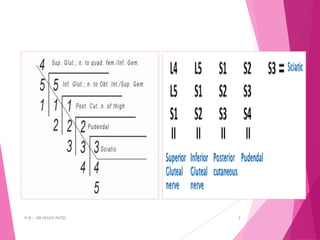
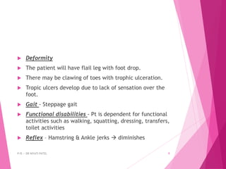
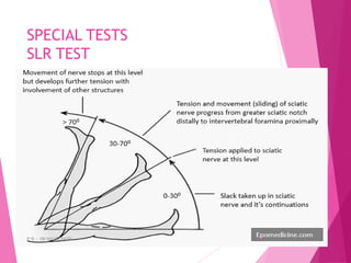
SCIATIC
NEUROPATHY
P/B :- DR NIYATI PATEL 1
LUMBOSACRAL PLEXUS
P/B :- DR NIYATI PATEL 2
P/B :- DR NIYATI PATEL 3
ANATOMY
 It is the thickest nerve of the body. Terminated from lumbosacral
plexus
 ROOT VALUE:- L4,L5,S1,S2
 Ventral division of ventral rami of L4,L5,S1,S2  TIBIAL NERVE
 Dorsal division of ventral rami of L4,L5,S1,S2  COMMON
PERONEAL NERVE
 MOTOR SUPPLY
 1. Hamstring (biceps femoris, semitendinosus, semimembranous,
ischial part of adductor magnus)
 2. Tibial nerve muscles – gastrocnemius, plantaris, soleus,
popliteus, tibialis posterior, flexor digitorum longus, flexor
halluces longus
 3. Common peroneal nerve muscles – tibialis anterior, extensor
halluces longus, extensor digitorum longus, extensor digitorum
brevis, peroneus tertius, peroneus longus, peroneus brevis
P/B :- DR NIYATI PATEL 4
P/B :- DR NIYATI PATEL 5
CAUSES
 • Penetrating wounds around the pelvis
 • Fractures of the pelvis and femur
 • Dislocation of the hip joint
 • Badly placed intramuscular injections in the
gluteal region
 • Compression within the pelvis by a neoplasm or
foetal head
 PIRIFORMIS SYNDROME - Nerve may undergo
entrapment or compression by piriformis muscle
as it traverses the sciatic notch.
P/B :- DR NIYATI PATEL 6
SIGN & SYMPTOMS
 Sensory
 There will be complete loss of sensation below the knee
except for the area that is supplied by the femoral
nerve (saphenous nerve).
 The autonomous zone for the sciatic nerve is the heel,
the skin over the metatarsal head in the sole, the
dorsum of the feet as far as medially up to the second
metatarsal as well as a small strip of the lateral aspect
of the leg.
P/B :- DR NIYATI PATEL 7
 Motor
 The muscles that will be paralyzed are biceps femoris,
semimembranous, semi tendinous, hamstring part of
adductor magnus.
 All the muscles supplied by the tibial and common
peroneal which are the branches of the sciatic nerve
will also be paralyzed.
P/B :- DR NIYATI PATEL 8
 Deformity
 The patient will have flail leg with foot drop.
 There may be clawing of toes with trophic ulceration.
 Tropic ulcers develop due to lack of sensation over the
foot.
 Gait - Steppage gait
 Functional disabilities - Pt is dependent for functional
activities such as walking, squatting, dressing, transfers,
toilet activities
 Reflex – Hamstring & Ankle jerks  diminishes
P/B :- DR NIYATI PATEL 9
INVESTIGATION
 RADIOGRAPH :- shows whether there is presence of fracture
 MRI :- To delineate complete avulsion of nerve roots
 SD CURVE:- abnormality in conduction can be verified.
Sharp curve, long chronaxie, low rheobase and the absence
of contraction with repetitive stimuli indicates
denervation. If it is done 2-3 weeks after injury, it shows
the sign of denervation and to find out whether it is
moderate or severe injury
 NCV:- To find out the severance of nerve fibers with
wallerian degeneration.
 EMG:- it will help to find out reversible and irreversible
nerve damage and will help map out whether it pre
ganglionic/ post ganglionic lesion
P/B :- DR NIYATI PATEL 10
TYPES OF INJURIES
 In Neuropraxia  pain, numbness, muscle
weakness, minimal muscle wasting is present.
Recovery occurs within minutes to days
 In Axonotmesis  there is pain, evident
muscle wasting, complete loss of motor,
sensory and sympathetic functions. Recovery
time– months (axon regeneration at 1-1.5
mm/day)
 In Neurotmesis  no pain, complete loss of
motor, sensory and sympathetic functions.
Recovery time – months and only with
surgery
P/B :- DR NIYATI PATEL 11
SPECIAL TESTS
SLR TEST
P/B :- DR NIYATI PATEL 12
TREATMENTS
 IG stimulation to the paralysed muscles
 Passive movements
 TA stretching
 Splintage: Night splints such as L splints may be given
mainly to prevent foot drop and contractures of the
plantar flexors. As the patient has intact quadriceps knee
stability is not affected hence below knee caliper such as
ankle foot orthosis may be prescribed that will help the
patient to be ambulatory in a much comfortable manner
 Padded foot wear or microcellular rubber foot wear
 Metatarsal bar may be given to the foot wear to prevent
metatarsal drop
 Care of anaesthetic foot
P/B :- DR NIYATI PATEL 13

7.SCIATIC NERVE.pdf

  • 1.
  • 2.
    LUMBOSACRAL PLEXUS P/B :-DR NIYATI PATEL 2
  • 3.
    P/B :- DRNIYATI PATEL 3
  • 4.
    ANATOMY  It isthe thickest nerve of the body. Terminated from lumbosacral plexus  ROOT VALUE:- L4,L5,S1,S2  Ventral division of ventral rami of L4,L5,S1,S2  TIBIAL NERVE  Dorsal division of ventral rami of L4,L5,S1,S2  COMMON PERONEAL NERVE  MOTOR SUPPLY  1. Hamstring (biceps femoris, semitendinosus, semimembranous, ischial part of adductor magnus)  2. Tibial nerve muscles – gastrocnemius, plantaris, soleus, popliteus, tibialis posterior, flexor digitorum longus, flexor halluces longus  3. Common peroneal nerve muscles – tibialis anterior, extensor halluces longus, extensor digitorum longus, extensor digitorum brevis, peroneus tertius, peroneus longus, peroneus brevis P/B :- DR NIYATI PATEL 4
  • 5.
    P/B :- DRNIYATI PATEL 5
  • 6.
    CAUSES  • Penetratingwounds around the pelvis  • Fractures of the pelvis and femur  • Dislocation of the hip joint  • Badly placed intramuscular injections in the gluteal region  • Compression within the pelvis by a neoplasm or foetal head  PIRIFORMIS SYNDROME - Nerve may undergo entrapment or compression by piriformis muscle as it traverses the sciatic notch. P/B :- DR NIYATI PATEL 6
  • 7.
    SIGN & SYMPTOMS Sensory  There will be complete loss of sensation below the knee except for the area that is supplied by the femoral nerve (saphenous nerve).  The autonomous zone for the sciatic nerve is the heel, the skin over the metatarsal head in the sole, the dorsum of the feet as far as medially up to the second metatarsal as well as a small strip of the lateral aspect of the leg. P/B :- DR NIYATI PATEL 7
  • 8.
     Motor  Themuscles that will be paralyzed are biceps femoris, semimembranous, semi tendinous, hamstring part of adductor magnus.  All the muscles supplied by the tibial and common peroneal which are the branches of the sciatic nerve will also be paralyzed. P/B :- DR NIYATI PATEL 8
  • 9.
     Deformity  Thepatient will have flail leg with foot drop.  There may be clawing of toes with trophic ulceration.  Tropic ulcers develop due to lack of sensation over the foot.  Gait - Steppage gait  Functional disabilities - Pt is dependent for functional activities such as walking, squatting, dressing, transfers, toilet activities  Reflex – Hamstring & Ankle jerks  diminishes P/B :- DR NIYATI PATEL 9
  • 10.
    INVESTIGATION  RADIOGRAPH :-shows whether there is presence of fracture  MRI :- To delineate complete avulsion of nerve roots  SD CURVE:- abnormality in conduction can be verified. Sharp curve, long chronaxie, low rheobase and the absence of contraction with repetitive stimuli indicates denervation. If it is done 2-3 weeks after injury, it shows the sign of denervation and to find out whether it is moderate or severe injury  NCV:- To find out the severance of nerve fibers with wallerian degeneration.  EMG:- it will help to find out reversible and irreversible nerve damage and will help map out whether it pre ganglionic/ post ganglionic lesion P/B :- DR NIYATI PATEL 10
  • 11.
    TYPES OF INJURIES In Neuropraxia  pain, numbness, muscle weakness, minimal muscle wasting is present. Recovery occurs within minutes to days  In Axonotmesis  there is pain, evident muscle wasting, complete loss of motor, sensory and sympathetic functions. Recovery time– months (axon regeneration at 1-1.5 mm/day)  In Neurotmesis  no pain, complete loss of motor, sensory and sympathetic functions. Recovery time – months and only with surgery P/B :- DR NIYATI PATEL 11
  • 12.
    SPECIAL TESTS SLR TEST P/B:- DR NIYATI PATEL 12
  • 13.
    TREATMENTS  IG stimulationto the paralysed muscles  Passive movements  TA stretching  Splintage: Night splints such as L splints may be given mainly to prevent foot drop and contractures of the plantar flexors. As the patient has intact quadriceps knee stability is not affected hence below knee caliper such as ankle foot orthosis may be prescribed that will help the patient to be ambulatory in a much comfortable manner  Padded foot wear or microcellular rubber foot wear  Metatarsal bar may be given to the foot wear to prevent metatarsal drop  Care of anaesthetic foot P/B :- DR NIYATI PATEL 13