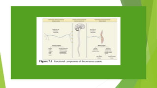
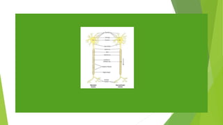
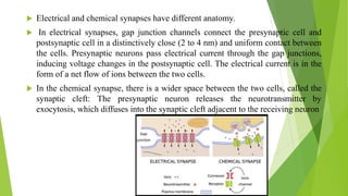
The nervous system consists of the brain, spinal cord and nerves. It detects changes inside and outside the body and responds through electrical signals called nerve impulses. Neurons conduct these impulses while neuroglia provide support. There are two main types of synapses - electrical and chemical. At chemical synapses, a neurotransmitter is released from the presynaptic neuron and binds to receptors on the postsynaptic neuron.