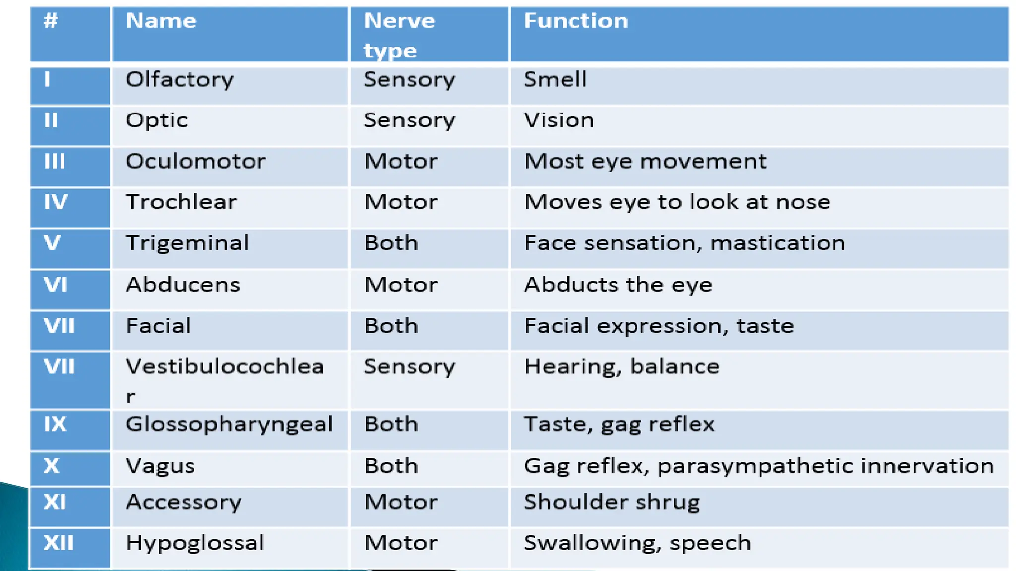
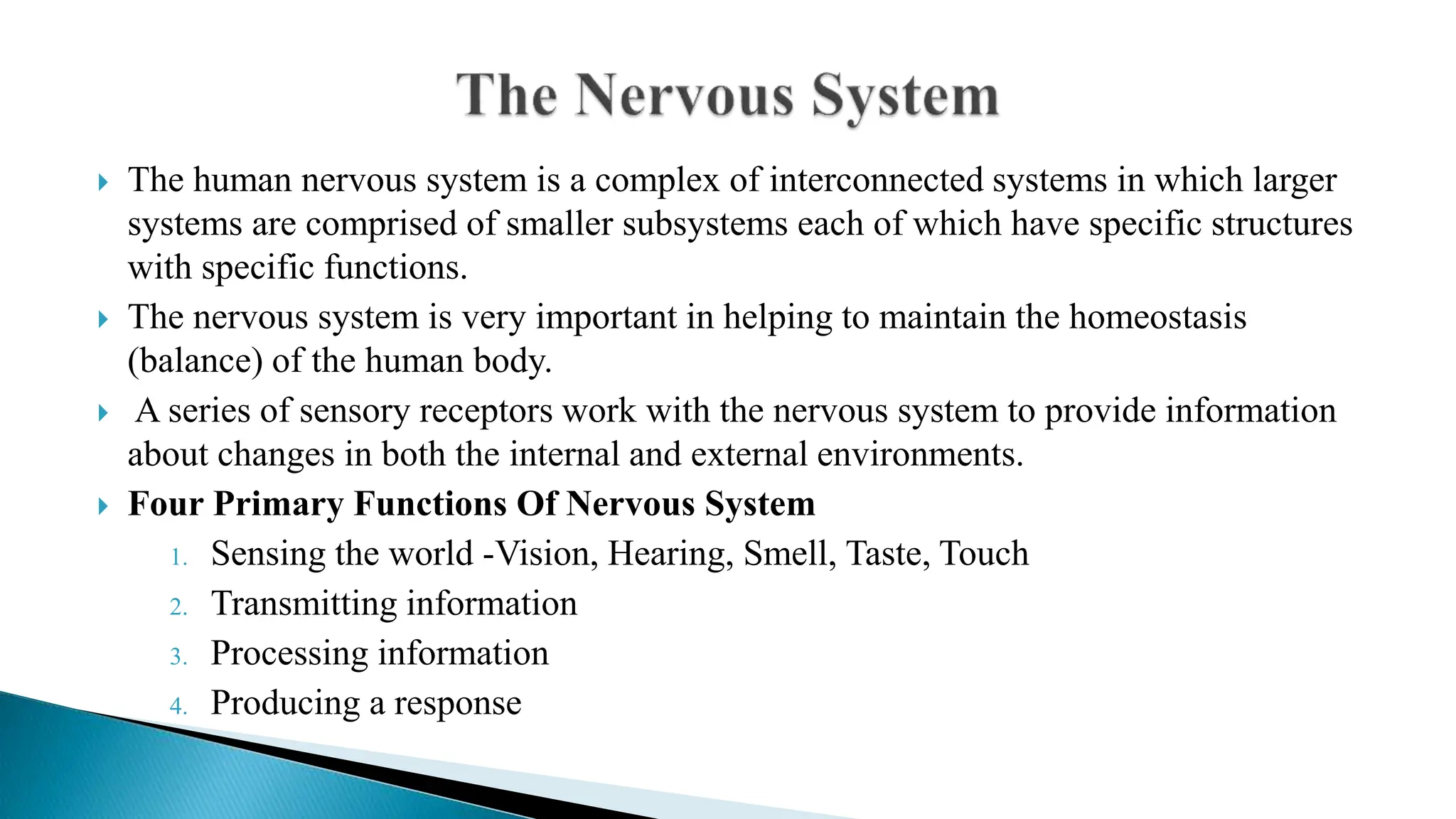
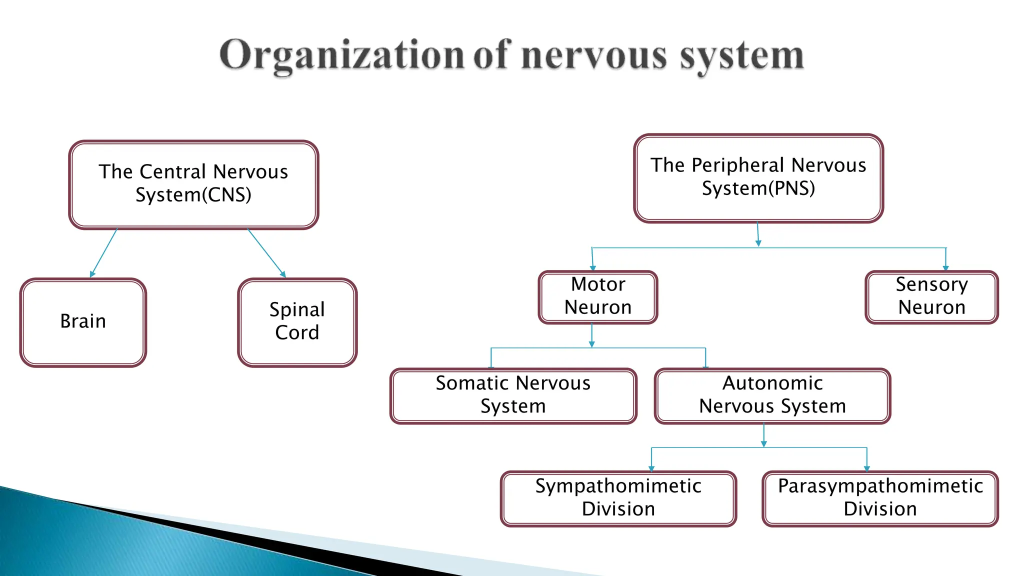
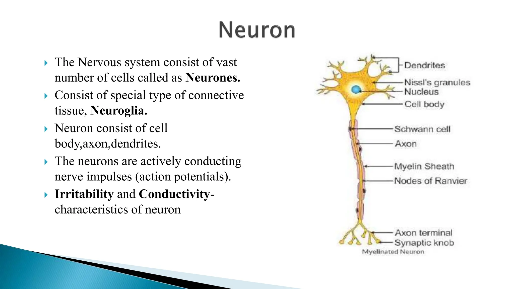
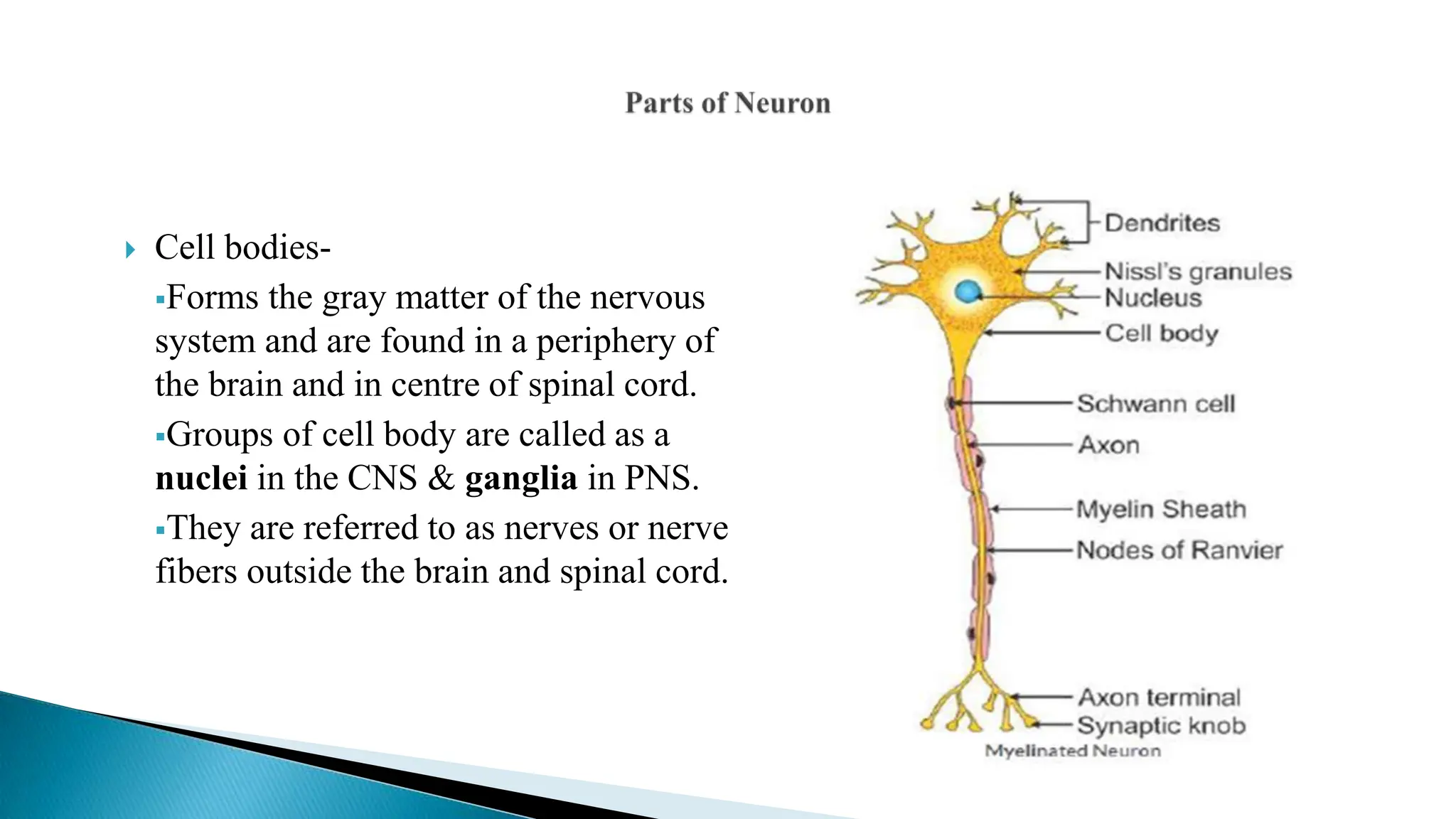
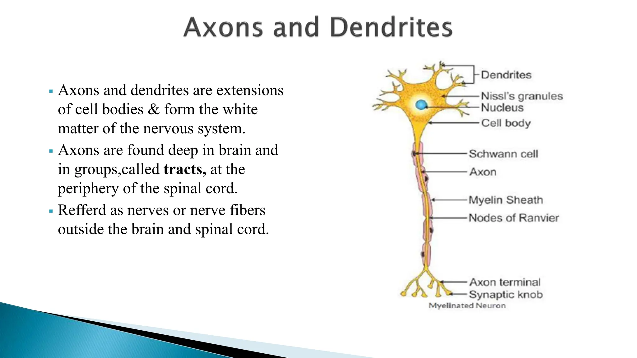
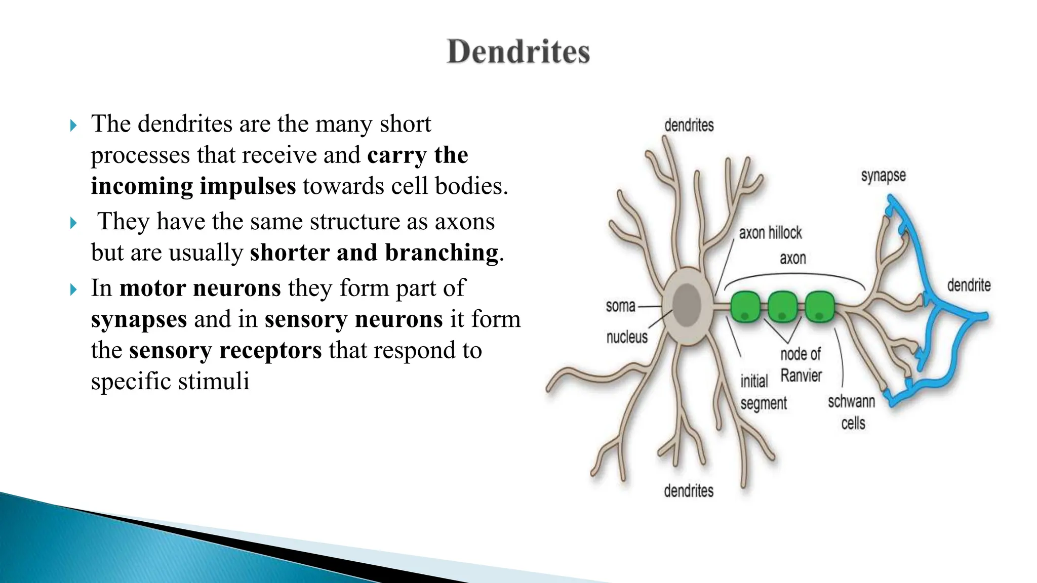
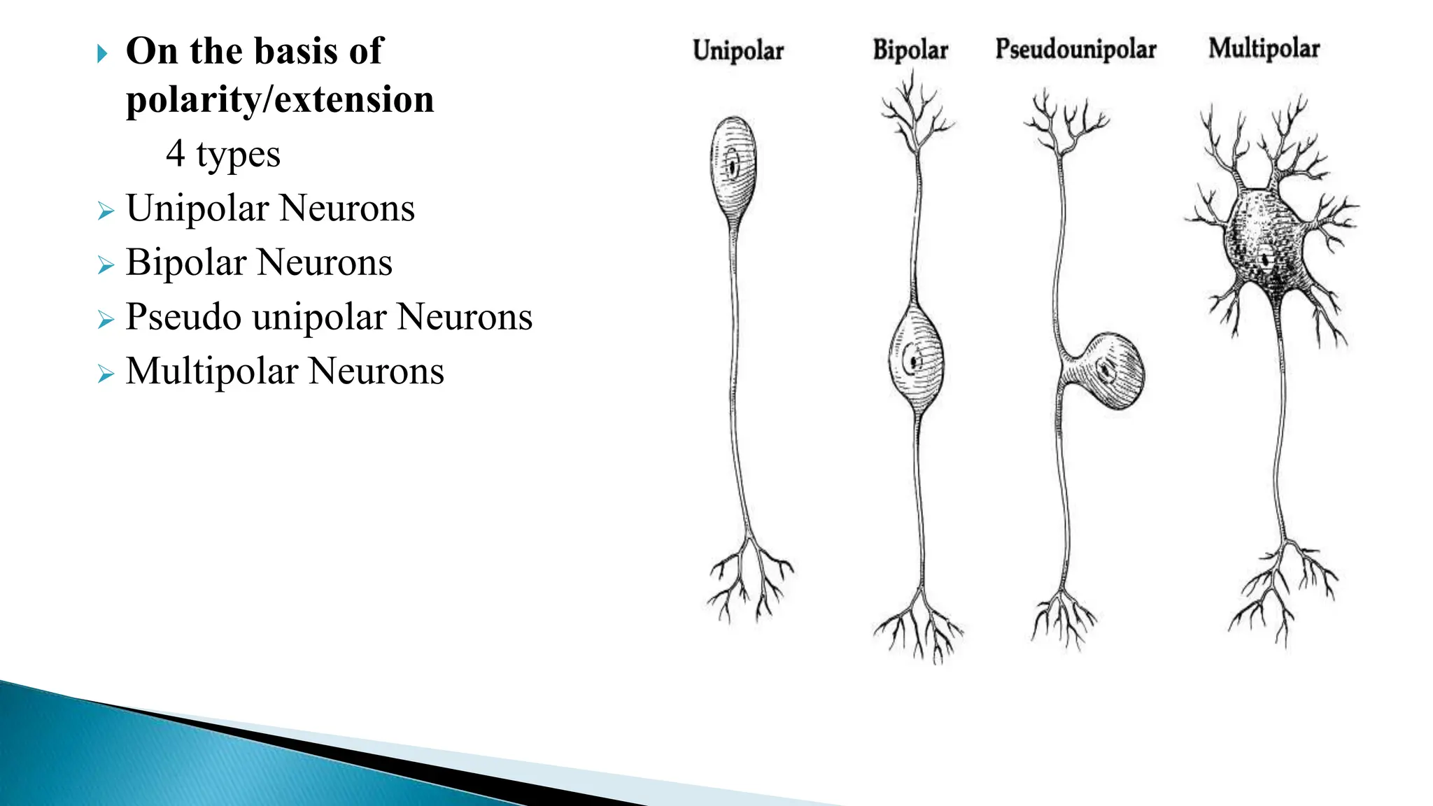
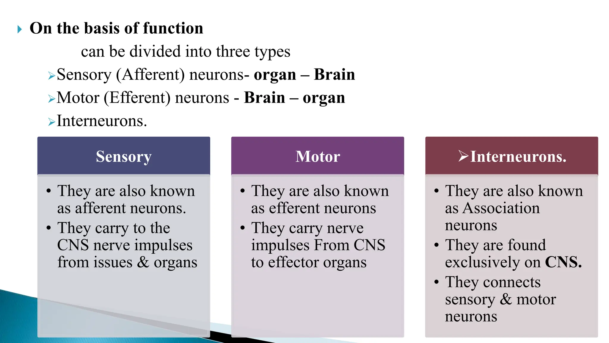
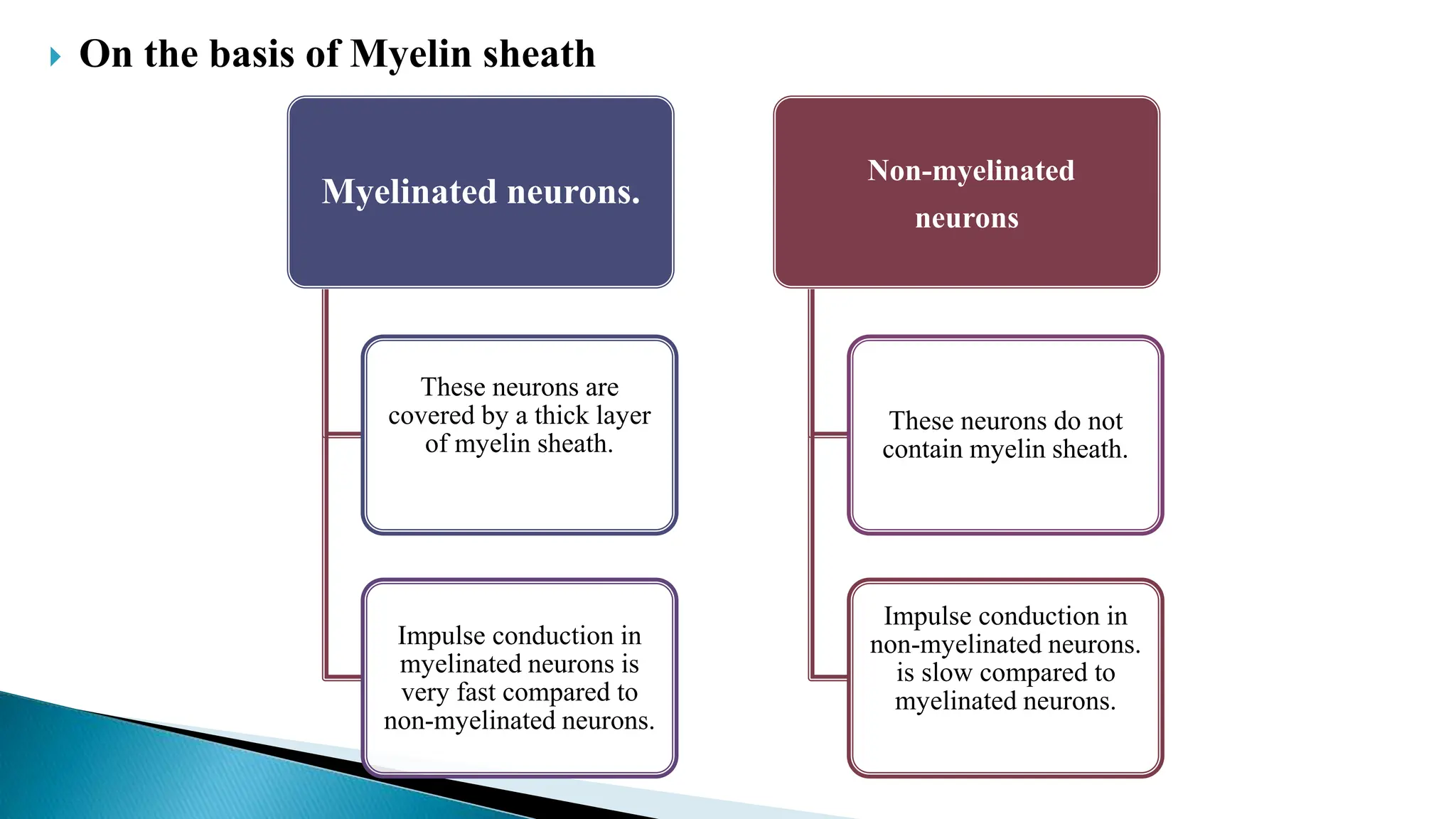
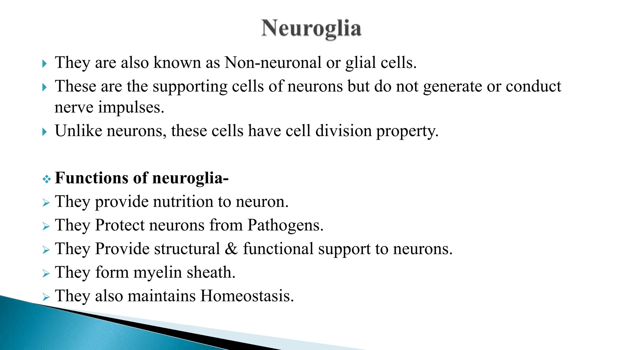
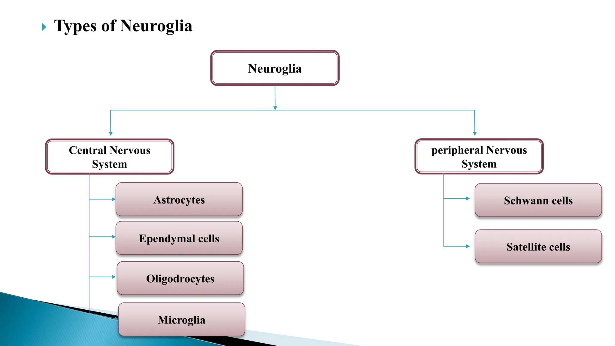
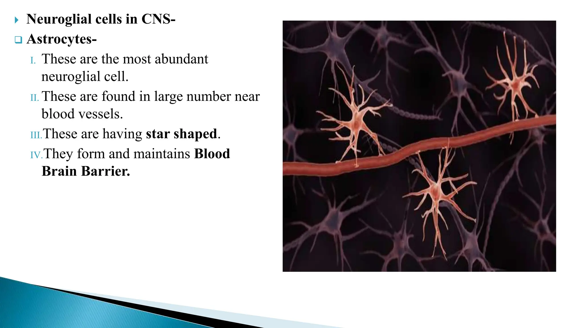
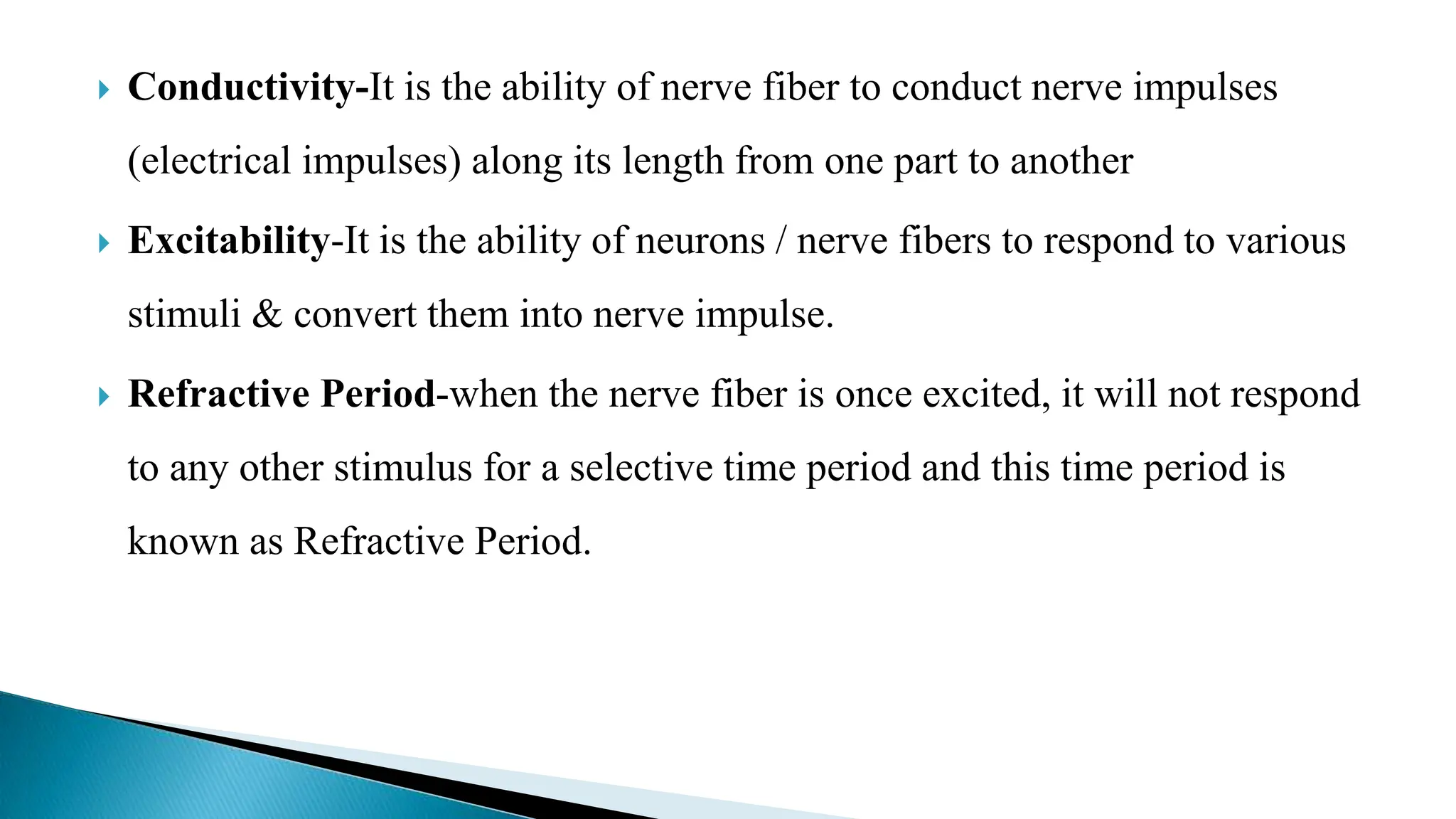
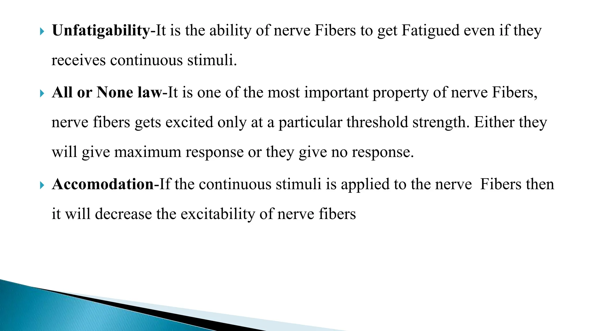
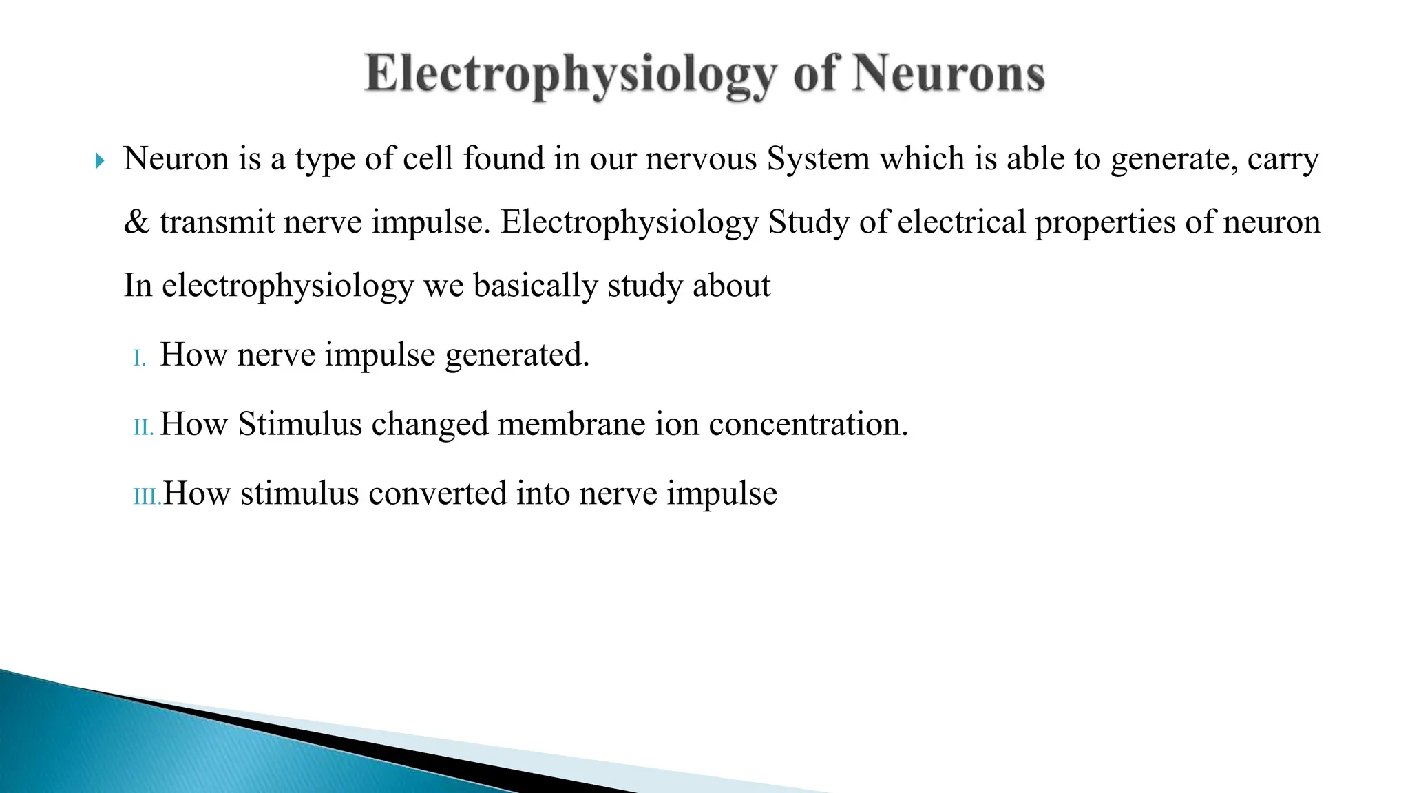
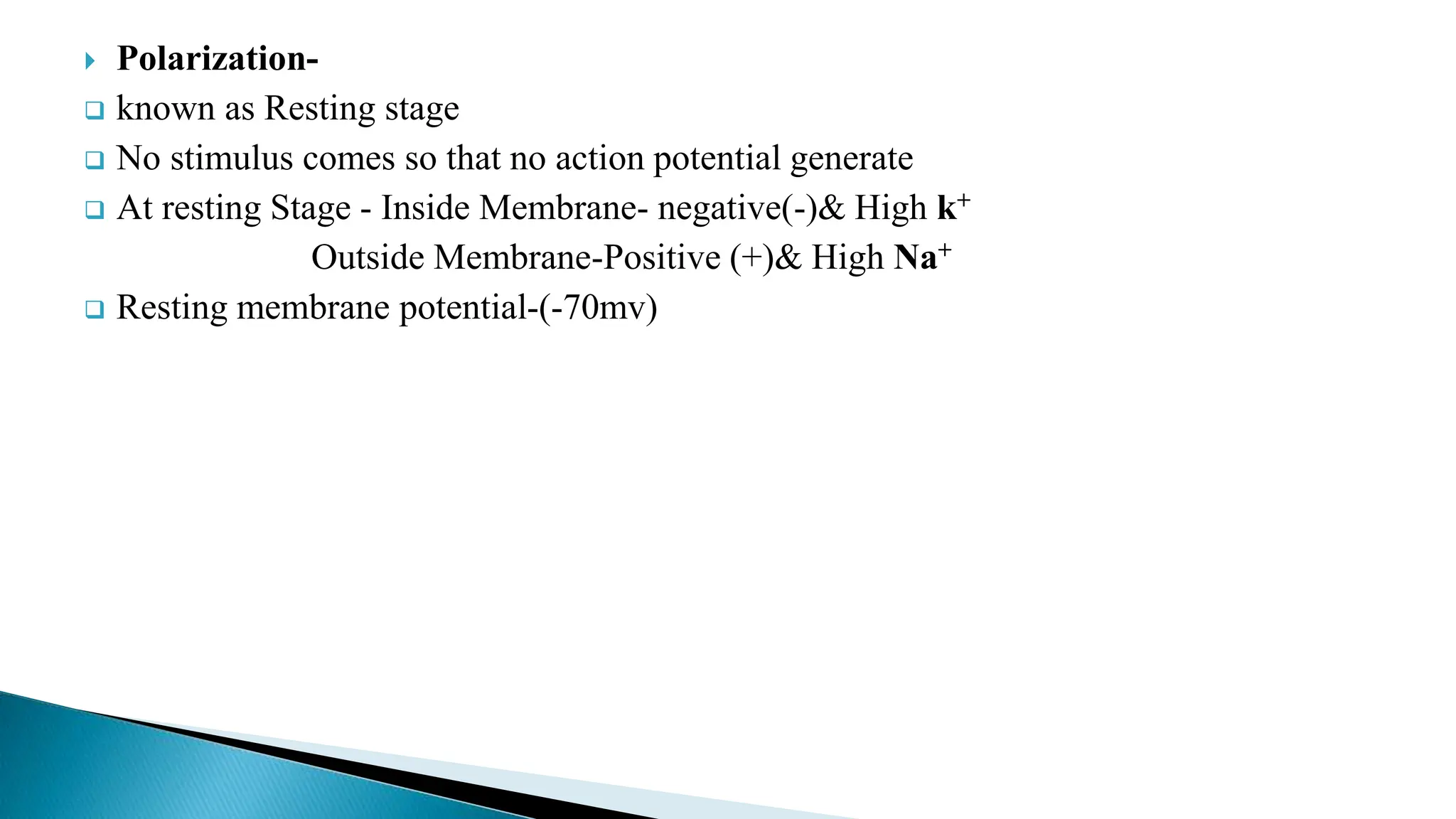
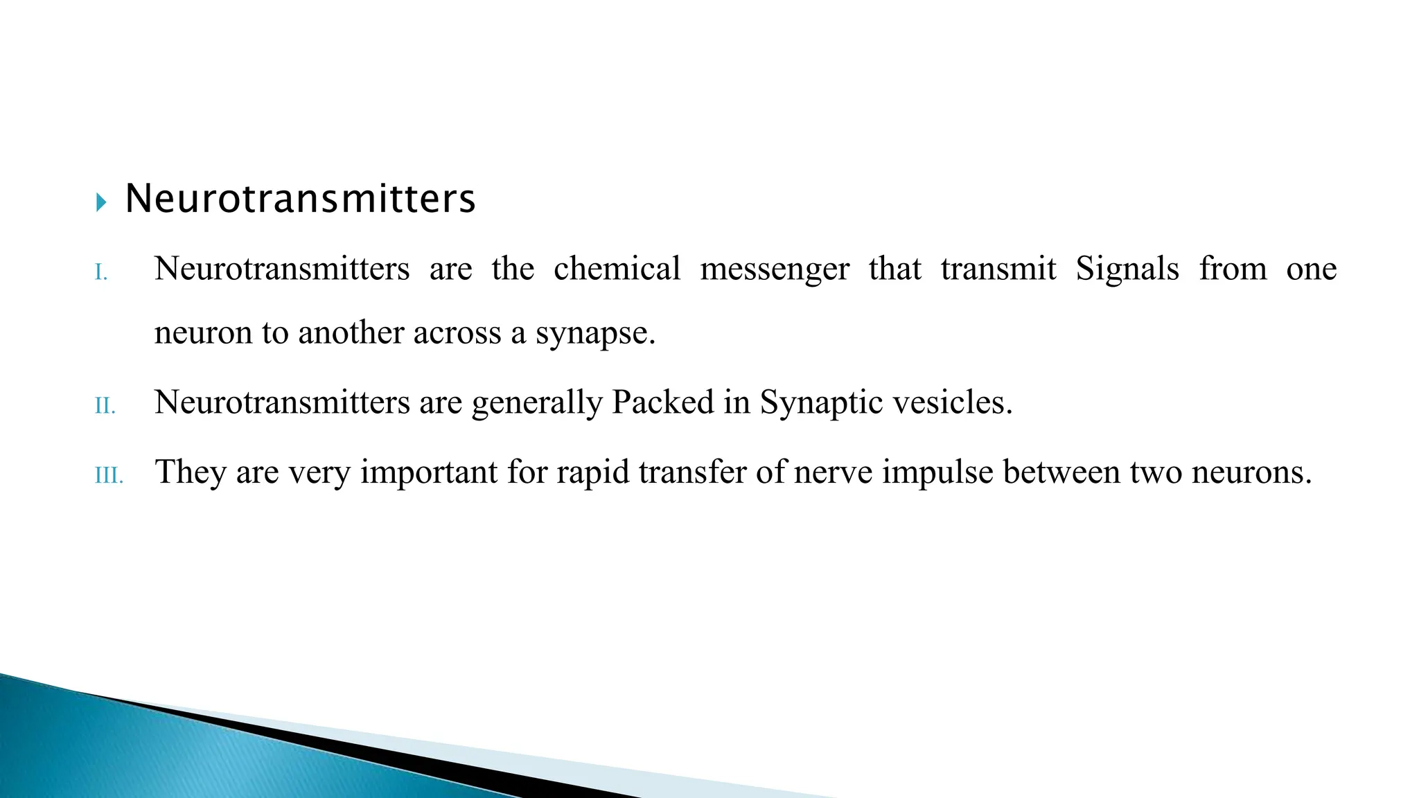
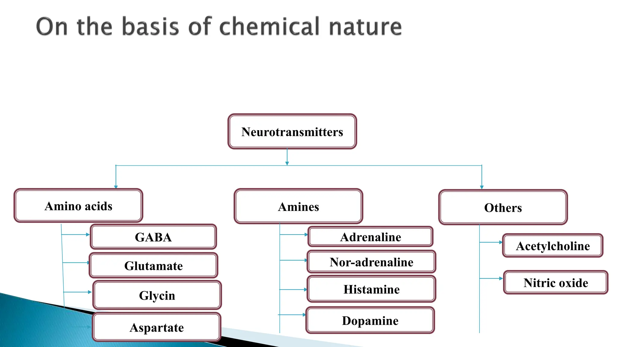
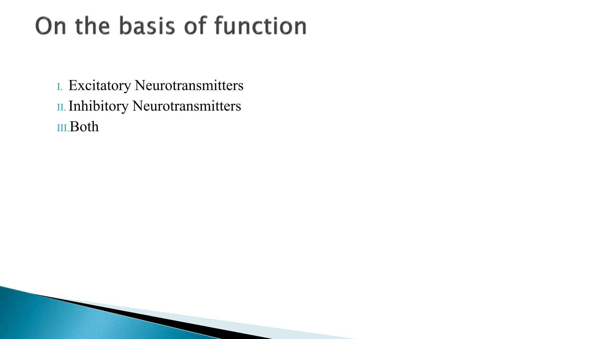
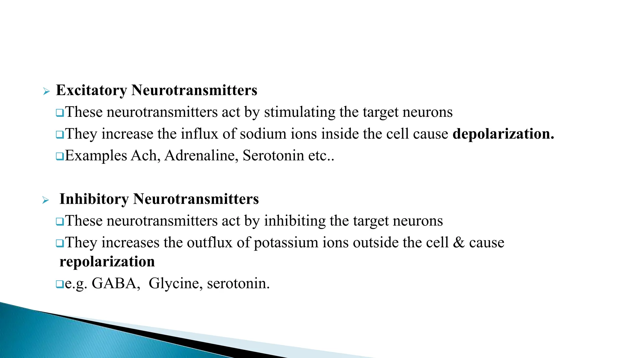
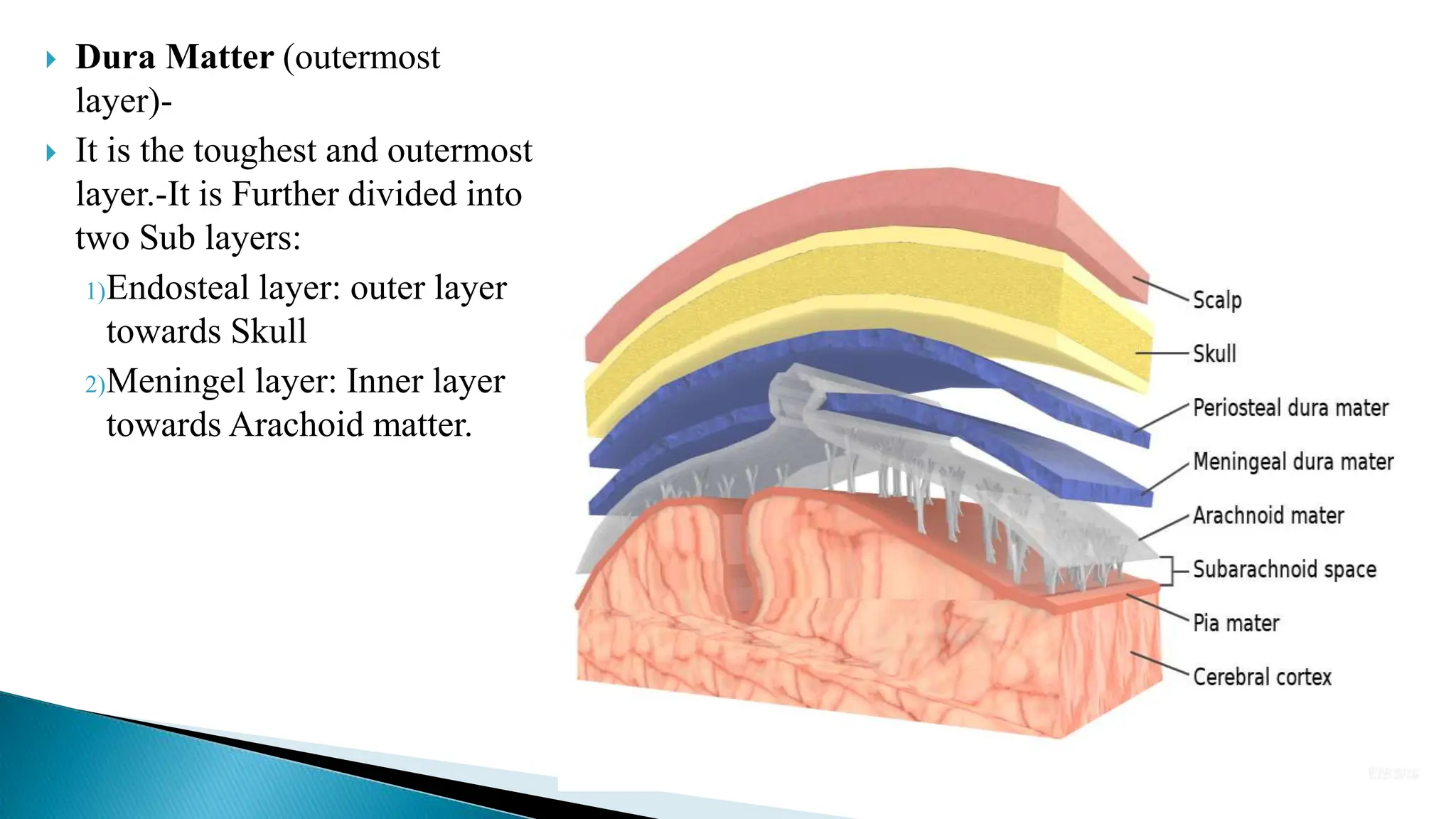
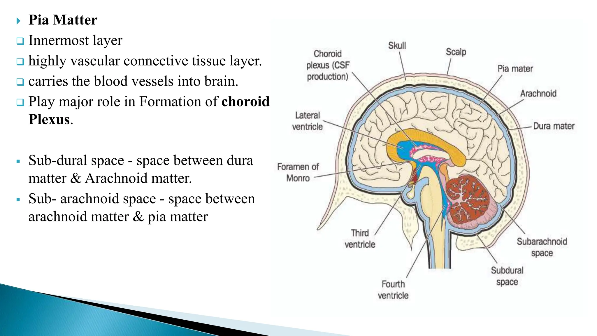
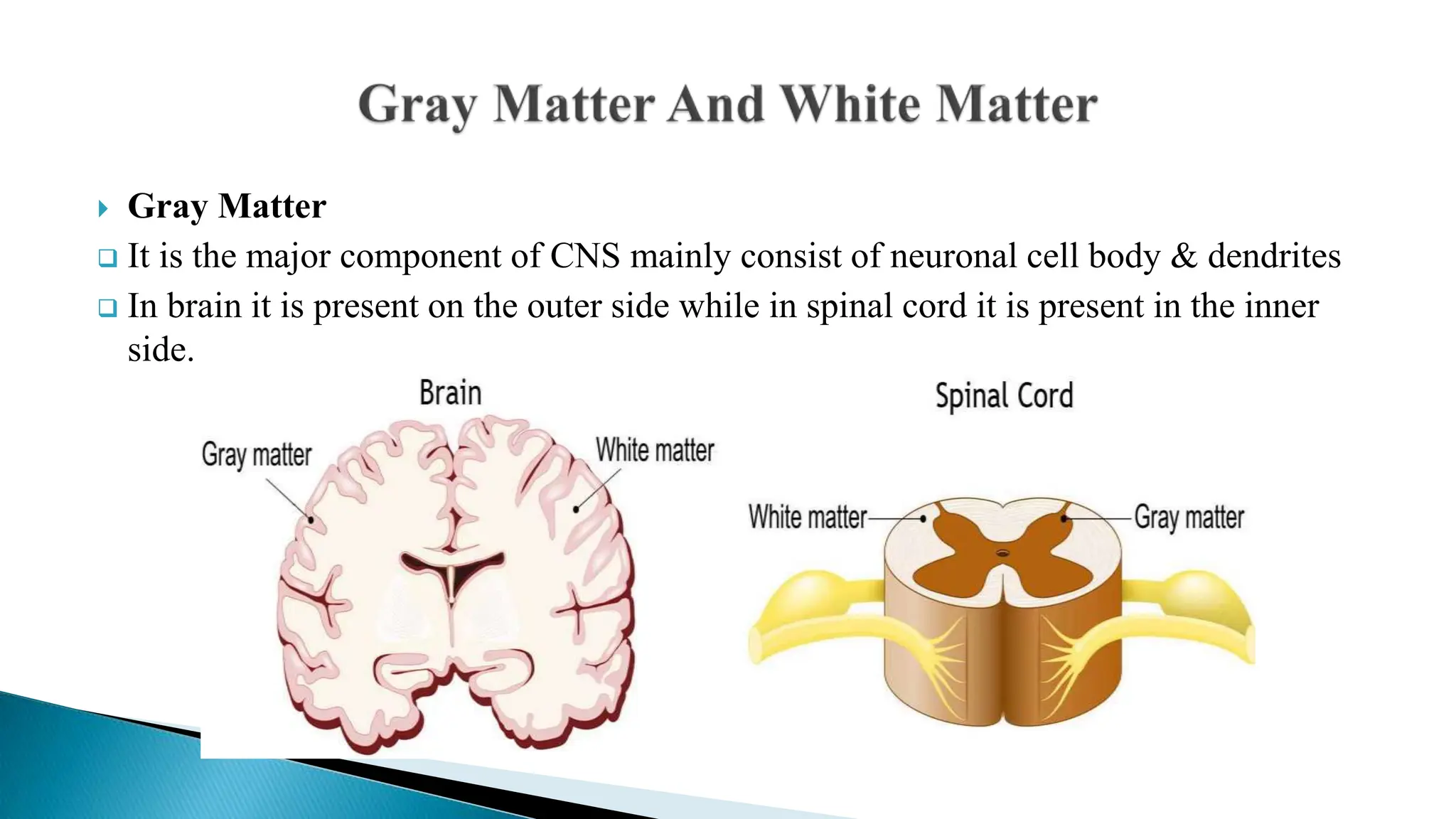
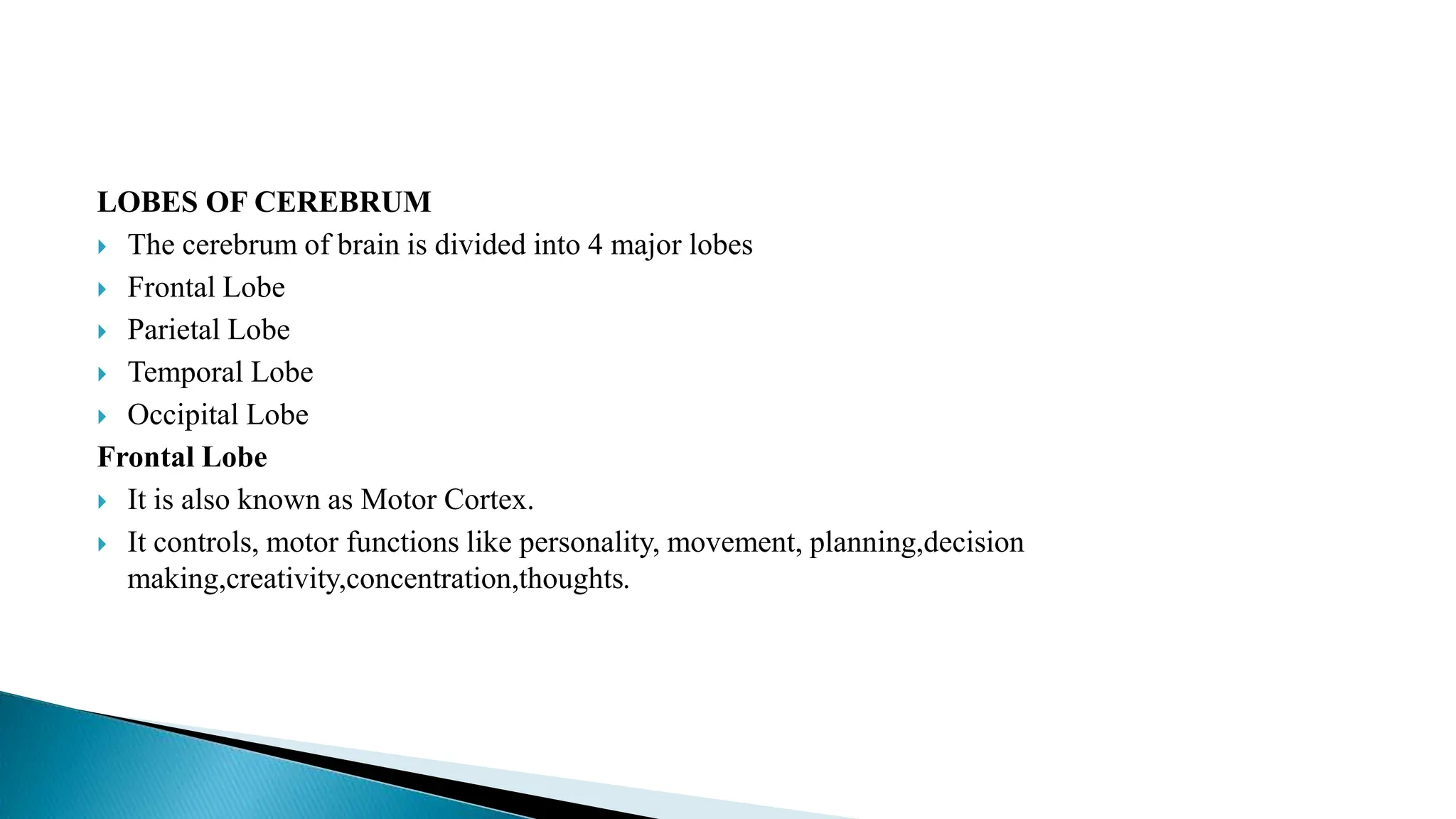
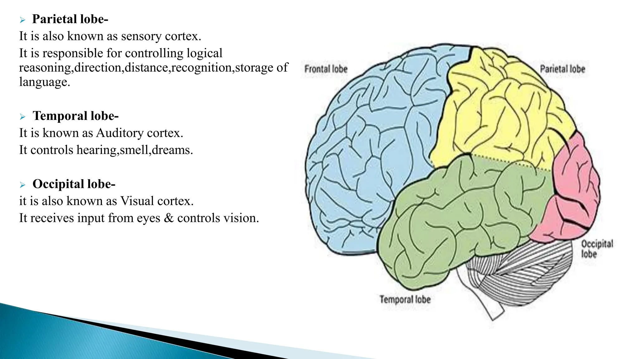
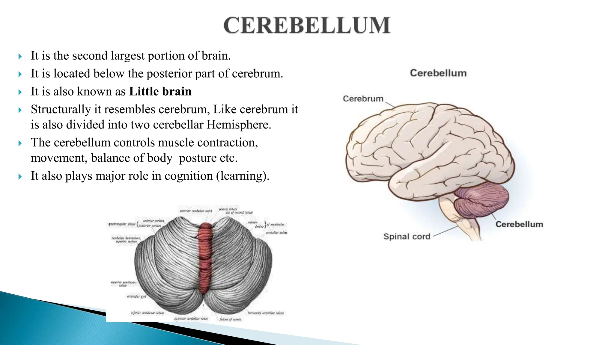
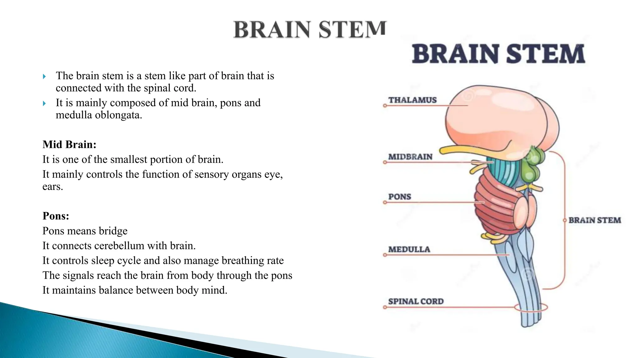
The document provides an extensive overview of the human nervous system, detailing its complex structure and functions, including the roles of various types of neurons, neuroglia, and the central and peripheral nervous systems. It discusses the basic properties of neurons, including action potentials and neurotransmission, along with the protective structures such as meninges and the cerebral spinal fluid. Additionally, it elaborates on the functional aspects of different brain regions and their associated roles in sensory and motor functions.


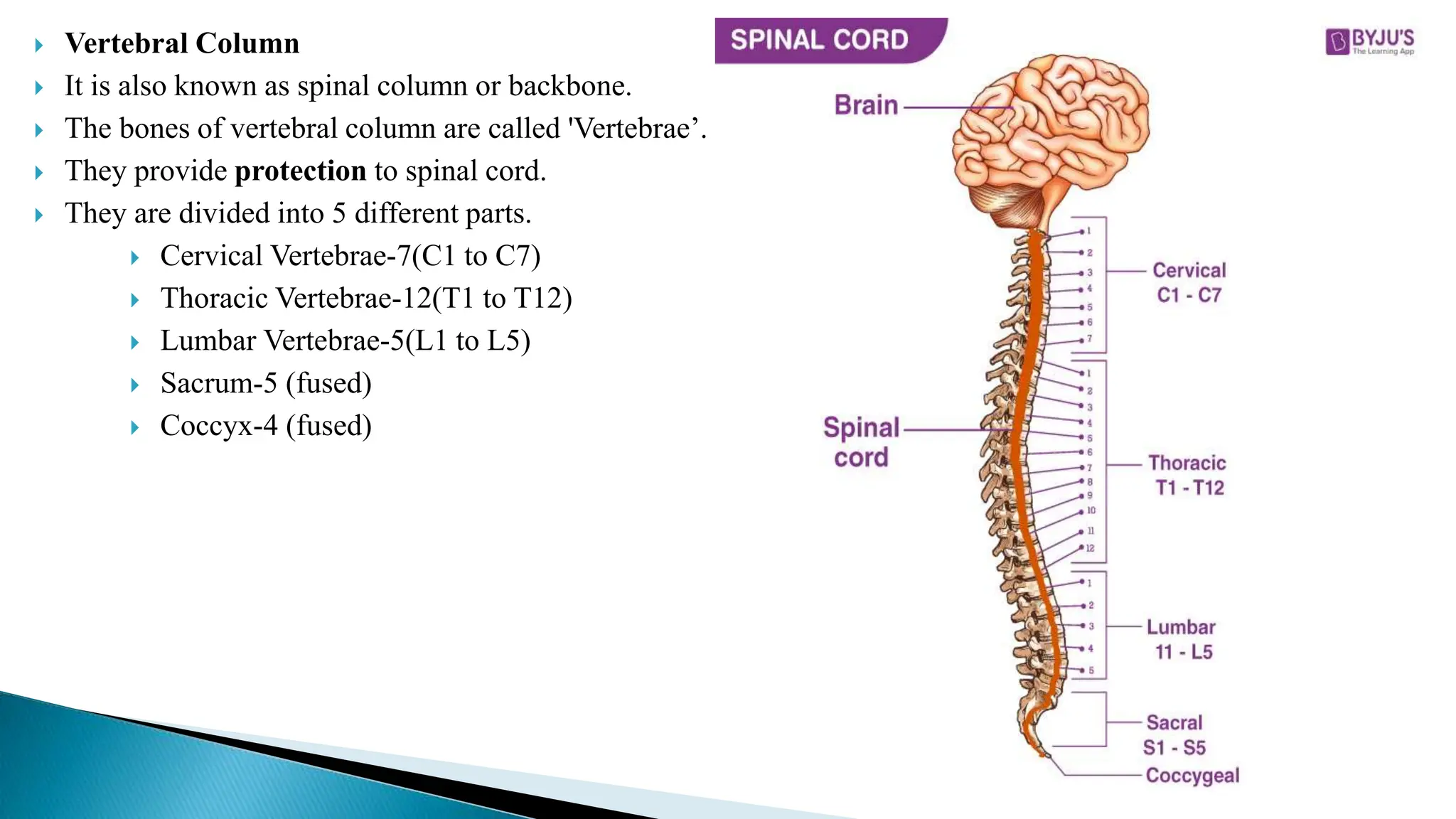
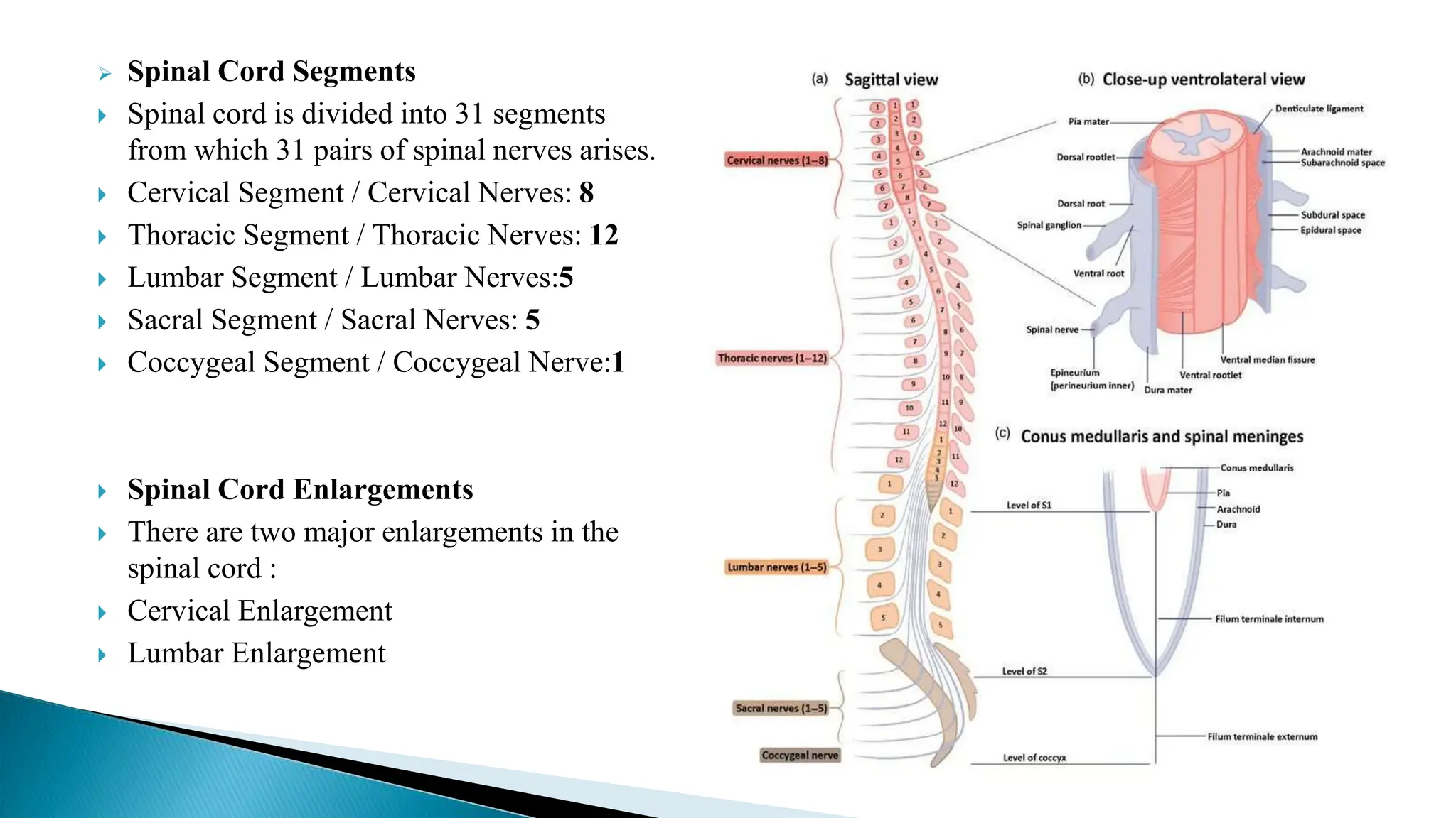
![ Cervical Enlargement-It is the superior
enlargement extends from 4th cervical vertebrae to
the 1st thoracic vertebrae [C3-T2].
Lumbar Enlargement-It is the inferior
enlargement extends from 9th to 12th thoracic
vertebrae [T9-T12]](https://image.slidesharecdn.com/unit1nervoussystem-240614105726-9d451133/75/Unit-1-Nervous-System-pptx-by-Nutan-Kamble-67-2048.jpg)