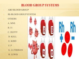
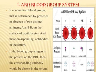
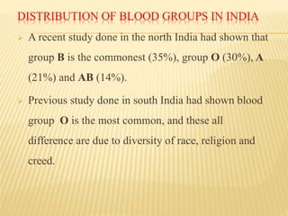
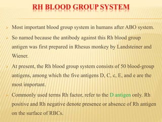
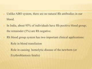
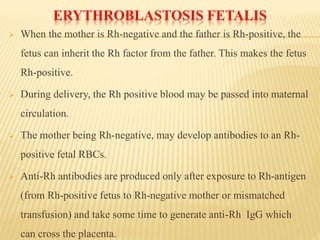
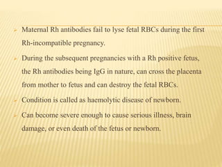
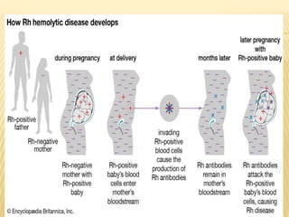
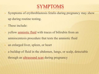
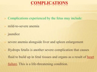
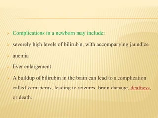
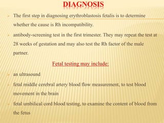
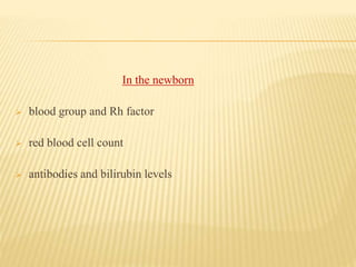
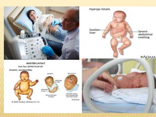
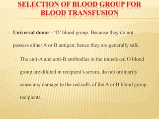
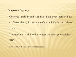
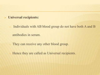
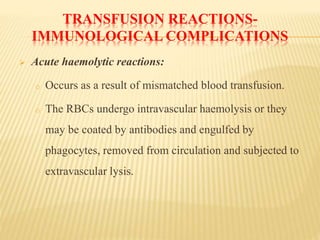
DR. MANISH
TIWARI
TUTOR
MICROBIOLOGY
INTRODUCTION
 Immunoheamatology= immuno+ heam+ logy = immune
system, heam= blood, logy= study.
 It is the study of blood group, blood transfusion, blood group
antigen, antigen- antibody reaction and the blood group
disorder is called immunoheamatolgy.
 Ehrlich and Morgenroth was 1st described blood group in goat.
 Karl Landsteiner,a Viennese pathologist , successfully
identified the ABO blood group in human.
APPLICATION IN ....
1. Transfusion of blood & its components
2. Diagnosis, prevention & management of immunization
associated with pregnancy
3. Leukocyte testing for organ transplantation
4. Laboratory resolution of parentage problems
AGGLUTINATION
 Agglutination reactions involve
particulate antigens capable of
binding antibody molecules.
 Since antibody molecules are
multivalent, suspended particulate
antigens form large clumps or
aggregates, easily visible without
magnification, when exposed to
specific antibodies.
COMPLETE OR INCOMPLETE ANTIBODIES
1. Complete antibody: which is
capable of agglutinating cells
in physiological saline.
2. Incomplete antibody: it is
bind to erythrocyte or bacteria
but does not produce
agglutination.
BLOOD GROUP SYSTEMS
1. ABO BLOOD GROUP
2. Rh BLOOD GROUP SYSTEM
3. OTHERS
A. MNS
B. I/i
C. DUFFY
D. KELL
E. KIDD
F. P
G. LUTHERAN
H. LEWIS
1. ABO BLOOD GROUP SYSTEM
 It contain four blood groups,
that is determined by presence
or absence of two distinct
antigens, A and B, on the
surface of erythrocytes. And
there crossponding antibodies
in the serum.
 If the blood group antigen is
the present on the RBC then
the crossponding antibody
would be absent in the serum.
GROUP- A
 Express A antigen on RBC surface
 Genotypes AA or AO
 Have naturally occurring, clinically significant, predominantly IgM
(with a small amount of IgG) antibodies against type B (anti-B)
Subgroups:
A1 (80%)
A2 (20%)
 Significance: some with A2 have antibodies against the A1
subgroup (anti-A1)
GROUP -B
 Express B Ag on RBC surface
 Genotypes BB or BO
 Have naturally occurring
clinically significant,
predominantly IgM (with a
small amount of IgG)
antibodies against type A cells
GROUP- O
 Have neither A nor B antigens on their RBC
 Genotype OO (“universal donors”)
 Have naturally occurring, clinically significant, very high titer,
anti-A, anti-B and anti-A,B antibodies
 Maternal anti-A,B can cross the placenta to cause hemolytic
disease of the newborn
 Group O cells have the most H antigen
GROUP -AB
 Express A and B Ag on
RBC surface
 Genotypes A1B or A2B
 have no ABO antibodies
(“universal recipients”)
OTHER BLOOD GROUP SYSTEMS:
 Kell (K,k,Kx)
 Duffy (Fy)
 Kidd (Jka, Jkb)
 MNSs
 P
 Lewis (Le)
 Lutheran (Lu)
NATURAL ISOANTIBODIES
 Anti a and anti b isoantibodies are called natural antibodies
because they are seen to arise without antigenic stimulation.
 They are igM in nature and it produced by the age of 6
months.
 ABO in other animal:- it is also present in the some other
animals for example rodents and apes, such as chimpanzees,
and gorillas.
H- ANTIGEN
 Seen on RBC of ABO-Universal distributed
 BOMBAY (Oh)PHENOTYPE:
 Patients lack the H gene and therefore cannot make H antigen, A
or B antigen on their red cells
 Have very strong anti-A, anti-B, and anti-H and can only receive
cells from a Bombay donor
DISTRIBUTION OF BLOOD GROUPS IN INDIA
 A recent study done in the north India had shown that
group B is the commonest (35%), group O (30%), A
(21%) and AB (14%).
 Previous study done in south India had shown blood
group O is the most common, and these all
difference are due to diversity of race, religion and
creed.
RH BLOOD GROUP SYSTEM
 Most important blood group system in humans after ABO system.
 So named because the antibody against this Rh blood group
antigen was first prepared in Rhesus monkey by Landsteiner and
Wiener.
 At present, the Rh blood group system consists of 50 blood-group
antigens, among which the five antigens D, C, c, E, and e are the
most important.
 Commonly used terms Rh factor, refer to the D antigen only. Rh
positive and Rh negative denote presence or absence of Rh antigen
on the surface of RBCs.
 Unlike ABO system, there are no natural Rh antibodies in our
blood.
 In India, about 95% of individuals have Rh positive blood group;
the remainder (5%) are Rh negative.
 Rh blood group system has two important clinical applications:
o Role in blood transfusion
o Role in causing hemolytic disease of the newborn (or
Erythroblastosis fetalis)
ERYTHROBLASTOSIS FETALIS
 When the mother is Rh-negative and the father is Rh-positive, the
fetus can inherit the Rh factor from the father. This makes the fetus
Rh-positive.
 During delivery, the Rh positive blood may be passed into maternal
circulation.
 The mother being Rh-negative, may develop antibodies to an Rh-
positive fetal RBCs.
 Anti-Rh antibodies are produced only after exposure to Rh-antigen
(from Rh-positive fetus to Rh-negative mother or mismatched
transfusion) and take some time to generate anti-Rh IgG which
can cross the placenta.
 Maternal Rh antibodies fail to lyse fetal RBCs during the first
Rh-incompatible pregnancy.
 During the subsequent pregnancies with a Rh positive fetus,
the Rh antibodies being IgG in nature, can cross the placenta
from mother to fetus and can destroy the fetal RBCs.
 Condition is called as haemolytic disease of newborn.
 Can become severe enough to cause serious illness, brain
damage, or even death of the fetus or newborn.
SYMPTOMS
 Symptoms of erythroblastosis fetalis during pregnancy may show
up during routine testing.
 These include:
 yellow amniotic fluid with traces of bilirubin from an
amniocentesis procedure that tests the amniotic fluid
 an enlarged liver, spleen, or heart
 a buildup of fluid in the abdomen, lungs, or scalp, detectable
through an ultrasound scan during pregnancy
 Newborns with the condition may display visible symptoms as
well as some that show up on scans, such as:
 pale skin
 yellow amniotic fluid, umbilical cord, skin, or eyes, either at birth
or within 24 to 36 hours of delivery
 spleen or liver enlargement
COMPLICATIONS
 Complications experienced by the fetus may include:
 mild-to-severe anemia
 jaundice
 severe anemia alongside liver and spleen enlargement
 Hydrops fetalis is another severe complication that causes
fluid to build up in fetal tissues and organs as a result of heart
failure. This is a life-threatening condition.
 Complications in a newborn may include:
 severely high levels of bilirubin, with accompanying jaundice
 anemia
 liver enlargement
 A buildup of bilirubin in the brain can lead to a complication
called kernicterus, leading to seizures, brain damage, deafness,
or death.
DIAGNOSIS
 The first step in diagnosing erythroblastosis fetalis is to determine
whether the cause is Rh incompatibility.
 antibody-screening test in the first trimester. They may repeat the test at
28 weeks of gestation and may also test the Rh factor of the male
partner.
Fetal testing may include:
 an ultrasound
 fetal middle cerebral artery blood flow measurement, to test blood
movement in the brain
 fetal umbilical cord blood testing, to examine the content of blood from
the fetus
In the newborn
 blood group and Rh factor
 red blood cell count
 antibodies and bilirubin levels
TREATMENT
 Treatment may include a fetal blood transfusion and delivery of
the fetus between 32 and 37 weeks gestation.
 Treatment options for newborns with the condition include:
 blood transfusion
 intravenous (IV) fluids
 managing breathing problems
 IV immunoglobulin (IVIG)
 The goal of IVIG antibody therapy is to reduce the breakdown of
red blood cells and levels of circulating bilirubin.
PREVENTION
 Erythroblastosis fetalis is a preventable condition.
 A medication called Rh immunoglobulin (Rhig), also known as
RhoGAM, can help prevent Rh sensitization.
 This medication prevents the pregnant woman from developing
Rh-positive antibodies.
 Women at risk for Rh sensitization should receive RhoGAM doses
at specific times during their pregnancy and after delivery.
 at 28 weeks of gestation
 72 hours following delivery, if the newborn is Rh-positive
 within 72 hours of a miscarriage, abortion, or ectopic pregnancy
 following an invasive prenatal test, such as an amniocentesis or
CVS
 after any vaginal bleeding
 If a woman has a pregnancy that extends beyond 40 weeks, the
doctor may recommend an additional dose of RhoGAM.
SAFE BLOOD TRANSFUSION PRACTICES
 The recipient's plasma should not contain any antibodies that
will damage the donor's RBCs.
 The donor plasma should not have any antibodies that will
damage the recipient's RBCs.
 The donor red cells should not have any antigen that is lacking
in the recipient RBCs. If the transfused cells possess a 'foreign
antigen' it will stimulate an immune response in the recipient.
SELECTION OF BLOOD GROUP FOR
BLOOD TRANSFUSION
 Universal donor - ‘O’ blood group. Because they do not
possess either A or B antigen; hence they are generally safe.
o The anti-A and anti-B antibodies in the transfused O blood
group are diluted in recipient’s serum, do not ordinarily
cause any damage to the red cells of the A or B blood group
recipients.
 Dangerous O group:
o Observed that if the anti-A and anti-B antibody titers are high
(1 :200 or above) in the serum of the individuals with O blood
group.
o Transfusion of such blood may result in damage to recipient’s
RBCs.
o Should not be used for transfusion.
 Universal recipients:
o Individuals with AB blood group do not have both A and B
antibodies in serum.
o They can receive any other blood group.
o Hence they are called as Universal recipients.
TRANSFUSION REACTIONS-
IMMUNOLOGICAL COMPLICATIONS
 Acute haemolytic reactions:
o Occurs as a result of mismatched blood transfusion.
o The RBCs undergo intravascular haemolysis or they
may be coated by antibodies and engulfed by
phagocytes, removed from circulation and subjected to
extravascular lysis.
o Symptoms - fever, chills, chest pain, back pain,
haemorrhage, increased heart rate, dyspnoea, hypotension
and rarely kidney injury.
o When transfusion reaction is suspected, transfusion should
be stopped immediately, and blood should be sent for tests
to evaluate for presence of hemolysis.
o Treatment is supportive.
 Delayed hemolytic reactions - occur in some cases of mismatched
blood transfusion. Mechanism similar to acute hemolytic reactions,
but they are milder and occur late.
 Febrile non-hemolytic reactions - most common type, occur due to
the release of inflammatory chemical signals by WBCs present in
stored donor blood.
 Allergic reactions occur when the recipient has preformed
antibodies to certain chemicals in the donor blood, and it does not
require prior exposure to blood transfusions.
 Post transfusion thrombocytopenia may occur following
transfusion containing platelets possessing surface protein Human
platelet antigen-1a (HPA-1a).
 Transfusion-associated acute lung injury (TRALI)-
Characterized by acute respiratory distress; probably mediated by
anti-HLA antibodies. It is more common in pregnancy
TRANSFUSION REACTIONS-
NON-IMMUNOLOGICAL COMPLICATIONS
Infections and iron overload. Bacteria
Infections: • Treponema pallidum
Viruses: • Leptospira interrogans
 Human immunodeficiency viruses
(HIV)
• Borrelia burgdorferi
 Hepatitis B virus (HBV) • Skin flora
 Hepatitis C virus, rarely hepatitis D
and hepatitis A viruses
Protozoa
 Cytomegalovirus (CMV) • Plasmodium species
 Human T-Lymphotropic Virus • Babesia species
 Slow virus causing variant
Creutzfeldt-Jakob disease
• Leishmania donovani
• Toxoplasma gondii
• Trypanosoma cruzi
COMMON TESTS USED IN
IMMUNOHEMATOLOGY
1. Coombs Test (Antiglobulin Test)
2. RBC typing
3. Cross matching
4. Antibody Screening
5. Compatibility Testing
MCQ
1. Which of the following is not true about blood group B?
a) Universal blood group
b) Have igM
c) Have B antigen
d) Have a antiA antibody
2. Which of the following antibody are responsible for hemolytic
disease of newborn?
a) igA
b) igD
c) igM
d) igG
3. Which of the following statement is true about Rh blood
group?
a) It determine the negative & positive.
b) It consist of 50 blood group antigen
c) Only A
d) A & B
4. Which of the following are immunological complication of
transfusion reaction?
a) Delayed hemolytic reactions
b) Allergic reactions
c) Transfusion-associated acute lung injury
d) All
5. If the blood of two different groups is mixed together, what
problem is observed?
a) Coagulation
b) Agglutination
c) Only B
d) A& B
6. Which of the following is known as universal recipient?
a) A
b) B
c) AB
d) O
7. Which of the following combination of Rh group of mother
and fetus is lethal for fetus?
a) Rh (-) mother & Rh (+) fetus
b) Rh (+) mother & Rh (+) fetus
c) Rh (+) mother & Rh (-) fetus
d) Rh (-) mother & Rh (-) fetus
8. On what basis of blood group is classified?
a) Antigen
b) Antibody
c) Rh factor
d) A&B
9. Which of the following infectious agents are responsible for
transfusion reaction?
a. HIV
b. Toxoplasma
c. CMV
d. All
10. Rh factor of the blood was discovered by:
a) Louis Pasteur
b) Landsteiner and Weiner
c) Janskey
d) None of these
11. Which of the following statement is NOT true?
a) About 95% of individuals have Rh positive blood group; the
remainder (5%) are Rh negative.
b) ABO and Rh groups are based on antigen-antibody reactions
c) People having A-antigen on RBC surface have anti-A antibody in
their plasma.
d) The ABO system was first discovered in 1950s.
WRITE SHORT NOTES ON:
1. Hemolytic disease of newborn (7marks)
Immunohematology

Immunohematology

  • 1.
  • 2.
    INTRODUCTION  Immunoheamatology= immuno+heam+ logy = immune system, heam= blood, logy= study.  It is the study of blood group, blood transfusion, blood group antigen, antigen- antibody reaction and the blood group disorder is called immunoheamatolgy.  Ehrlich and Morgenroth was 1st described blood group in goat.  Karl Landsteiner,a Viennese pathologist , successfully identified the ABO blood group in human.
  • 3.
    APPLICATION IN .... 1.Transfusion of blood & its components 2. Diagnosis, prevention & management of immunization associated with pregnancy 3. Leukocyte testing for organ transplantation 4. Laboratory resolution of parentage problems
  • 4.
    AGGLUTINATION  Agglutination reactionsinvolve particulate antigens capable of binding antibody molecules.  Since antibody molecules are multivalent, suspended particulate antigens form large clumps or aggregates, easily visible without magnification, when exposed to specific antibodies.
  • 5.
    COMPLETE OR INCOMPLETEANTIBODIES 1. Complete antibody: which is capable of agglutinating cells in physiological saline. 2. Incomplete antibody: it is bind to erythrocyte or bacteria but does not produce agglutination.
  • 6.
    BLOOD GROUP SYSTEMS 1.ABO BLOOD GROUP 2. Rh BLOOD GROUP SYSTEM 3. OTHERS A. MNS B. I/i C. DUFFY D. KELL E. KIDD F. P G. LUTHERAN H. LEWIS
  • 7.
    1. ABO BLOODGROUP SYSTEM  It contain four blood groups, that is determined by presence or absence of two distinct antigens, A and B, on the surface of erythrocytes. And there crossponding antibodies in the serum.  If the blood group antigen is the present on the RBC then the crossponding antibody would be absent in the serum.
  • 8.
    GROUP- A  ExpressA antigen on RBC surface  Genotypes AA or AO  Have naturally occurring, clinically significant, predominantly IgM (with a small amount of IgG) antibodies against type B (anti-B) Subgroups: A1 (80%) A2 (20%)  Significance: some with A2 have antibodies against the A1 subgroup (anti-A1)
  • 9.
    GROUP -B  ExpressB Ag on RBC surface  Genotypes BB or BO  Have naturally occurring clinically significant, predominantly IgM (with a small amount of IgG) antibodies against type A cells
  • 10.
    GROUP- O  Haveneither A nor B antigens on their RBC  Genotype OO (“universal donors”)  Have naturally occurring, clinically significant, very high titer, anti-A, anti-B and anti-A,B antibodies  Maternal anti-A,B can cross the placenta to cause hemolytic disease of the newborn  Group O cells have the most H antigen
  • 11.
    GROUP -AB  ExpressA and B Ag on RBC surface  Genotypes A1B or A2B  have no ABO antibodies (“universal recipients”)
  • 12.
    OTHER BLOOD GROUPSYSTEMS:  Kell (K,k,Kx)  Duffy (Fy)  Kidd (Jka, Jkb)  MNSs  P  Lewis (Le)  Lutheran (Lu)
  • 13.
    NATURAL ISOANTIBODIES  Antia and anti b isoantibodies are called natural antibodies because they are seen to arise without antigenic stimulation.  They are igM in nature and it produced by the age of 6 months.  ABO in other animal:- it is also present in the some other animals for example rodents and apes, such as chimpanzees, and gorillas.
  • 14.
    H- ANTIGEN  Seenon RBC of ABO-Universal distributed  BOMBAY (Oh)PHENOTYPE:  Patients lack the H gene and therefore cannot make H antigen, A or B antigen on their red cells  Have very strong anti-A, anti-B, and anti-H and can only receive cells from a Bombay donor
  • 15.
    DISTRIBUTION OF BLOODGROUPS IN INDIA  A recent study done in the north India had shown that group B is the commonest (35%), group O (30%), A (21%) and AB (14%).  Previous study done in south India had shown blood group O is the most common, and these all difference are due to diversity of race, religion and creed.
  • 16.
    RH BLOOD GROUPSYSTEM  Most important blood group system in humans after ABO system.  So named because the antibody against this Rh blood group antigen was first prepared in Rhesus monkey by Landsteiner and Wiener.  At present, the Rh blood group system consists of 50 blood-group antigens, among which the five antigens D, C, c, E, and e are the most important.  Commonly used terms Rh factor, refer to the D antigen only. Rh positive and Rh negative denote presence or absence of Rh antigen on the surface of RBCs.
  • 17.
     Unlike ABOsystem, there are no natural Rh antibodies in our blood.  In India, about 95% of individuals have Rh positive blood group; the remainder (5%) are Rh negative.  Rh blood group system has two important clinical applications: o Role in blood transfusion o Role in causing hemolytic disease of the newborn (or Erythroblastosis fetalis)
  • 18.
    ERYTHROBLASTOSIS FETALIS  Whenthe mother is Rh-negative and the father is Rh-positive, the fetus can inherit the Rh factor from the father. This makes the fetus Rh-positive.  During delivery, the Rh positive blood may be passed into maternal circulation.  The mother being Rh-negative, may develop antibodies to an Rh- positive fetal RBCs.  Anti-Rh antibodies are produced only after exposure to Rh-antigen (from Rh-positive fetus to Rh-negative mother or mismatched transfusion) and take some time to generate anti-Rh IgG which can cross the placenta.
  • 19.
     Maternal Rhantibodies fail to lyse fetal RBCs during the first Rh-incompatible pregnancy.  During the subsequent pregnancies with a Rh positive fetus, the Rh antibodies being IgG in nature, can cross the placenta from mother to fetus and can destroy the fetal RBCs.  Condition is called as haemolytic disease of newborn.  Can become severe enough to cause serious illness, brain damage, or even death of the fetus or newborn.
  • 21.
    SYMPTOMS  Symptoms oferythroblastosis fetalis during pregnancy may show up during routine testing.  These include:  yellow amniotic fluid with traces of bilirubin from an amniocentesis procedure that tests the amniotic fluid  an enlarged liver, spleen, or heart  a buildup of fluid in the abdomen, lungs, or scalp, detectable through an ultrasound scan during pregnancy
  • 22.
     Newborns withthe condition may display visible symptoms as well as some that show up on scans, such as:  pale skin  yellow amniotic fluid, umbilical cord, skin, or eyes, either at birth or within 24 to 36 hours of delivery  spleen or liver enlargement
  • 23.
    COMPLICATIONS  Complications experiencedby the fetus may include:  mild-to-severe anemia  jaundice  severe anemia alongside liver and spleen enlargement  Hydrops fetalis is another severe complication that causes fluid to build up in fetal tissues and organs as a result of heart failure. This is a life-threatening condition.
  • 24.
     Complications ina newborn may include:  severely high levels of bilirubin, with accompanying jaundice  anemia  liver enlargement  A buildup of bilirubin in the brain can lead to a complication called kernicterus, leading to seizures, brain damage, deafness, or death.
  • 25.
    DIAGNOSIS  The firststep in diagnosing erythroblastosis fetalis is to determine whether the cause is Rh incompatibility.  antibody-screening test in the first trimester. They may repeat the test at 28 weeks of gestation and may also test the Rh factor of the male partner. Fetal testing may include:  an ultrasound  fetal middle cerebral artery blood flow measurement, to test blood movement in the brain  fetal umbilical cord blood testing, to examine the content of blood from the fetus
  • 26.
    In the newborn blood group and Rh factor  red blood cell count  antibodies and bilirubin levels
  • 28.
    TREATMENT  Treatment mayinclude a fetal blood transfusion and delivery of the fetus between 32 and 37 weeks gestation.  Treatment options for newborns with the condition include:  blood transfusion  intravenous (IV) fluids  managing breathing problems  IV immunoglobulin (IVIG)  The goal of IVIG antibody therapy is to reduce the breakdown of red blood cells and levels of circulating bilirubin.
  • 29.
    PREVENTION  Erythroblastosis fetalisis a preventable condition.  A medication called Rh immunoglobulin (Rhig), also known as RhoGAM, can help prevent Rh sensitization.  This medication prevents the pregnant woman from developing Rh-positive antibodies.  Women at risk for Rh sensitization should receive RhoGAM doses at specific times during their pregnancy and after delivery.
  • 30.
     at 28weeks of gestation  72 hours following delivery, if the newborn is Rh-positive  within 72 hours of a miscarriage, abortion, or ectopic pregnancy  following an invasive prenatal test, such as an amniocentesis or CVS  after any vaginal bleeding  If a woman has a pregnancy that extends beyond 40 weeks, the doctor may recommend an additional dose of RhoGAM.
  • 31.
    SAFE BLOOD TRANSFUSIONPRACTICES  The recipient's plasma should not contain any antibodies that will damage the donor's RBCs.  The donor plasma should not have any antibodies that will damage the recipient's RBCs.  The donor red cells should not have any antigen that is lacking in the recipient RBCs. If the transfused cells possess a 'foreign antigen' it will stimulate an immune response in the recipient.
  • 32.
    SELECTION OF BLOODGROUP FOR BLOOD TRANSFUSION  Universal donor - ‘O’ blood group. Because they do not possess either A or B antigen; hence they are generally safe. o The anti-A and anti-B antibodies in the transfused O blood group are diluted in recipient’s serum, do not ordinarily cause any damage to the red cells of the A or B blood group recipients.
  • 33.
     Dangerous Ogroup: o Observed that if the anti-A and anti-B antibody titers are high (1 :200 or above) in the serum of the individuals with O blood group. o Transfusion of such blood may result in damage to recipient’s RBCs. o Should not be used for transfusion.
  • 34.
     Universal recipients: oIndividuals with AB blood group do not have both A and B antibodies in serum. o They can receive any other blood group. o Hence they are called as Universal recipients.
  • 35.
    TRANSFUSION REACTIONS- IMMUNOLOGICAL COMPLICATIONS Acute haemolytic reactions: o Occurs as a result of mismatched blood transfusion. o The RBCs undergo intravascular haemolysis or they may be coated by antibodies and engulfed by phagocytes, removed from circulation and subjected to extravascular lysis.
  • 36.
    o Symptoms -fever, chills, chest pain, back pain, haemorrhage, increased heart rate, dyspnoea, hypotension and rarely kidney injury. o When transfusion reaction is suspected, transfusion should be stopped immediately, and blood should be sent for tests to evaluate for presence of hemolysis. o Treatment is supportive.
  • 37.
     Delayed hemolyticreactions - occur in some cases of mismatched blood transfusion. Mechanism similar to acute hemolytic reactions, but they are milder and occur late.  Febrile non-hemolytic reactions - most common type, occur due to the release of inflammatory chemical signals by WBCs present in stored donor blood.  Allergic reactions occur when the recipient has preformed antibodies to certain chemicals in the donor blood, and it does not require prior exposure to blood transfusions.
  • 38.
     Post transfusionthrombocytopenia may occur following transfusion containing platelets possessing surface protein Human platelet antigen-1a (HPA-1a).  Transfusion-associated acute lung injury (TRALI)- Characterized by acute respiratory distress; probably mediated by anti-HLA antibodies. It is more common in pregnancy
  • 39.
    TRANSFUSION REACTIONS- NON-IMMUNOLOGICAL COMPLICATIONS Infectionsand iron overload. Bacteria Infections: • Treponema pallidum Viruses: • Leptospira interrogans  Human immunodeficiency viruses (HIV) • Borrelia burgdorferi  Hepatitis B virus (HBV) • Skin flora  Hepatitis C virus, rarely hepatitis D and hepatitis A viruses Protozoa  Cytomegalovirus (CMV) • Plasmodium species  Human T-Lymphotropic Virus • Babesia species  Slow virus causing variant Creutzfeldt-Jakob disease • Leishmania donovani • Toxoplasma gondii • Trypanosoma cruzi
  • 40.
    COMMON TESTS USEDIN IMMUNOHEMATOLOGY 1. Coombs Test (Antiglobulin Test) 2. RBC typing 3. Cross matching 4. Antibody Screening 5. Compatibility Testing
  • 41.
    MCQ 1. Which ofthe following is not true about blood group B? a) Universal blood group b) Have igM c) Have B antigen d) Have a antiA antibody 2. Which of the following antibody are responsible for hemolytic disease of newborn? a) igA b) igD c) igM d) igG
  • 42.
    3. Which ofthe following statement is true about Rh blood group? a) It determine the negative & positive. b) It consist of 50 blood group antigen c) Only A d) A & B 4. Which of the following are immunological complication of transfusion reaction? a) Delayed hemolytic reactions b) Allergic reactions c) Transfusion-associated acute lung injury d) All
  • 43.
    5. If theblood of two different groups is mixed together, what problem is observed? a) Coagulation b) Agglutination c) Only B d) A& B 6. Which of the following is known as universal recipient? a) A b) B c) AB d) O
  • 44.
    7. Which ofthe following combination of Rh group of mother and fetus is lethal for fetus? a) Rh (-) mother & Rh (+) fetus b) Rh (+) mother & Rh (+) fetus c) Rh (+) mother & Rh (-) fetus d) Rh (-) mother & Rh (-) fetus 8. On what basis of blood group is classified? a) Antigen b) Antibody c) Rh factor d) A&B
  • 45.
    9. Which ofthe following infectious agents are responsible for transfusion reaction? a. HIV b. Toxoplasma c. CMV d. All 10. Rh factor of the blood was discovered by: a) Louis Pasteur b) Landsteiner and Weiner c) Janskey d) None of these
  • 46.
    11. Which ofthe following statement is NOT true? a) About 95% of individuals have Rh positive blood group; the remainder (5%) are Rh negative. b) ABO and Rh groups are based on antigen-antibody reactions c) People having A-antigen on RBC surface have anti-A antibody in their plasma. d) The ABO system was first discovered in 1950s.
  • 47.
    WRITE SHORT NOTESON: 1. Hemolytic disease of newborn (7marks)