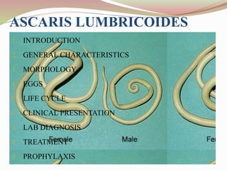
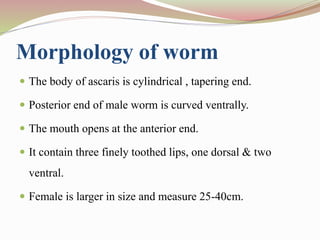
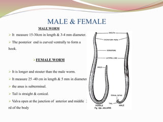
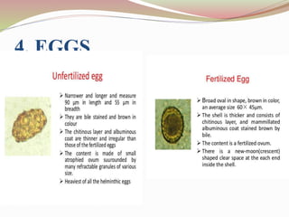
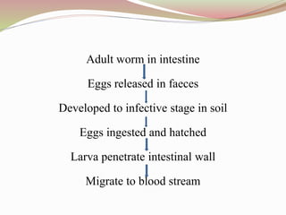
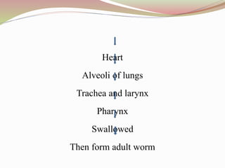
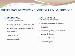
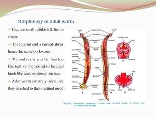
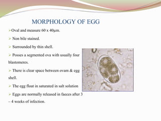
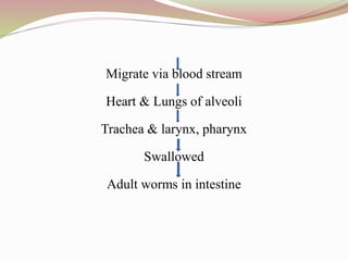
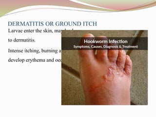
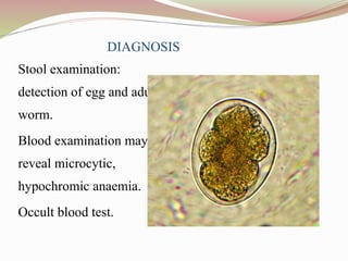
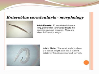
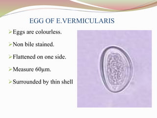
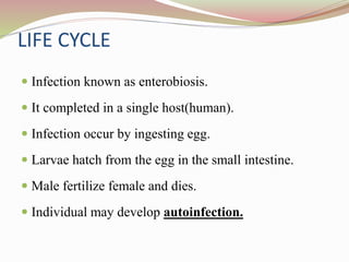
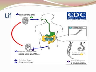
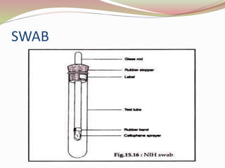
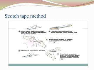
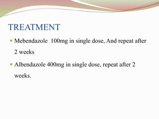
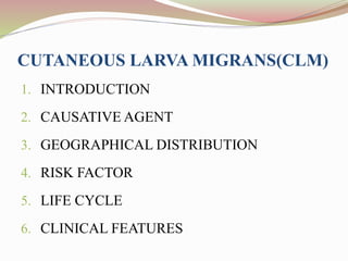
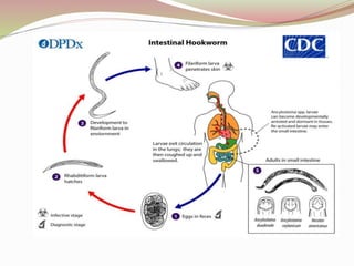
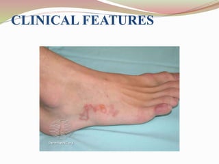
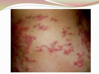
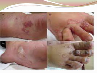
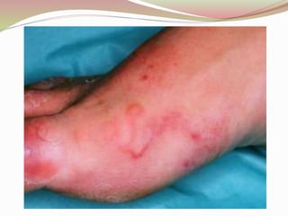
INTESTINAL NEMATODES
By- Dr. Manish Tiwari
Mail – mprabhat111@gmail.com
INTESTINAL NEMATODES
CHARACTERISTICS
 It belongs to phylum Nemathelminthes.
 They are elongated, cylindrical, bilaterally symmetrical.
 Unsegmented worm with tapering ends.
 The nematode means thread like structure.
 It has no suckers, no hooks.
 Body cavity is present.
 Body is covered with tough cuticle.
 Sexes are separate.
 Male is generally smaller than female.
 Fertilization is internal.
 They pass their life cycle in one host except filarial
nematode.
DIVISION OF FEMALE NEMATODE
1. Oviparous – Nematode which lay eggs.
 un-segmented egg (Ascaris lumbricoides)
 segmented egg (Hook worms)
1. Viviparous - Nematode which give the birth to
larvae.(W. bancrofti, B. malayi)
2. Ovo-viviparous – lay eggs containing
larvae.(strongyloides stercoralis)
CON……
Small intestine Large intestine
 Ascaris lumbricoides
 A. duodenale
 N. americanus
 T. spiralis
 Capillaria Spp.
 Enterobius vermicularis
 Trichuris trichiura
ASCARIS LUMBRICOIDES
1. INTRODUCTION
2. GENERAL CHARACTERISTICS
3. MORPHOLOGY
4. EGGS
5. LIFE CYCLE
6. CLINICAL PRESENTATION
7. LAB DIAGNOSIS
8. TREATMENT
9. PROPHYLAXIS
1.INTRODUCTION
 Most common helminthic human infection.
 Largest nematode to infect the human intestine.
 High prevalence in underdeveloped countries.
 It is estimated that ˃250 million people worldwide are
infected by this parasite
 It has simple life cycle.
. GENERAL
CHARACTERISTICS
 Family is ascaridae.
 Common name roundworm.
 Infective stage is embryonated egg containing
rhabditiform larva.
 Habitat small intestine particularly the jejunum of
man.
Morphology of worm
 The body of ascaris is cylindrical , tapering end.
 Posterior end of male worm is curved ventrally.
 The mouth opens at the anterior end.
 It contain three finely toothed lips, one dorsal & two
ventral.
 Female is larger in size and measure 25-40cm.
MALE & FEMALE
MALE WORM
 It measure 15-30cm in length & 3-4 mm diameter.
 The posterior end is curved ventrally to form a
hook.
FEMALE WORM
 It is longer and stouter than the male worm.
 It measure 25 -40 cm in length & 5 mm in diameter.
 the anus is subterminal.
 Tail is straight & conical.
 Valva open at the junction of anterior and middle 3
rd of the body
4. EGGS
5. LIFE CYCLE
 The life cycle of A. lumbricoides is passed in only one host
(man).
 No intermediate host is required.
 Worm reside in the small intestine
 Fertilized eggs contain unsegmented ova which are passed
faeces in the soil.
 Rhabditiform larva developed from unsegmented ovum.
Adult worm in intestine
Eggs released in faeces
Developed to infective stage in soil
Eggs ingested and hatched
Larva penetrate intestinal wall
Migrate to blood stream
Heart
Alveoli of lungs
Trachea and larynx
Pharynx
Swallowed
Then form adult worm
6. CLINICAL FEATURES
7. LABORATORY DIAGNOSIS
 Macroscopic examination
Adult worm passed though anus, mouth, nose, ear.
 Demonstration of larvae
Detection in sputum during larvaal migration.
 Stool microscopy
Identification of fertilized and un-fertilized eggs.
 Serodiagnosis
Ascaris antibody can be detected By indirect
immunofluorescence test(IFA), indirect
haemagglutination assay (IHA).
 Eosinophilia
During the larval migration.
8. TREATMENT
Albendazole 400mg : single dose
Mebendazole 100mg : twice daily for 3 days
Pyrantel pamoate
piperazine
9. PROPHYLAXIS
Proper disposal of human faeces
Avoidance of eating raw vegetables and salads.
Periodic treatment with an effective anthelminthic, in
communities that lack sanitization facilities.
ANCYLOSTOMA
DUODENALE
 Old world hook worm
 Habitat – small intestine in man particularly in the
jejunum and rarely in duodenum & ileum.
 Remain attached to the intestinal wall.
 Ancylostomiasis
DIFFERENCE BETWEEN A.DUODENALE& N. AMERICANUS.
A. DUODENALE N. AMERICANUS
 Old world hook worm
 Anterior end bends in same direction.
 Six teeth, four on ventral surface &
two on dorsal.
 Larger & thicker.
 More pathogenic.
 New world hook worm.
 Anterior end bends in opposite
direction.
 Four cutting plates, two each on
ventral & dorsal.
 Smaller & thinner.
 Less pathogenic.
Morphology of adult worm
They are small , pinkish & fusiform in
shape
 The anterior end is curved dorsally ,
hence the name hookworm.
 The oral cavity provide four hook
like teeth on the ventral surface and two
knob like teeth on dorsal surface.
 Adult worm are rarely seen , because
they attached to the intestinal mucosa.
MORPHOLOGY OF EGG
Oval and measure 60 x 40µm.
 Non bile stained.
 Surrounded by thin shell.
 Posses a segmented ova with usually four
blastomeres.
 There is clear space between ovam & egg
shell.
 The egg float in saturated in salt solution
 Eggs are normally released in faeces after 3
– 4 weeks of infection.
TRANSMISSION
 Penetration of skin.
 Ingestion of larva.
 Trans-placental transmission.
LIFE CYCLE
 Human are the only natural host.
 Adult worm lives in small intestine.
 Eggs passed in feaces.
 Rhabditiform larvae hatch out from the egg in the soil.
 Grows in size by feeding on bacteria and organic matter.
 Third larval stage is filariform larva.
 When a person walk barefooted, they penetrate the
skin.
 Larvae enter the venules and reaches right heart and
lungs.
 Crawl over epiglottis to pharynx and are swallowed.
 Get matured and life cycle completed.
Adult worm reside in intestine
Eggs discharge in faeces
hatch in soil
Rhabditiform larva developed (non infective)
Filariform larva developed ( infective stage)
Then penetrate skin
Migrate via blood stream
Heart & Lungs of alveoli
Trachea & larynx, pharynx
Swallowed
Adult worms in intestine
DERMATITIS OR GROUND ITCH
Larvae enter the skin, may lead
to dermatitis.
Intense itching, burning and
develop erythema and oedema.
RESPIRATORY
MANIFESTATION
Low grade fever.
Cough.
Dyspnea.
Pneumonia with pulmonary consolidation.
Bronchitis.
INTESTINAL MANIFESTION
Low grade fever
Iron deficiency anaemia (hookworm ingest blood)
Nausea
Vomiting
Diarrhea
Abdominal discomfort
DIAGNOSIS
Stool examination:
detection of egg and adult
worm.
Blood examination may
reveal microcytic,
hypochromic anaemia.
Occult blood test.
TREATMENT
 Mebendazole
 Thiabendazole
 Treatment of iron deficiency anaemia with
replacement iron therapy.
 Folic acid and vitamin B12.
ENTEROBIUS VERMICULARIS
1. GENERAL CHARACTERISTICS
2. MORPHOLOGY
3. EGG OF E. VERMICULARIS
4. LIFE CYCLE
5. CLINICAL FEATURE
6. LAB DIAGNOSIS
7. TREATMENT
GENERAL CHARACTERISTICS
 Common name - Pin worm , Thread worm and seat
worm
 Habitat - Caecum, appendix
 Infective stage - Embryonated egg
MORPHOLOGY
EGG OF E.VERMICULARIS
Eggs are colourless.
Non bile stained.
Flattened on one side.
Measure 60µm.
Surrounded by thin shell
LIFE CYCLE
 Infection known as enterobiosis.
 It completed in a single host(human).
 Infection occur by ingesting egg.
 Larvae hatch from the egg in the small intestine.
 Male fertilize female and dies.
 Individual may develop autoinfection.
Life Cycle
CLINICAL PRESENTATION
 The clinical feature of pinworm infection, also called
enterobiasis, includes:-
 itching or prickling pain in the anal area (usually at
night time or in the early morning), intense anal
itching,
 difficulty sleeping,
 rarely abdominal discomfort
 loss of appetite.
PATHOGENICITY
 Pin worm infection most frequent in school age
children.
 Children suffering sleepless night due to itching at the
perianal region.
 May show irritability, enuresis and weight loss.
 Scratching predispose to secondry bacterial infection.
LABORATORY DIAGNOSIS
1. Detection of adult worm: may be
detected in the perianal region or at the
surface of stool.
2. Demonstration of egg:
Egg not discharge into faeces.
Non bile stained & flattened on one
side
It is contain fully developed larva.
NIH swab
3. No serological test are available.
SWAB
Scotch tape method
TREATMENT
 Mebendazole 100mg in single dose, And repeat after
2 weeks
 Albendazole 400mg in single dose, repeat after 2
weeks.
CUTANEOUS LARVA MIGRANS(CLM)
1. INTRODUCTION
2. CAUSATIVE AGENT
3. GEOGRAPHICAL DISTRIBUTION
4. RISK FACTOR
5. LIFE CYCLE
6. CLINICAL FEATURES
INTRODUCTION
 Cutaneous larva migrans (CLM) is a parasitic skin infection.
 It is caused by hook worm larva that usually infect the cats,
dogs and other animals.
 Human can be infected with the larva by walking barefoot
on soft soil has been contaminated with animal faeces.
 It is also called creeping eruption.
2. CAUSATIVE AGENT
 There are many types of hook worm can causes cutaneous larva migran.
 It is caused by:
1. Ankylostoma braziliense: it is hookworm of wild and domestic dogs and
cats
2. Ankylostoma caninum: it dog hookworm that found in australia.
3. Uncinaria stenocephala: it is alo dog hookworm that found in europe.
4. Bunostomum phlebotomum: it is cattle hookworm.
 NOTE:- it is also caused by ankylostoma duodenale,
nector americanus and strongyloides sterocalis.
3. GEOGRAPHICAL DISTRIBUTION:- it is most
commonly found in the tropical and subtropical country .
 RISK FACTOR:- People of all ages and sex can be
affected by cutaneous larva migrans if they have been
exposed to hookworm larvae.
 Groups at risk include those with occupations or hobbies
that contact with warm, moist, sandy soil. These may
include:
 Barefoot beachcombers and sunbathers
 Children in sandpits
 Farmers
 Gardeners
 Plumbers
 Hunters
 Electricians
 Carpenters
 Pest exterminators.
CLINICAL FEATURES
TREATMENT
 Anthelmintics such as thiabendazole, albendazole,
mebendazole and ivermectin are used.
Intestinal nematodes

Intestinal nematodes

  • 1.
    INTESTINAL NEMATODES By- Dr.Manish Tiwari Mail – mprabhat111@gmail.com
  • 2.
    INTESTINAL NEMATODES CHARACTERISTICS  Itbelongs to phylum Nemathelminthes.  They are elongated, cylindrical, bilaterally symmetrical.  Unsegmented worm with tapering ends.  The nematode means thread like structure.  It has no suckers, no hooks.  Body cavity is present.
  • 3.
     Body iscovered with tough cuticle.  Sexes are separate.  Male is generally smaller than female.  Fertilization is internal.  They pass their life cycle in one host except filarial nematode.
  • 4.
    DIVISION OF FEMALENEMATODE 1. Oviparous – Nematode which lay eggs.  un-segmented egg (Ascaris lumbricoides)  segmented egg (Hook worms) 1. Viviparous - Nematode which give the birth to larvae.(W. bancrofti, B. malayi) 2. Ovo-viviparous – lay eggs containing larvae.(strongyloides stercoralis)
  • 5.
    CON…… Small intestine Largeintestine  Ascaris lumbricoides  A. duodenale  N. americanus  T. spiralis  Capillaria Spp.  Enterobius vermicularis  Trichuris trichiura
  • 6.
    ASCARIS LUMBRICOIDES 1. INTRODUCTION 2.GENERAL CHARACTERISTICS 3. MORPHOLOGY 4. EGGS 5. LIFE CYCLE 6. CLINICAL PRESENTATION 7. LAB DIAGNOSIS 8. TREATMENT 9. PROPHYLAXIS
  • 7.
    1.INTRODUCTION  Most commonhelminthic human infection.  Largest nematode to infect the human intestine.  High prevalence in underdeveloped countries.  It is estimated that ˃250 million people worldwide are infected by this parasite  It has simple life cycle.
  • 8.
    . GENERAL CHARACTERISTICS  Familyis ascaridae.  Common name roundworm.  Infective stage is embryonated egg containing rhabditiform larva.  Habitat small intestine particularly the jejunum of man.
  • 9.
    Morphology of worm The body of ascaris is cylindrical , tapering end.  Posterior end of male worm is curved ventrally.  The mouth opens at the anterior end.  It contain three finely toothed lips, one dorsal & two ventral.  Female is larger in size and measure 25-40cm.
  • 10.
    MALE & FEMALE MALEWORM  It measure 15-30cm in length & 3-4 mm diameter.  The posterior end is curved ventrally to form a hook. FEMALE WORM  It is longer and stouter than the male worm.  It measure 25 -40 cm in length & 5 mm in diameter.  the anus is subterminal.  Tail is straight & conical.  Valva open at the junction of anterior and middle 3 rd of the body
  • 11.
  • 12.
    5. LIFE CYCLE The life cycle of A. lumbricoides is passed in only one host (man).  No intermediate host is required.  Worm reside in the small intestine  Fertilized eggs contain unsegmented ova which are passed faeces in the soil.  Rhabditiform larva developed from unsegmented ovum.
  • 13.
    Adult worm inintestine Eggs released in faeces Developed to infective stage in soil Eggs ingested and hatched Larva penetrate intestinal wall Migrate to blood stream
  • 14.
    Heart Alveoli of lungs Tracheaand larynx Pharynx Swallowed Then form adult worm
  • 15.
  • 17.
    7. LABORATORY DIAGNOSIS Macroscopic examination Adult worm passed though anus, mouth, nose, ear.  Demonstration of larvae Detection in sputum during larvaal migration.  Stool microscopy Identification of fertilized and un-fertilized eggs.
  • 18.
     Serodiagnosis Ascaris antibodycan be detected By indirect immunofluorescence test(IFA), indirect haemagglutination assay (IHA).  Eosinophilia During the larval migration.
  • 19.
    8. TREATMENT Albendazole 400mg: single dose Mebendazole 100mg : twice daily for 3 days Pyrantel pamoate piperazine
  • 20.
    9. PROPHYLAXIS Proper disposalof human faeces Avoidance of eating raw vegetables and salads. Periodic treatment with an effective anthelminthic, in communities that lack sanitization facilities.
  • 21.
    ANCYLOSTOMA DUODENALE  Old worldhook worm  Habitat – small intestine in man particularly in the jejunum and rarely in duodenum & ileum.  Remain attached to the intestinal wall.  Ancylostomiasis
  • 22.
    DIFFERENCE BETWEEN A.DUODENALE&N. AMERICANUS. A. DUODENALE N. AMERICANUS  Old world hook worm  Anterior end bends in same direction.  Six teeth, four on ventral surface & two on dorsal.  Larger & thicker.  More pathogenic.  New world hook worm.  Anterior end bends in opposite direction.  Four cutting plates, two each on ventral & dorsal.  Smaller & thinner.  Less pathogenic.
  • 23.
    Morphology of adultworm They are small , pinkish & fusiform in shape  The anterior end is curved dorsally , hence the name hookworm.  The oral cavity provide four hook like teeth on the ventral surface and two knob like teeth on dorsal surface.  Adult worm are rarely seen , because they attached to the intestinal mucosa.
  • 24.
    MORPHOLOGY OF EGG Ovaland measure 60 x 40µm.  Non bile stained.  Surrounded by thin shell.  Posses a segmented ova with usually four blastomeres.  There is clear space between ovam & egg shell.  The egg float in saturated in salt solution  Eggs are normally released in faeces after 3 – 4 weeks of infection.
  • 25.
    TRANSMISSION  Penetration ofskin.  Ingestion of larva.  Trans-placental transmission.
  • 26.
    LIFE CYCLE  Humanare the only natural host.  Adult worm lives in small intestine.  Eggs passed in feaces.  Rhabditiform larvae hatch out from the egg in the soil.  Grows in size by feeding on bacteria and organic matter.  Third larval stage is filariform larva.
  • 27.
     When aperson walk barefooted, they penetrate the skin.  Larvae enter the venules and reaches right heart and lungs.  Crawl over epiglottis to pharynx and are swallowed.  Get matured and life cycle completed.
  • 28.
    Adult worm residein intestine Eggs discharge in faeces hatch in soil Rhabditiform larva developed (non infective) Filariform larva developed ( infective stage) Then penetrate skin
  • 29.
    Migrate via bloodstream Heart & Lungs of alveoli Trachea & larynx, pharynx Swallowed Adult worms in intestine
  • 31.
    DERMATITIS OR GROUNDITCH Larvae enter the skin, may lead to dermatitis. Intense itching, burning and develop erythema and oedema.
  • 32.
  • 33.
    INTESTINAL MANIFESTION Low gradefever Iron deficiency anaemia (hookworm ingest blood) Nausea Vomiting Diarrhea Abdominal discomfort
  • 34.
    DIAGNOSIS Stool examination: detection ofegg and adult worm. Blood examination may reveal microcytic, hypochromic anaemia. Occult blood test.
  • 35.
    TREATMENT  Mebendazole  Thiabendazole Treatment of iron deficiency anaemia with replacement iron therapy.  Folic acid and vitamin B12.
  • 36.
    ENTEROBIUS VERMICULARIS 1. GENERALCHARACTERISTICS 2. MORPHOLOGY 3. EGG OF E. VERMICULARIS 4. LIFE CYCLE 5. CLINICAL FEATURE 6. LAB DIAGNOSIS 7. TREATMENT
  • 37.
    GENERAL CHARACTERISTICS  Commonname - Pin worm , Thread worm and seat worm  Habitat - Caecum, appendix  Infective stage - Embryonated egg
  • 38.
  • 39.
    EGG OF E.VERMICULARIS Eggsare colourless. Non bile stained. Flattened on one side. Measure 60µm. Surrounded by thin shell
  • 40.
    LIFE CYCLE  Infectionknown as enterobiosis.  It completed in a single host(human).  Infection occur by ingesting egg.  Larvae hatch from the egg in the small intestine.  Male fertilize female and dies.  Individual may develop autoinfection.
  • 41.
  • 42.
    CLINICAL PRESENTATION  Theclinical feature of pinworm infection, also called enterobiasis, includes:-  itching or prickling pain in the anal area (usually at night time or in the early morning), intense anal itching,  difficulty sleeping,  rarely abdominal discomfort  loss of appetite.
  • 43.
    PATHOGENICITY  Pin worminfection most frequent in school age children.  Children suffering sleepless night due to itching at the perianal region.  May show irritability, enuresis and weight loss.  Scratching predispose to secondry bacterial infection.
  • 44.
    LABORATORY DIAGNOSIS 1. Detectionof adult worm: may be detected in the perianal region or at the surface of stool. 2. Demonstration of egg: Egg not discharge into faeces. Non bile stained & flattened on one side It is contain fully developed larva. NIH swab 3. No serological test are available.
  • 45.
  • 46.
  • 47.
    TREATMENT  Mebendazole 100mgin single dose, And repeat after 2 weeks  Albendazole 400mg in single dose, repeat after 2 weeks.
  • 48.
    CUTANEOUS LARVA MIGRANS(CLM) 1.INTRODUCTION 2. CAUSATIVE AGENT 3. GEOGRAPHICAL DISTRIBUTION 4. RISK FACTOR 5. LIFE CYCLE 6. CLINICAL FEATURES
  • 49.
    INTRODUCTION  Cutaneous larvamigrans (CLM) is a parasitic skin infection.  It is caused by hook worm larva that usually infect the cats, dogs and other animals.  Human can be infected with the larva by walking barefoot on soft soil has been contaminated with animal faeces.  It is also called creeping eruption.
  • 50.
    2. CAUSATIVE AGENT There are many types of hook worm can causes cutaneous larva migran.  It is caused by: 1. Ankylostoma braziliense: it is hookworm of wild and domestic dogs and cats 2. Ankylostoma caninum: it dog hookworm that found in australia. 3. Uncinaria stenocephala: it is alo dog hookworm that found in europe. 4. Bunostomum phlebotomum: it is cattle hookworm.
  • 51.
     NOTE:- itis also caused by ankylostoma duodenale, nector americanus and strongyloides sterocalis. 3. GEOGRAPHICAL DISTRIBUTION:- it is most commonly found in the tropical and subtropical country .  RISK FACTOR:- People of all ages and sex can be affected by cutaneous larva migrans if they have been exposed to hookworm larvae.  Groups at risk include those with occupations or hobbies that contact with warm, moist, sandy soil. These may include:
  • 52.
     Barefoot beachcombersand sunbathers  Children in sandpits  Farmers  Gardeners  Plumbers  Hunters  Electricians  Carpenters  Pest exterminators.
  • 54.
  • 58.
    TREATMENT  Anthelmintics suchas thiabendazole, albendazole, mebendazole and ivermectin are used.