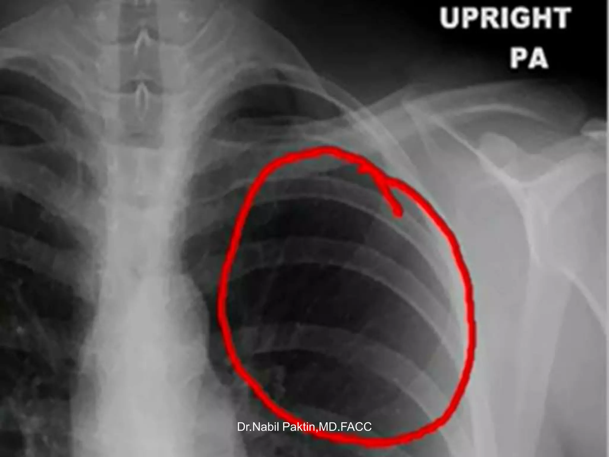
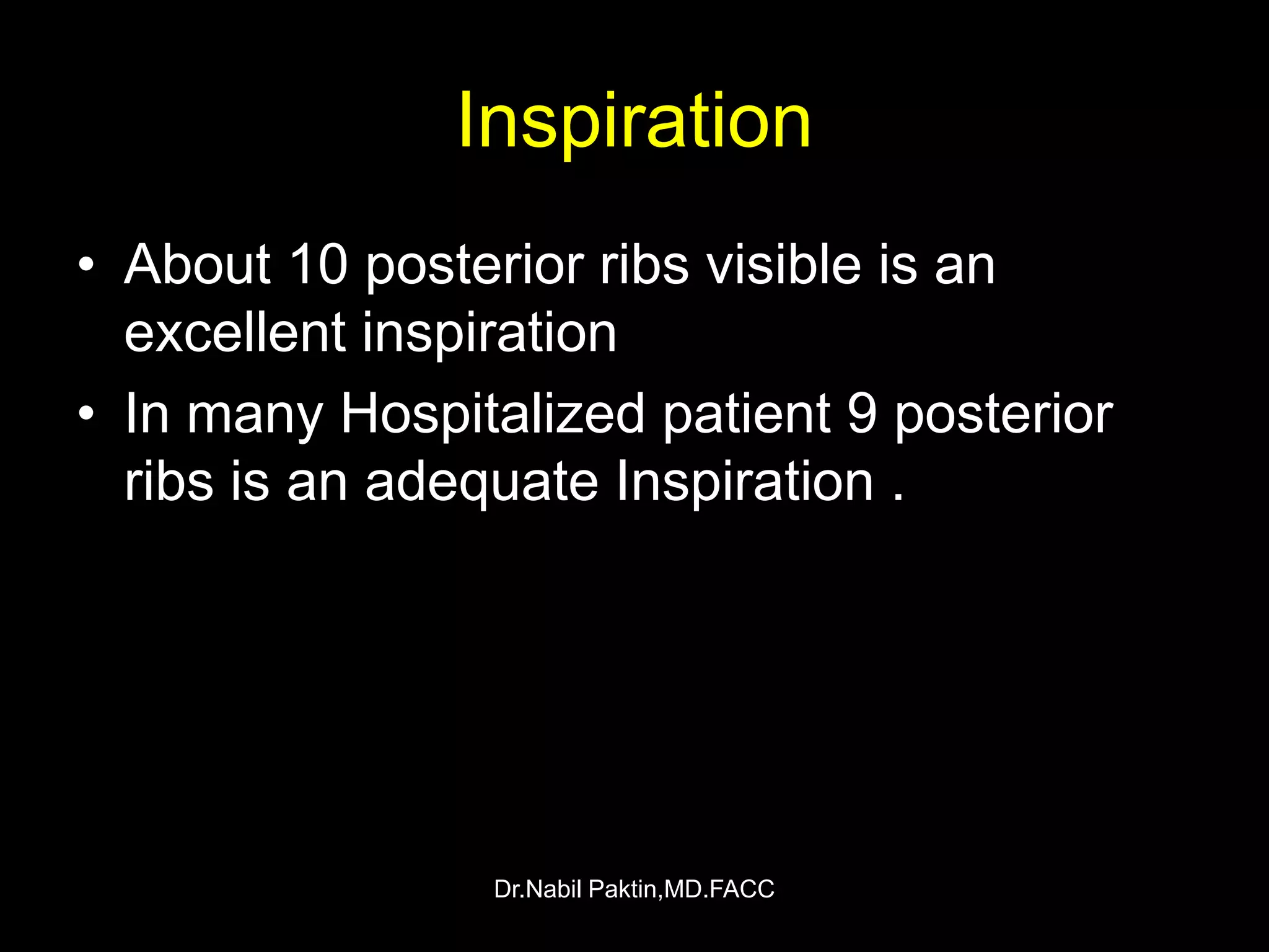
Dr.Nabil Paktin,MD,FACC. Trainer Specialist of Clinical Cardiology Postgraduate Program of Cardiology Afghanistan Cardiovascular(cardiology) society Lecture series
Radio Frequency Ablation (RFA Treatment ) -Modern Technology for management l...SafeMedTrip
Safemedtrip india's number one company of medical treatment, which provide low cost treatment package for international patient. if you have any problem Please scan and email your medical reports to us at hospitalindia@gmail.com or help@safemedtrip.com or call us at +91-9899993637 and we shall get you a Free, No Obligation Opinion from India's leading Specialist Doctors.
SHORT TALK ABOUT DIFFERENTIAL DIAGNOSIS ABOUT UNILATERAL HYPERLUCENT HEMITHORAX , COMMON AND LESS COMMON CAUSES WITH CLUES TO DIAGNOSIS AND SOME EXAMPLES
HOPPING YOU LIKE IT
DR HISHAM ALKHATIB
CONSULTANT RADIOLOGIST
SELDINGER TECHNIQUE & INTERVENTIONAL RADIOLOGYRiyas M K
its a basic introduction about Seldinger technique and Intervetional radiology.In interventional radiology, procedures generally start with the Seldinger technique to access the vasculature, using a needle through which a guidewire is inserted, followed by navigation of catheters within the vessels.
Basic Chest X ray Views - AP, PA & Lateral etc . pptxDr Abna J
PA PROJECTION
Sit or stand upright.
Positioned to minimize magnification of the anteriorly positioned heart and consequent obscuration of the lungs.
Make sure the patient is standing straight and is equally distributing the weight of the body on both feet.
The upright position is preferred for the following reasons: It prevents engorgement (an excess of blood) of pulmonary vessels.
It allows full expansion of the lungs
To visualize possible air and fluid levels in the chest.
An upright chest film is preferred over an upright abdominal film for the diagnosis of pneumoperitoneum (free air in the abdominal cavity).
Ask the patient to move the shoulders forward and downward, so that the chest wall and both shoulders are in contact with the cassette. This helps to carry the clavicles below the lung apices.
It is very important to minimize breast shadows.
Ask the patient to pull the breasts upward and laterally (outwards), then remove her hands as she leans against the cassette holder to keep them in position.
Rotation
Even a small degree of rotation distorts the mediastinal borders, and the lung nearest the film will appear less translucent.
The following points should be stressed to obtain a true PA view (without rotation):
Ensure that the patient is standing evenly on both feet.
Both shoulders should be rolled forward and downward.
The chest radiograph should be well centred so that the medial ends of the clavicle are equidistant from the vertebral spinous processes at T4/5.
CENTRAL RAY
Over T7 vertebra
SID: 72 inches
Central ray
Film holder (image receptor) placement
The horizontal dimension of an average chest is greater than the vertical dimension.
This requires that a 14 x 17-inch film holder or image receptor (IR) be placed crosswise.
Or lengthwise depending on body type.
Collimation
The upper border of the illuminated field should be at the level of vertebra prominence (4 cm above the apex of lungs).
This will result in a lower collimation border of 1-2 inches below the costophrenic angle, if the central ray was correctly centred.
A general rule for average adult patients is to show a minimum of 10 ribs on a good PA chest radiograph.
Evaluation criteria for a good PA projection
Entire lung fields from apices to costophrenic angles should be clearly demonstrated.
No rotation. (both the right and left sternal ends of the clavicle will be the same distance from the center line of the spine.)
The direction of rotation can be determined by which sternal end of the clavicle is closest to the spine.
Trachea is visible in midline.
Scapula projected outside the lung fields.
Ten posterior ribs are visible above the diaphragm.
There is a sharp outline of the heart and diaphragm.
A faint shadow of the ribs and superior thoracic vertebrae is visible through the heart shadow.
Lung markings are visible from the hilum to the periphery of the lung.
Variations
An expiratory film may be helpful under some circumstances.
in this tutorial i am speaking about chest x-ray quality that include :
1- Inclusion
2- inspiration/lung
3- volume
4- projection
5- penetration
6- Rotation
7- artifact
i try to make it easy and simple for medical students and junior doctors to help them in clinical life.
New Directions in Targeted Therapeutic Approaches for Older Adults With Mantl...i3 Health
i3 Health is pleased to make the speaker slides from this activity available for use as a non-accredited self-study or teaching resource.
This slide deck presented by Dr. Kami Maddocks, Professor-Clinical in the Division of Hematology and
Associate Division Director for Ambulatory Operations
The Ohio State University Comprehensive Cancer Center, will provide insight into new directions in targeted therapeutic approaches for older adults with mantle cell lymphoma.
STATEMENT OF NEED
Mantle cell lymphoma (MCL) is a rare, aggressive B-cell non-Hodgkin lymphoma (NHL) accounting for 5% to 7% of all lymphomas. Its prognosis ranges from indolent disease that does not require treatment for years to very aggressive disease, which is associated with poor survival (Silkenstedt et al, 2021). Typically, MCL is diagnosed at advanced stage and in older patients who cannot tolerate intensive therapy (NCCN, 2022). Although recent advances have slightly increased remission rates, recurrence and relapse remain very common, leading to a median overall survival between 3 and 6 years (LLS, 2021). Though there are several effective options, progress is still needed towards establishing an accepted frontline approach for MCL (Castellino et al, 2022). Treatment selection and management of MCL are complicated by the heterogeneity of prognosis, advanced age and comorbidities of patients, and lack of an established standard approach for treatment, making it vital that clinicians be familiar with the latest research and advances in this area. In this activity chaired by Michael Wang, MD, Professor in the Department of Lymphoma & Myeloma at MD Anderson Cancer Center, expert faculty will discuss prognostic factors informing treatment, the promising results of recent trials in new therapeutic approaches, and the implications of treatment resistance in therapeutic selection for MCL.
Target Audience
Hematology/oncology fellows, attending faculty, and other health care professionals involved in the treatment of patients with mantle cell lymphoma (MCL).
Learning Objectives
1.) Identify clinical and biological prognostic factors that can guide treatment decision making for older adults with MCL
2.) Evaluate emerging data on targeted therapeutic approaches for treatment-naive and relapsed/refractory MCL and their applicability to older adults
3.) Assess mechanisms of resistance to targeted therapies for MCL and their implications for treatment selection
Title: Sense of Taste
Presenter: Dr. Faiza, Assistant Professor of Physiology
Qualifications:
MBBS (Best Graduate, AIMC Lahore)
FCPS Physiology
ICMT, CHPE, DHPE (STMU)
MPH (GC University, Faisalabad)
MBA (Virtual University of Pakistan)
Learning Objectives:
Describe the structure and function of taste buds.
Describe the relationship between the taste threshold and taste index of common substances.
Explain the chemical basis and signal transduction of taste perception for each type of primary taste sensation.
Recognize different abnormalities of taste perception and their causes.
Key Topics:
Significance of Taste Sensation:
Differentiation between pleasant and harmful food
Influence on behavior
Selection of food based on metabolic needs
Receptors of Taste:
Taste buds on the tongue
Influence of sense of smell, texture of food, and pain stimulation (e.g., by pepper)
Primary and Secondary Taste Sensations:
Primary taste sensations: Sweet, Sour, Salty, Bitter, Umami
Chemical basis and signal transduction mechanisms for each taste
Taste Threshold and Index:
Taste threshold values for Sweet (sucrose), Salty (NaCl), Sour (HCl), and Bitter (Quinine)
Taste index relationship: Inversely proportional to taste threshold
Taste Blindness:
Inability to taste certain substances, particularly thiourea compounds
Example: Phenylthiocarbamide
Structure and Function of Taste Buds:
Composition: Epithelial cells, Sustentacular/Supporting cells, Taste cells, Basal cells
Features: Taste pores, Taste hairs/microvilli, and Taste nerve fibers
Location of Taste Buds:
Found in papillae of the tongue (Fungiform, Circumvallate, Foliate)
Also present on the palate, tonsillar pillars, epiglottis, and proximal esophagus
Mechanism of Taste Stimulation:
Interaction of taste substances with receptors on microvilli
Signal transduction pathways for Umami, Sweet, Bitter, Sour, and Salty tastes
Taste Sensitivity and Adaptation:
Decrease in sensitivity with age
Rapid adaptation of taste sensation
Role of Saliva in Taste:
Dissolution of tastants to reach receptors
Washing away the stimulus
Taste Preferences and Aversions:
Mechanisms behind taste preference and aversion
Influence of receptors and neural pathways
Impact of Sensory Nerve Damage:
Degeneration of taste buds if the sensory nerve fiber is cut
Abnormalities of Taste Detection:
Conditions: Ageusia, Hypogeusia, Dysgeusia (parageusia)
Causes: Nerve damage, neurological disorders, infections, poor oral hygiene, adverse drug effects, deficiencies, aging, tobacco use, altered neurotransmitter levels
Neurotransmitters and Taste Threshold:
Effects of serotonin (5-HT) and norepinephrine (NE) on taste sensitivity
Supertasters:
25% of the population with heightened sensitivity to taste, especially bitterness
Increased number of fungiform papillae
ARTIFICIAL INTELLIGENCE IN HEALTHCARE.pdfAnujkumaranit
Artificial intelligence (AI) refers to the simulation of human intelligence processes by machines, especially computer systems. It encompasses tasks such as learning, reasoning, problem-solving, perception, and language understanding. AI technologies are revolutionizing various fields, from healthcare to finance, by enabling machines to perform tasks that typically require human intelligence.
Ethanol (CH3CH2OH), or beverage alcohol, is a two-carbon alcohol
that is rapidly distributed in the body and brain. Ethanol alters many
neurochemical systems and has rewarding and addictive properties. It
is the oldest recreational drug and likely contributes to more morbidity,
mortality, and public health costs than all illicit drugs combined. The
5th edition of the Diagnostic and Statistical Manual of Mental Disorders
(DSM-5) integrates alcohol abuse and alcohol dependence into a single
disorder called alcohol use disorder (AUD), with mild, moderate,
and severe subclassifications (American Psychiatric Association, 2013).
In the DSM-5, all types of substance abuse and dependence have been
combined into a single substance use disorder (SUD) on a continuum
from mild to severe. A diagnosis of AUD requires that at least two of
the 11 DSM-5 behaviors be present within a 12-month period (mild
AUD: 2–3 criteria; moderate AUD: 4–5 criteria; severe AUD: 6–11 criteria).
The four main behavioral effects of AUD are impaired control over
drinking, negative social consequences, risky use, and altered physiological
effects (tolerance, withdrawal). This chapter presents an overview
of the prevalence and harmful consequences of AUD in the U.S.,
the systemic nature of the disease, neurocircuitry and stages of AUD,
comorbidities, fetal alcohol spectrum disorders, genetic risk factors, and
pharmacotherapies for AUD.
MANAGEMENT OF ATRIOVENTRICULAR CONDUCTION BLOCK.pdfJim Jacob Roy
Cardiac conduction defects can occur due to various causes.
Atrioventricular conduction blocks ( AV blocks ) are classified into 3 types.
This document describes the acute management of AV block.
Title: Sense of Smell
Presenter: Dr. Faiza, Assistant Professor of Physiology
Qualifications:
MBBS (Best Graduate, AIMC Lahore)
FCPS Physiology
ICMT, CHPE, DHPE (STMU)
MPH (GC University, Faisalabad)
MBA (Virtual University of Pakistan)
Learning Objectives:
Describe the primary categories of smells and the concept of odor blindness.
Explain the structure and location of the olfactory membrane and mucosa, including the types and roles of cells involved in olfaction.
Describe the pathway and mechanisms of olfactory signal transmission from the olfactory receptors to the brain.
Illustrate the biochemical cascade triggered by odorant binding to olfactory receptors, including the role of G-proteins and second messengers in generating an action potential.
Identify different types of olfactory disorders such as anosmia, hyposmia, hyperosmia, and dysosmia, including their potential causes.
Key Topics:
Olfactory Genes:
3% of the human genome accounts for olfactory genes.
400 genes for odorant receptors.
Olfactory Membrane:
Located in the superior part of the nasal cavity.
Medially: Folds downward along the superior septum.
Laterally: Folds over the superior turbinate and upper surface of the middle turbinate.
Total surface area: 5-10 square centimeters.
Olfactory Mucosa:
Olfactory Cells: Bipolar nerve cells derived from the CNS (100 million), with 4-25 olfactory cilia per cell.
Sustentacular Cells: Produce mucus and maintain ionic and molecular environment.
Basal Cells: Replace worn-out olfactory cells with an average lifespan of 1-2 months.
Bowman’s Gland: Secretes mucus.
Stimulation of Olfactory Cells:
Odorant dissolves in mucus and attaches to receptors on olfactory cilia.
Involves a cascade effect through G-proteins and second messengers, leading to depolarization and action potential generation in the olfactory nerve.
Quality of a Good Odorant:
Small (3-20 Carbon atoms), volatile, water-soluble, and lipid-soluble.
Facilitated by odorant-binding proteins in mucus.
Membrane Potential and Action Potential:
Resting membrane potential: -55mV.
Action potential frequency in the olfactory nerve increases with odorant strength.
Adaptation Towards the Sense of Smell:
Rapid adaptation within the first second, with further slow adaptation.
Psychological adaptation greater than receptor adaptation, involving feedback inhibition from the central nervous system.
Primary Sensations of Smell:
Camphoraceous, Musky, Floral, Pepperminty, Ethereal, Pungent, Putrid.
Odor Detection Threshold:
Examples: Hydrogen sulfide (0.0005 ppm), Methyl-mercaptan (0.002 ppm).
Some toxic substances are odorless at lethal concentrations.
Characteristics of Smell:
Odor blindness for single substances due to lack of appropriate receptor protein.
Behavioral and emotional influences of smell.
Transmission of Olfactory Signals:
From olfactory cells to glomeruli in the olfactory bulb, involving lateral inhibition.
Primitive, less old, and new olfactory systems with different path
Knee anatomy and clinical tests 2024.pdfvimalpl1234
This includes all relevant anatomy and clinical tests compiled from standard textbooks, Campbell,netter etc..It is comprehensive and best suited for orthopaedicians and orthopaedic residents.
Basavarajeeyam is an important text for ayurvedic physician belonging to andhra pradehs. It is a popular compendium in various parts of our country as well as in andhra pradesh. The content of the text was presented in sanskrit and telugu language (Bilingual). One of the most famous book in ayurvedic pharmaceutics and therapeutics. This book contains 25 chapters called as prakaranas. Many rasaoushadis were explained, pioneer of dhatu druti, nadi pareeksha, mutra pareeksha etc. Belongs to the period of 15-16 century. New diseases like upadamsha, phiranga rogas are explained.
- Video recording of this lecture in English language: https://youtu.be/lK81BzxMqdo
- Video recording of this lecture in Arabic language: https://youtu.be/Ve4P0COk9OI
- Link to download the book free: https://nephrotube.blogspot.com/p/nephrotube-nephrology-books.html
- Link to NephroTube website: www.NephroTube.com
- Link to NephroTube social media accounts: https://nephrotube.blogspot.com/p/join-nephrotube-on-social-media.html
Hemodialysis: Chapter 3, Dialysis Water Unit - Dr.Gawad
Chest-X ray , How to read a CXR
1. The Chest X-Ray Basics
How to read A CXR ?
NABIL PAKTIN, M.D.,F.A.C.C.
Trainer Specialist of Postgraduate Medial Education
Afghanistan – Kabul
8/10/11
Dr.Nabil Paktin,MD.FACC
21. 5 steps to CXR interpretation
• 1- assess the lung expansion
• 2- assess the pleura
• 3- look for infiltrate
• 4- look at the mediastinum
• 5-Assess the abdomen
Dr.Nabil Paktin,MD.FACC
30. Techniques
• Volume of PE and whether it’s
mobile or loculated.
• Sensitive method for detecting
small quantity of PF(50-
100ml).
• Nondependent hemithorax to
confirm a pneumothorax in a
patient who could not be
examined erect .
• if the layering fluid is 1 cm
thick, indicates an effusion of
greater than 200 mL that is
amenable to thoracentesis
Dr.Nabil Paktin,MD.FACC
32. Technical consideration
Inspiration
• The patient should be
in full inspiration .
• Shows better
intrapulmonary
abnormalities
• The diaphragm fount
at about the level of
the 8th -10th posterior
ribs or 5th -6th anterior
rib on good inspiration.
Dr.Nabil Paktin,MD.FACC
33. • On a good PA film ,
the thoracic spine
Penetration disk spaces should
be barely visible
through the heart
but bony details of
the spine are not
usually be seen
through the heart .
• On the lateral view ,
proper penetration
and inspiration is
seen through the
spine appears to
darken as you move
caudally . This is
due to more air in
lung in the lower
lobes and less
Dr.Nabil Paktin,MD.FACC
chest wall .
34. Penetration cont…• There is no
adequate
lung detail
• Absence of
peripheral
vasculature
• See
vertebrae
extending
down into
the
abdominal
region.
• Underpenetrated • overpenetrated
Dr.Nabil Paktin,MD.FACC
35. • The patient must
Rotation be flat against the
cassette , if there
is rotation of the
patient , the
mediastinum may
look very unusual .
• Clavicular heads
whether they are
in equal distance
from the spinous
process of the
thoracic vertebral
Dr.Nabil Paktin,MD.FACC
bodies .
36. Rotation cont…
• See the rotation heads of the clavicles
and the spinous processes .
Dr.Nabil Paktin,MD.FACC
37. Recognizing a technically
adequate Chest x ray
• Factors to evaluate :
1- Penetration
2- Inspiration
3- Rotation
4- Angulation
Dr.Nabil Paktin,MD.FACC
38. Penetration
• You should be
able to just see the
thoracic spine
through the Heart .
Dr.Nabil Paktin,MD.FACC
40. Inspiration
• About 10 posterior ribs visible is an
excellent inspiration
• In many Hospitalized patient 9 posterior
ribs is an adequate Inspiration .
Dr.Nabil Paktin,MD.FACC
41. Anterior Vs. Posterior ribs
• Anterior ribs
• Posterior
will be
ribs are
visible but
those that
are harder
are most
to see .
apparent on
They run
the chest x
more or less
ray .they
rum more or at a 45
degree
less
angle
horizontally.
downward
to ward the
• How to tell the difference between the feet ,
anterior and posterior ribs .
Dr.Nabil Paktin,MD.FACC
42. • Ten posterior ribs showing is an excellent inspiration
Dr.Nabil Paktin,MD.FACC
43. Pitfall due to poor inspiration
• Poor inspiration will crowd lung marking and make it
appear as though the Paktin,MD.FACC airspace disease
Dr.Nabil patient has
44. Same Patient
• Better Inspiration and the disease at the lung bases has
cleared Dr.Nabil Paktin,MD.FACC
45. Rotation
• If the spinous
process of
the vertebral
body is
equidistant
from the
medial ends
of each
clavicle.
There is no
rotation
Dr.Nabil Paktin,MD.FACC
48. Pitfall due to marked rotation
• Severe rotation may make the pulmonary arteries
Dr.Nabil Paktin,MD.FACC
appear larger on the side farther from film .
49. Angulation
• If the X- ray beam is angle toward the
head ( mostly because the patient is semi-
recumbent ) . The fils so obtained is called
an “ apical lordotic” view .
• Anterior structure ( like the clavicles) will
be projected higher on the film than
posterior structures .
Dr.Nabil Paktin,MD.FACC
50. Pitfall due to angulation
• A film which is apical lordotic ( beam is angled up toward
head) will have an unusually shaped heart and the sharp
border of the left hemidiaphragm will be absent .
Dr.Nabil Paktin,MD.FACC
51. Important Points
• The factors to evaluate the quality of a
chest x-ray are :
- Penetration – see spine through the heart
- Inspiration – at least 8-9 posterior ribs
- Rotation – spinous process between
clavicles
- Angulation – clavicle over 3rd rib
Dr.Nabil Paktin,MD.FACC
52. What is most wrong with this image ( click any
that apply )?
• Penetration
• Inspiration
• Rotation
• Angulation
Dr.Nabil Paktin,MD.FACC
53. Correct
• The image is apical
lordotic look at the
high position of the
clavicles . It is also
underpenetrated .
You can’t tell if its
rotated and the
degree of
inspiration is
adequate
Dr.Nabil Paktin,MD.FACC
55. Can’t tell
• You may be right but you can’t tell from
the image given .
Dr.Nabil Paktin,MD.FACC
56. Correct
• The image is apical
lordotic look at the
high position of the
clavicles . It is also
underpenetrated .
You can’t tell if its
rotated and the
degree of
inspiration is
adequate
Dr.Nabil Paktin,MD.FACC
57. What is most wrong with this
image ( click any that apply )?
• Penetration
• Inspiration
• Rotation
• Angulation
Dr.Nabil Paktin,MD.FACC
59. Correct
• The patient has
taken a poor
inspiration . He
is also rotated
toward his own
right . Is
slightly
underpenetrate
d and he is not
angulated .
Dr.Nabil Paktin,MD.FACC
60. Correct
• The patient has
taken a poor
inspiration . He
is also rotated
toward his own
right . Is
slightly
underpenetrate
d and he is not
angulated .
Dr.Nabil Paktin,MD.FACC
62. What is most wrong with this
image ( click any that apply )?
• Penetration
• Inspiration
• Rotation
• Angulation
Dr.Nabil Paktin,MD.FACC
63. Correct
• The film is
underpenetrated .you
can’t see the heart
through the spine .
The degree of
inspiration is
probably adequate .
Rotation can not be
evaluated and there
is a slight amount of
Angulation
.incidentally there is a
large bronghogenic
ca in the left lung .
Dr.Nabil Paktin,MD.FACC
65. Can’t tell
• You may be right but you can’t tell from
the image given .
Dr.Nabil Paktin,MD.FACC
66. What is most wrong with this
image ( click any that apply )?
• Penetration
• Inspiration
• Rotation
• Angulation
Dr.Nabil Paktin,MD.FACC
67. Correct
• The primary technical
problem here is the
patient is rotated
considerably toward
her own left side.
Notice how the
hemidiaphragm
appears elevated on
the side to which the
patient is rotated ( red
arrow )
Dr.Nabil Paktin,MD.FACC
90. Pitfalls Due to Under Penetration
• If the films is
underpenetrated , the left
hemidiaphragm ) and left
lung base ) will not be
visible and they
pulmonary marking will
appear more prominent
than they actually are .
Dr.Nabil Paktin,MD.FACC
91. AP Versus PA
the effect of magnification
• In al PA film the heart is closer to the film
and thus less magnified .
- the standard chest X-ray is a PA film .
• In a AP film , the heart is farther from the
films and is more magnified .
- Portable chest X-ray are almost always
done AP.
Dr.Nabil Paktin,MD.FACC
92. AP Versus PA
the effect of magnification
• AP portable film make the • On this patient the PA film
does .
heart look larger than itDr.Nabil Paktin,MD.FACC
is done one hour later .
96. Mediastinum Cont…
• The lobes of
the lungs
forming the
margins of
the lungs
along the
mediastinum
and chest
wall .
Dr.Nabil Paktin,MD.FACC
97. Hila • Composed of
pulmonary artery
and it’s branches ,
and adjacent and
pulmonary veins .
• The pulmonary
arteries and upper
lobe veins
significantly
contribute to the
hilar shadow on
plain Chest X-ray .
• Left hilum is slightly
at a higher position (
0.5-2cm) than the
Dr.Nabil Paktin,MD.FACC
right hilum.
98. • The arteries and
Pulmonary Vessels veins branch out
from the Hila .
Becoming smaller
toward the
periphery .
• The larger central
vessels are better
seen . In the upright
position , the lower
lung vessels are
larger than the
upper lung vessels
due to gravitational
effects on flow . If
the patient is supine
Dr.Nabil Paktin,MD.FACC , this called
Cephalization .
99. • Angle of contact with
Diaphragm the chest wall is
acute and sharp.
• Blunting of the angle
is sometimes
normally seen in
athletes.
• Normally right
hemidiaphragm is
1.5-3.5cm higher
than the left
difference of more
than 3 cm is
considered
abnormal .
• In 3% of population .
Left hemidiaphragm
is at a higher level
Dr.Nabil Paktin,MD.FACC
than the right .
100. Diaphragm cont…
• Check for doming of diaphragm by drawing a line prependicular from the mid
point of the dome to a line joining costopherenic and cardiophrenic angles .
Dr.Nabil Paktin,MD.FACC
• The distance is :>1.5cm less than that consider flattened .
103. 1- scalloping
Normal Variant
2- muscle slips
3- diaphragm
hump and
dromedary
diaphragm
4-eventration
5-accessory
diaphragm.
Dr.Nabil Paktin,MD.FACC
104. Interpretation
How to look at a chest PA view
• Comparison with previous X-rays
- every effort should be made to obtain previous film
for comparison with the current film.
- The easiest way to identify a new abnormality is to
note its absence on a previous film!!
- The key to successfully interpreting any radiograph
is to be systemic .
- Examine all parts of the film in an orderly manner
and do this consistently .
Dr.Nabil Paktin,MD.FACC
105. Side marker
• The position of side marker allows the
radiograph to be oriented correctly for reading .
Dr.Nabil Paktin,MD.FACC
106. Technique
• Next concentrate on the technical factors :
1- is the examination complete ?
2- Are all the requested views included?
3- Is the entire anatomical area included on the
film :
1- Positioning
2- Inspiration
3- Exposure
4- Rotation
Dr.Nabil Paktin,MD.FACC
107. Systematic analysis
• 1- soft tissue including breast , chest wall ,
companion shadow .
• 2- bones – shoulder girdles , spine and rib
cage .
• 3- diaphragm position . Shape ,
subdiaphragmatic abnormalities .
• 4- review abdomen for bowel gas , organ
size , abnormal calcification , free air .
• 5- plastic – ETT . Lines , tubes .
Dr.Nabil Paktin,MD.FACC
108. Systematic analysis cont…
• 6- review mediastinum:
- Overal size and shape
- Trachea : position , carina , the trachea
should be central .
- Margins :SVC ascending aorta , right
atrium , left subclavian artery , aortic arch ,
main pulmonary artery , left ventricle .
- Lines and stripes : paratracheal ,
paraspinal , paraesophageal (
azygoesophageal) , paraaortic .
Dr.Nabil Paktin,MD.FACC
- Retrosternal clear space .
109. Systematic analysis cont…
• 7- heart size , shape : the width of the heart should be no greater than 50%
of the width of the cage .
• 8-Review hila :
• A- normal relationships
• B- size
• 9- parenchyma : now finally ready to examine the lungs!! Mentally divide the
entire chest into upper , middle and lower thirds . Then , methodically
compare the right and left sides of each lung section looking for asymmetry .
• The easiest way to identify an abnormality is to confirm that it does not exist
on the other side ! .
• Compare lung sizes , aeration , vascular distinctness and abnormal
opacities .
• 10- pleura : costopherenic and cardiophrenic angles , thickening fissures –
major and minor – if seen .
Dr.Nabil Paktin,MD.FACC
111. Hidden Areas !!!
1- supraclavicular regions
.
2- Ends of ribs
3- retroclavicular regions
4-posterior mediastnal
and paravertebral
regions .
Dr.Nabil Paktin,MD.FACC
113. • One of the most useful sign in
chest radiology is the
silhouette sign .
Silhouette sign
• The silhouette sign is actually
elimination of the silhouette or
loss of lung/sot tissue interface
caused by a mass or fluid in
the normally air filled lung .
• For example . If an
intrathoracic opacity is in
anatomic contact with the heart
border , then the opacity will
obscure that border .
• The sign is commonly applied
to the heart , aorta , chest wall • Two objects of with the same
, and diaphragm . radiographic density touch each
• The location of this other , the border between them
abnormality can help to disappear .
determine the location
anatomically .
Dr.Nabil Paktin,MD.FACC
127. Air bronchogram
• Air bronchogram is a
tubular outline of an
airway made visible by
filling of the surrounding
alveoli by fluid or
inflammatory exudates .
• Normal bronchi not
usually visualized due to
thin wall and an air – air
interface .
• Consolidation , pul.edema
, nonobstrucutive
pulmonary atelectasis ,
severe interstitial disease
, neoplasm and normal Dr.Nabil Paktin,MD.FACC
expiration .
128. Consolidation
• Defined as a process
in which air in the
alveoli is replaced by
products of disease .
• The bronchi to the
consolidated area are
usually widely patent .
• In most instances ,
alveolar filling is
patchy,i.e. not all acini
are involved .
• The radiographic
opacity is therefore
nonhomogeneous ,
sometimes with are
Dr.Nabil Paktin,MD.FACC
bronchogram .
129. Collapse ( atelectasis )
• Atelectasis is volume loss due to alveolar
collapse or failure to expand causing increased
opacification of radiograph.
• Collapse may affect a whole lung or a
subdivision ( lobe , segment ) .
• Types
- Obstructive
- Compressive
- Cicatrization
- Adhesive
- Passive Dr.Nabil Paktin,MD.FACC
130. General features of lobar collapse
• Shift of fissures • Other signs :
• Area of increased opacity • A hilar mass , which also
• Crowding of vessels suggest carcinoma as the
• Tracheal displacement cause .
toward the side of the • The presence of a foreign
collapse body
• Hilar shift • The presence of an
• Mediastnal shift toward endotracheal tube , is it
the side of the collapse sited too low ?
• Elevation of the • Other evidence of
hemidiaphragm malignant disease ( e.g.
• Herniation of the opposite rib metastases , effusion )
lung across the midline .
Dr.Nabil Paktin,MD.FACC
132. Signs of right upper lobe
collapse
• Minor fissures move upward with concavity inferiorly
.
• An area of opacity that lies against apex of
mediastinum
• Tracheal shift to the right
• Rihgt hilum is elevated and the intermediate
bronchus assumes horizontal position .
• Loss of right paratracheal stripe ( silhouette sign ) .
Dr.Nabil Paktin,MD.FACC
140. Signs of right middle lobe collapse
• This is often not immediately obvious on the
frontal film .
• Ill defined shadowing is evident adjacent to the
right heart border , which becomes indistinct .
• Right heart border is silhouetted .
• Minor fissure moves downward .
• Collapse of right middle lobe more obvious on
lateral view
• In lateral view , collapsed lobe has triangular
shape with apex at the hilum
• Also best seen inDr.Nabil Paktin,MD.FACC .
lordotic view
141. Right lower lobe collapse
• Right lower lobe zone shadowing is combined with obliteration
of the hemidiaphragm ( silhouette sign ) .
• The right heart border , which is anterior is usually still clearly
seen ( silhouette sign again ) .
• The oblique fissure lies more horizontally and may become
visible , giving a sharp upper margin to the shadowing .
• If the lobe is collapsed completely it may appear as a
triangular opacity being anterior will still be clearly seen .
• On lateral ,Abnormally increased density over the lower
thoracic spine due to the triangular opacity of the collapsed
Dr.Nabil Paktin,MD.FACC
lobe.
143. Right lower
lobe collapse.
Loss of volume
in the right
lung, the right
hemithorax is
hyper
translucent.
Dr.Nabil Paktin,MD.FACC
144. Left upper lobe collapse
• The left lung lacks a middle lobe and there fore a minor fissure ,
so left upper lobe atelectasis presents a different picture from
that of the right upper lobe collapse .
• The result is predominantly anterior shift of the upper lobe in
the left upper lobe collapse , with loss of the left upper cardiac
border .
• It casts a veil like opacity over the left hemithorax normally
more dense toward the apex .
• The expanded lower lobe will migrate to a location both
superior and posterior to the upper lobe in order to occupy the
vacated space and so the aortic knuckle characteristically
Dr.Nabil Paktin,MD.FACC
remains clearly visible .
148. Left lower lobe collapse
• The left lower lobe collapse medially and posteriorly to
lie behind the heart .
• It classically displays a triangular opacity which may be
visible through the cardiac shadow or may overlie it ,
giving the heart an unusually straight lateral border .
• The hemidiaphragm may be obscured where the opacity
lies against it .
• In the lateral film there is abnormally increased density
over the lower thoracic spine due to the triangular
opacity of the collapse lobe .
Dr.Nabil Paktin,MD.FACC
152. Total collapse to the lung
• When the obstruction
within the main stem
bronchus .
• The appearance is one of
total opacification of the
affected hemithorax .
• The volume loss causes
deviation of the trachea
and shift of the
mediastinum to the
affected side .
• An effusion will produce
midline shift in the
opposite direction ,
however , collapse and
effusion often coexist in
which case there may be Dr.Nabil Paktin,MD.FACC
minimal shift .
177. Diaphragm
• Eventration
it’s caused due to absence of a
part of muscle in the
diaphragm which is replaced
by a thin layer of connective
tissue .
It is usually associated with
trisomies 13,18, pulmonary
hypoplasia , congenital CMV .
Eventration is more common on
the left side .
Radiological features include:
- Hemidiaphragm not
visualized .
- Multicystic mass in the chest .
- Mediastnal shift to opposite .
Dr.Nabil Paktin,MD.FACC
178. ?
Medicine is notorious for throwing surprises especially for non curious and not
experienced doctors !!!
Dr.Nabil Paktin,MD.FACC
180. Basics of cardiac diagnosis from chest X-ray
• The first observation usually made is that of
the heart size : the CARDIOTHORACIC RATIO
• HEART SIZE
• The cardiothoracic ratio is the maximum transverse
diameter of the heart divided by the greatest
internal diameter of the thoracic cage ( from inside
of rib to inside of rib ) .
• In normal people , the cardiothoracic ration is
usually less than 50% . Therefore , the
cardiothoracic ratio is a handy way of separating
most normal hearts from most abnormal hearts .
Dr.Nabil Paktin,MD.FACC
182. Pitfalls for Cardiomegaly !!!
• Extra cardiac cause of
cardiac enlargement include
:
- Inability to take a deep
breath because of
• Obesity
• Pregnancy or
• Ascites
• Or abnormalities of the
chest that compress the
heart such as
• Pectus excavatum deformity
or
• Straight back syndrome
Dr.Nabil Paktin,MD.FACC
183. • Ascending aorta
Cardiac contours
• Double density of
left atrial
enlargement
• Right atrium
• Aortic knob
• Main or undivided
segment of the
pulmonary artery
• Left ventricle
•
Dr.Nabil Paktin,MD.FACC
185. If the heart is enlarged and the main pulmonary artery is large ( stick
out beyond the tangent line ) then the Cardiomegaly is made up of at
least right ventricular enlargement .
If the heart is enlarged and the aorta is prominent ( ascending , knob ,
descending ) , then the Cardiomegaly is made up of at least Left
Dr.Nabil Paktin,MD.FACC
ventricular enlargement .
203. CXR showing large well marginated opacity through which the
Dr.Nabil Paktin,MD.FACC
right hilum is wee seen ( case of anterior mediastnal cyst )
204. Lateral x-ray showing a well marginated calcified mass in
Dr.Nabil Paktin,MD.FACC
the anterior mediastinum
205. • CXR and axial CT section showing a large
heterogeneous mass in the superior and anterior
Dr.Nabil Paktin,MD.FACC
mediastinum case of large retrosternal goiter .
206. Masses situated predominantly in middle and posterior
compartment
• CXR showing a right cardiophrenic angle opacity
– case of pericardial cyst .
Dr.Nabil Paktin,MD.FACC
207. • CXR showing a loculated opacity ( arrow ) causing
widening of mediastinum – case of aortic arch aneurysm
Dr.Nabil Paktin,MD.FACC
208. • CXR showing a well marginated right upper zone opacity
( cause of neural tumor ) Paktin,MD.FACC
Dr.Nabil
214. Digital scanogram showing left pleural diffuse thickening
with calcification
Dr.Nabil Paktin,MD.FACC
215. Pleural effusion with large cardiac silhouette
• CXR showing enlarge heart with bilateral
pleural effusion more evident on the left side .
Dr.Nabil Paktin,MD.FACC
216. Pleural effusion without pulmonary disease
• Pleural effusion more evident on the right side
Dr.Nabil Paktin,MD.FACC
217. Pleural effusion with pulmonary disease
• Right lower zone pneumonitis with pleural effusion . Note
Dr.Nabil Paktin,MD.FACC
the left upper lobe fungal ball.
218. Right lower lobe abscess with pleural effusion . Note air fluid level ( arrow ) .
Dr.Nabil Paktin,MD.FACC
219. Localized opacity with segmental distribution
• Right upper lobe mass causing fissure
bulging ( arrowDr.Nabil Paktin,MD.FACC
)
220. Rihgt upper and left lobe consolidation
Dr.Nabil Paktin,MD.FACC
221. Cystic and cavitary disease
• Dr.Nabil Paktin,MD.FACC
CXR showing right mid zone thick walled cavity with adjacent satellite
lesions abscess
222. Right lower zone costopherenic angle cavity with fluid level
.
Dr.Nabil Paktin,MD.FACC
227. • Left lower lobe soft tissue opacity
Dr.Nabil Paktin,MD.FACC
228. • Right upper lobe calcified nodule
Dr.Nabil Paktin,MD.FACC
229. • Right mid and lower zone multiple calcified
Dr.Nabil Paktin,MD.FACC
nodules
230. • Bilateral multiple lung nodules – typical
Dr.Nabil Paktin,MD.FACC
features of metastasis
231. Diffuse disease with a predominantly air-space pattern
• ARDS CXR in a patient with history of toxic gas
inhalation showing bilateral diffuse parenchymal
Dr.Nabil Paktin,MD.FACC
opacities .
234. Fibrocavitary disease
pattern of tuberculosis
• Bilateral upper lobe Fibrocavitary disease . More evident on the left
Dr.Nabil Paktin,MD.FACC
side